EEG of the brain: symptoms, preparation, results
Contents:
- Indications for EEG
- The study is carried out in such cases:
- Several types of EEG recording are distinguished:
- EEG - video monitoring
- How is the electroencephalogram
- REG( rheoencephalography)
- Using REG REGISTRATION
- Preparation for EEG
- Results of the electroencephalogram
- Echo EG
- How is Echo-EG carried out?
EEG of the brain is one of the most accessible methods of diagnosis, thanks to which it is possible to determine the fluctuations in the state of cells and parts of the brain. Using high-tech devices makes it possible to receive information in a short time.

Diagnosis
EEG is a curve line that occurs as a result of recording the activity of the electric potential of the brain. Such a method shows a mosaic of the state of the brain. The results of a healthy person have a specific picture, which shows the normal state of the course of nervous processes. If a person suffers from any disease of the brain, then the processes are violated.
The electroencephalogram shows the important parameters of the nervous system, they are called the property of rhythm, they give an opportunity to see the consistency of the actions of all parts of the brain.
Indications for EEG
The study is conducted in such cases:
- Evaluation of the level of immaturity of children's brain;
- Insomnia or other sleep disorders;
- Diseases of cerebral vessels;
- Bruises, trauma;
- Mental disorders;
- Hepatic encephalopathy;
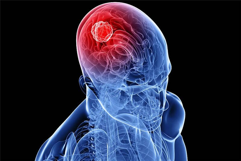
- Brain tumor;
- Nosological forms in which the brain is affected;
- Paroxysmal manifestations, epileptic or seizures associated with other abnormalities;
- Neuroinfections, inflammations, infectious neurotoxicosis;
- Poisoning by neurotoxic poison;
- Diencephalic syndrome;
- Neurosis;
- Degenerative, dysfunctional disorders;
- Comatose;
- Confirmation of brain death;
- Efficacy check and drug dosage selection for epilepsy.
Vibrations of brain activity in many cases occur intermittently, often episodes and outbreaks. Therefore, the longer the EEG record is recorded, the more accurate the results will be.
There are several types of EEG recording:
- Routine EEG is a study that is performed for the first time after examination of the treating doctor. From this stage, the diagnosis of the paroxysmal state begins. The method consists in fast recording( no more than fifteen minutes) of the biological potential of the human brain. Perform photo stimulation( load with the help of frequently flashing LEDs), hyperventilation( frequent breathing).This is necessary to detect any hidden changes.
- EEG with deprivation of night rest( deprivation) - the procedure is carried out according to the appointment of a specialist with insufficient data of the routine EEG.For the procedure with sleep deprivation, the patient needs to wake up a few hours before the EEG, or do not go to bed at all. Everything depends on the age category of the patient and the severity of the illness.
- A long( prolonged) electroencephalogram at which daytime sleep is recorded. The procedure is carried out with the probability of changes in the indicators during the sleep period.
- EEG of the brain at night sleep - the most informative method, a kind of standard. In this study, the sites are marked during waking, sleeping, waking and awakening. If necessary, the process is accompanied by the recording of video( video monitoring) in a dark room and the connection of special sensors( electro-oculogram, respiration recorder, electromyogram, electrocardiogram).
EEG - Video Monitoring
In the diagnosis of epilepsy, the parallel registration of the electroencephalogram and the video recording, where the behavioral activity of the patient is recorded, is very important, this method is called video monitoring. The duration and time( day, night) of the examination is prescribed by the treating doctor, it depends on the type and frequency of paroxysmal conditions, on the age category of the patient. Practice shows that convulsive condition is not always a sign of epilepsy. Sometimes the patient for a long time uses anticonvulsants without an accurate indication of this. To establish the correct diagnosis will help EEG - video monitoring.
How the electroencephalogram
is performed The EEG of the brain is a safe and painless study in which the patient relaxes, lies or sits with his eyes closed. The laboratory assistant wears a special cap, equipped with electrodes( a photo of the brain can be found on the net).
The electrodes are processed by the contact means, and the cap is connected to the registration device. The computer program registers the biological activity of the brain, comparing it with the video.

How the electroencephalogram
is performed A few days after the procedure the specialist issues the results of the study, which shows the brain eEG, which are recorded in the database for use in subsequent patient calls.
REG( rheoencephalography)
For the detection of dangerous pathologies, regulate and eeg of the brain are carried out. REG is a method by which the blood circulation in the brain is evaluated and information is obtained on the state of the vessels, blood circulation, and a specific part of the brain.
Also, re-cephalography helps to determine the viscosity of the blood, evaluate the latent stages, the speed and timing of the flow of blood, calculate the pulse wave speed, the severity of regional vessel reactions.
The procedure is carried out using a special recording apparatus - a rheograph. To conduct research, a person lays on his back, closes his eyes. The electrodes are attached to the head, which are fixed with rubber bands. For better conductivity, a special gel composition is applied to the electrodes. Further on the electrodes pass a weak discharge of current, with the help of which the state of the vessels in the brain is fixed.
The basis of REG is the difference between the scalp and the electrical conductivity of the blood, the change in pulse oscillations causes fluctuations in the electrical conductivity of the analyzed area.
Using REG REGISTRATION
Preparing for an EEG
What is the brain eag, we figured out, but how does it prepare for such a procedure? By prior arrangement with a specialist, it is required to cancel the use of anticonvulsants three days before the study. Hair on the head should be clean, it is not recommended to use various cosmetics for hair( gels, foams, varnishes, etc.).Dreadlocks and braids are required to dissolve, and, directly, before the procedure, you need to remove the earrings.
If the preparation for the brain eag is carried out for the child, then try to convince him that the procedure is safe, take with you the favorite toy of the baby. If he's scared, then practice at home, try to show him the procedure as a game. For a successful outcome of the examination, the baby should be calm, also it should be taken into account that the REG is not made for patients who have a runny nose, cough and other cold manifestations.
Results of the electroencephalogram
Results of the EEG study - a record in the computer's memory or on paper. How to decipher EEG of the brain? The paper fixes the curves, which the expert analyzes. He assesses the rhythm of waves, frequency and amplitude, reveals the elements, their distribution in time and space. Then the data is summarized, described in the conclusion, which is pasted into honey.card.

result of the EEG study
The result of the survey reflects the main characteristics of the electroencephalogram and includes three important sections:
- Typical wave affiliation and activity description. For example: "The alpha rhythm is recorded over the hemispheres. The average value of the amplitude is 58 μV on the right, 57 μV on the left, the frequency is dominated by 8.8 Hz. The occipital leads are dominated by alpha rhythm. "
- Conclusion on the description of EEG and interpretation. For example: "Signs of the irrigation of the middle structures of the brain and cortex. Paroxysmal activity and asymmetry between the hemispheres were not detected. "
- Evaluation of the correspondence of EEG results with clinical symptoms. For example: "There are fluctuations in the functional activity of the brain, which corresponds to the signs of epilepsy."
In the process of decoding, the following characteristics necessarily take into account:
- Spike activity;
- Basal rhythm;
- Changes in the background of tests( hyperventilation, closing and opening of the eyes, photostimulation);
- The level of symmetry of electro-activity of neurons of the right and left hemispheres.
- The final diagnosis is made, given the presence of specific signs that bother the patient.
Echo-EG
Echoencephalography is a brain examination for diagnosis, which is based on the reflection of ultrasound from the anatomical features of the head. ECHO-EG of the brain is an effective and simple method of examination in the process of emergency ambulance, in screening studies, for pre-diagnosis and under normal health conditions.
How is Echo-EG carried out?
Ultrasound without any obstacles gets through the bones of the skull and the external skin of the head, on the boundary between the separation of nerchime and cerebrospinal fluid( liquid and solid substances), the signal is reflected, and this is recorded by the apparatus.
In the absence of brain pathologies, the distance between the median structures and the superficial tissues of the head is the same from all sides. When the disease occurs, this distance changes.
The room for the procedure does not require specialized training. For example, shielding from electromagnetic rays, noise, sound insulation. The study can be carried out outside the walls of honey.in an outpatient setting, only an autonomous power supply is needed to connect the echoencephaloscope. During the examination, the patient is allowed to both lie and sit.
- The doctor, being behind the patient's head, sets the apparatus;
- Studying the history of the disease;
- Palpates and inspects the patient's head, noting the existing deformities of the skull, asymmetry, subcutaneous hematomas;
- A contact agent is applied to the skin of the head to enhance contact.
The examination is carried out in several modes of the apparatus: emission regime, orthogonal projection of the ventricle.
write the question in the form below: