Diagnosis of helminthiases
Diagnosis of bovine tapeworm is based on the finding of segments during defecation.Also conduct macroscopic feces, do anal scraping to detect segments of . Eggs and larvae( oncospheres) are not detected in the study of stool.
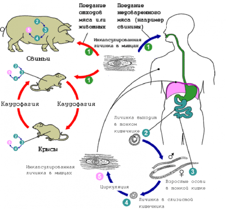
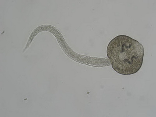
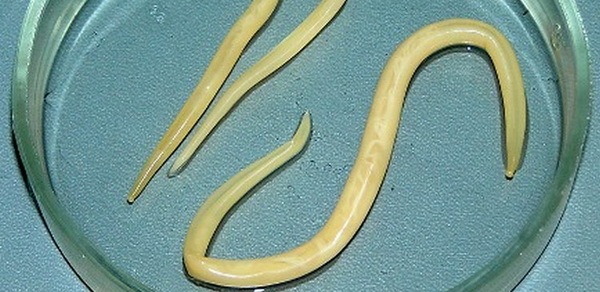
- Pork Chain - up to 3 m long, causes the to shade. An adult parasite lives in the small intestine, the detached segments are excreted with feces and enter the body of intermediate hosts - cats, dogs, pigs.Larvae( cysticerci) develop in meat, when consumed without proper culinary processing, and the person becomes infected.Pig-liver can occur in humans and in the larval stage, causing cysticercosis - flowing with brain damage and eyeballs.
The diagnosis is based on the presence of joints and eggs in the stool.When diagnosing pork thistle cysticerci determine in the eyes of an ophthalmoscope.They are also found in muscle tissue obtained from a biopsy.The study is subjected to cerebrospinal fluid.Relatively specific in the diagnosis of pork chain and the serological reaction of complement fixation.Sometimes cysticerci can also be found on roentgen.
- Dwarf catcher - up to 0.5 cm long. A person becomes infected through surrounding objects.In the gastrointestinal tract, the larva emerges from the egg( cysticercoid).In the intestine within 1-2 weeks it develops into an adult . Mostly sick children.
Diagnosis is made when eggs are detected in the stool.Since the eggs quickly break down in the external environment, stool for the diagnosis of the dwarf chain( as well as other types of worms) should be checked no more than 3 hours after the collection of material.
- Rat aspen - up to half a meter long.Rodents are the ultimate masters.The role of intermediate carriers is carried out by insects( butterflies, beetles, etc.).A person becomes infected when using unbaked flour products . The habitat of the parasite is the intestine .
Diagnosis of rat tartan is performed by examining the stool in which eggs are found.
- Pumpkin Aspen - slightly larger than rat, can reach a length of 0.7 m . The name was obtained because of the shape of the pumpkin seed of the parasite segment.Dogs get sick, a person becomes infected with an accidental ingestion of a flea.The parasite lives in the small intestine, causing dipilidiosis .
The diagnosis and detection of pumpkin tartar was made possible by microscopy of stool.
-
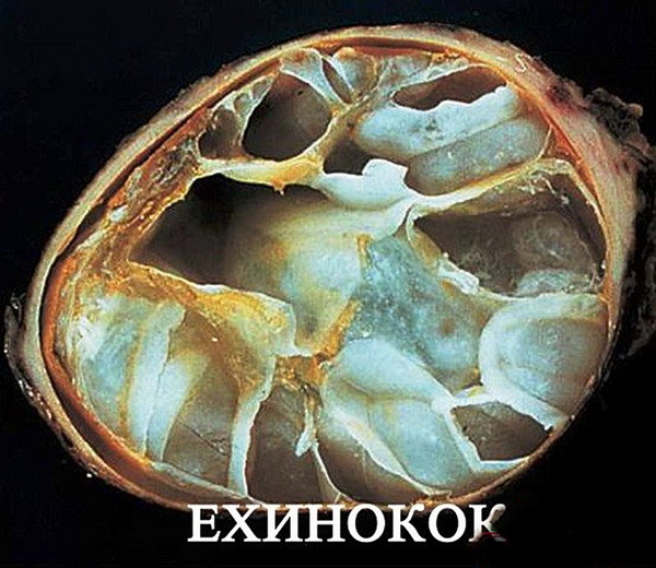 Echinococcus - The length of this small parasite does not exceed 6 mm.The segments of the helminth are secreted into the external environment by its final hosts from the family of catlike, canine.The person( the intermediate host) enters the small intestine through water and food, then the developing larvae penetrate the liver and lungs, but can be introduced into any organ.The disease develops - echinococcosis , anatomically representing a bubble filled with a liquid containing scolexes( larvae).The bubble grows rapidly, then the process slows down and the available formation can increase in size up to 20 years.Bubbles are both single and multiple.Complaints are caused by squeezing the bladder organs and the action of parasite toxins.
Diagnosis of echinococcosis and detection of echinococci is carried out to detect mature forms and eggs in animal faeces.Used in the diagnosis and clinical analysis of blood( it sharply increased eosinophils and ESR), a specific test is also done with the allergen( Casoni).Serological diagnosis of echinococci involves the reaction of latex agglutination.A puncture of the bladder is made, the contents of which are examined microscopically.In the study, scolexes are found.
- Alveococcus - helminthic parasite that causes alveococcosis .It is similar in structure to echinococcus.The final owner - foxes, foxes, dog-like.Intermediate - detachment of rodents.A person becomes infected when working with animal skins, infected water, berries, vegetables.The disease manifests itself in tuberous growths of the liver with growth in other organs.
Diagnosis of alveococcosis and the detection of alveococci are based on epidemiology, clinical picture, serological reactions, biochemistry.
Diagnosis of helminths of the class Trematode
Trematodes ( flukes).This variety of parasites of small size, using suckers for life inside the host.The general group of diseases is called trematodosis .The intermediate hosts of these helminths are mollusks, complementary - fish and crustaceans.The ultimate host is man, pets and some wild species.
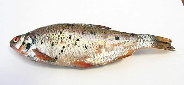
- Opisthorchis( cat litter) is the causative agent of of the opisthorchiasis .Parasite of biliary tract.Eggs of the helminth are excreted into the external environment with feces.The development goes on in reservoirs, where the larvae enter into the organisms of mollusks, then the fishes, when they are used, and the person becomes infected.There is a disease of allergy, stagnation of bile and inflammation of the gallbladder with ducts.
Diagnosis is made when cysts are detected in feces and duodenal contents.When diagnosing opisthorchiasis in a blood test - eosinophilia up to 80%, signs of anemia.
- Clonorhis( Chinese fluke) - calls clonorhoz .In all its manifestations, it resembles opisthorchiasis.
Diagnosis of a clonorhoz involves the examination of feces and contents of the duodenum.
- Hepatic fluke - pathogen of fasciola. Infection occurs when drinking water from water bodies and entering the intestine of water vegetation.Adolescaria( larvae) penetrate the liver.A rare disease that occurs in two phases of - is acute and chronic.The patient complains of a hepatic symptom, there are signs of inflammation of the pancreas and allergies.Perhaps the development of appendicitis, abscesses of the abdominal cavity, damage to the eyes, other organs.
The diagnosis of fascioliasis is based on the analysis of feces and the contents of the bile ducts entering the duodenum.In the material eggs of helminths of hepatic cleavage are determined.
- Rare forms of trematodes.To rare forms of flukes are dicrocemia ( parasite of cattle), pulmonary fluke - inhabiting in addition to humans in a variety of animals, including exotic( leopard, tiger, panther).The causative agent of this helminth is found in sputum.No less interesting are the metadonimus - a helminthic parasite that causes severe form of diarrhea in humans, and the nanofyte is a very small parasite.
In these cases, feces are also used to diagnose helminths and confirm the diagnosis.
- Group with schistosom ( blood flukes).The disease caused by these helminths is called schistosomiasis. Infection occurs when swimming in a reservoir containing larvae.A group of trematodes are: blood fluke, causative agent of intestinal schistosomiasis ( Manson), causative agent of Japanese schistosomiasis ( Katayama disease).
Depending on the type of parasite in the diagnosis of helminthiases, urine, cystoscopy, detection of eggs in stool, and serological methods are used.
Diagnosis of helminths of the group of Roundworms
- Pinworms are the most common helminths that settle in the human body.Call enterobiasis .The length of the parasites is up to 1 cm. They parasitize in the intestine.Life expectancy is up to a month.The female lays eggs around the anus.Complaints these parasites cause only with a large number of individuals.
Diagnosis of enterobiasis is based on the study of scrapings from the anal folds.In the material eggs of helminths are found.Analysis of stool in pinpointing pinworms is poorly informative, since it does not contain helminth eggs.
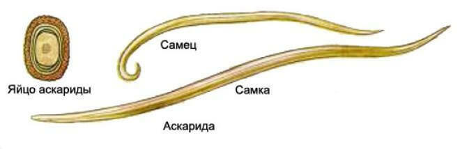
- Ascarids are the causative agents of ascaridosis .Round worms, which can reach a length of 40 cm. Infection occurs through the digestive system.Larvae penetrate into the blood, then into the lungs, the oral cavity and are swallowed by a man, getting into the intestine repeatedly already in the form of developing individuals.He lives ascarid for about a year.In the body causes severe allergic processes, can provoke intestinal obstruction.
Diagnosis of larval forms is almost impossible.With the help of X-ray diagnostics, it is possible to detect eosinophilic infiltrates in the lung tissues.The auxiliary method is RMP( micro-precipitation reaction).It is possible to detect ascarid larvae when examining sputum.When ascariasis is diagnosed in feces analysis, ascaris eggs are determined.
- Vlasoglavy.In the intestine parasitize the vaginal heads causing the trichocephalus .Complaints are nonspecific and are caused by mechanical difficulties of the intestine and allergies.
The diagnosis of trichocephaliasis and the detection of helminth withers are possible with the method of enrichment and isolation of eggs in stool.
- Trichinella - small-sized nematode , inhabiting the small intestinal mucosa of humans and animals , eating meat.The owner is one.Larvae from the wall of the intestine carry blood throughout the body.After 10 days, they penetrate into the muscles and there they are transformed into a capsular form, which can be in a viable condition for several years.There is a disease of allergic phenomena with fever, increased eosinophils, skin rashes and small vessels, hemorrhages in the intestines, muscle soreness.
 Diagnosis of trichinella is based on complaints and clinical manifestations, a study of muscle biopsies( microscopic examination of split muscle fibers for the presence of causative agent capsules).For this purpose, a special microscope-trichinelloscope and compressor are used.The diagnostic method of muscle digestion is widespread, in which the muscles are kept in a thermostat with artificial gastric juice.Under the microscope, larvae of Trichinella are found.In this form of helminthiasis, valuable data are given by serological reactions of precipitation, agglutination.Their only disadvantage is detection 2-3 weeks after infection.
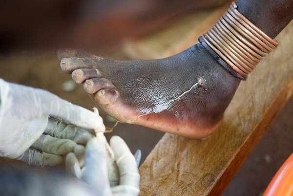
In addition to the round helminths examined, hookworms, strongyloids, filarias, rishty are common.These parasites we will not touch, because they cause diseases quite rare for our sites.
The most common methods for diagnosing helminths
We recommend to read:Among the methods for detecting parasitic forms are:
- Method of thick smear( Kato).Detection of eggs of helminths is improved when using malachite green and glycerin.Found eggs are clearly visible and identified by the type of pathogen.Especially informative is the method for diagnosing ascariasis, lentets, trematodes.
-
Enrichment method.Study of stool in a flotation solution that promotes the concentration of helminth eggs on the surface of the preparation.The formed film is studied microscopically.Several flotation solutions have been developed.It is used for identification of ascarids, vlasoglavov, lentetsov and other parasites.
- The Krasilnikov method( using detergents).Study of stool with the use of detergents - washing powders, which are able to separate the eggs from the stool and concentrate them in the formed sedimentary lump.
- Pararectal scrapings with a wooden spatula.Anal folds carefully scrape off with a spatula, pretreated with glycerin and soda solution, leaving eggs on the spatula.Then a microscopic examination of the scraping is carried out.The method is used in case of suspicion of enterobiasis and teniarinhoz.
- Berman's method is the detection and identification of larvae in feces.After centrifuging the mixture of stool and water, a sediment is examined in which larvae are found.It is used in the diagnosis of strongyloidiasis;
- Method( Harada-Mori) - cultivation on filter paper.This way you can find the larvae of the filaria.
- A method for studying the contents of the duodenum.In the process of duodenal probing, flakes are isolated from the secreted bile and, after mixing them with ether, they are centrifuged.The sediment is examined microscopically.The method is informative for opisthorchiasis, clonorchosis and other types of parasites.
Lotin Alexander