Ultrasound of the abdominal cavity organs: indications, preparation, interpretation of results
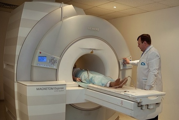
Ultrasound( short for ultrasound) is an informative, safe and relatively inexpensive method for diagnosing internal diseases in adults and children.It is based on the interaction of tissues with ultrasonic waves, which with the help of special programs is transformed into an image on the monitor of the ultrasound machine.
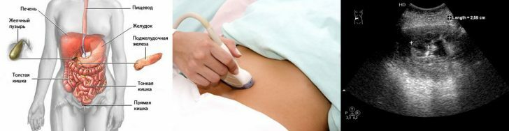
One of the areas of application of ultrasound diagnostics is the study of the state of organs located in the abdominal cavity. In particular, ultrasound can quickly and absolutely painlessly examine the liver, gallbladder, pancreas, spleen, bile duct .If necessary, you can even consider the lymph nodes, assess whether there is a free fluid in the abdominal cavity. Kidneys, , despite their retroperitoneal placement, are also being examined in conjunction with the aforementioned organs.
It is impossible to fully examine with the help of ultrasound scanning only the stomach and intestines.These structures of the digestive tract are filled with air, which is an obstacle to ultrasonic waves and a hindrance to ultrasound.
Table of contents: What shows ultrasound of the abdominal organs Indications for ultrasound Preparation for ultrasound of the abdominal cavity How does the procedureWhat does ultrasound of the abdominal cavity
Ultrasonic diagnosis gives the physician the following information about the organs under study:
- location detail;
- dimensions;
- form;
- the condition of the walls( this is important for the gallbladder and bile ducts);
- features parenchyma structure( homogeneous or not, are there any pathological inclusions, etc.);
- presence of concrements( stones) and neoplasms;
- condition of large veins( inferior hollow, splenic, portal).
Indications for ultrasound
The following symptoms and complaints of the patient may be indicative of ultrasound of the abdominal organs:
-
 pain and heaviness in the abdomen;
 - nausea, vomiting;
- stool disorder;
- flatulence;
- yellowness of skin and mucous membranes;
- belching;
- heartburn;
- change in biochemical blood test;
- general examination data indicating that there is a pathological formation in the abdominal cavity.
The organs of the abdominal cavity are always examined in a complex, because they are functionally related to each other - often pathological changes in one are reflected in the state of the others.Therefore, for example, it is inappropriate to perform targeted ultrasound examination of the liver, and do not pay attention to the condition of the pancreas or gall bladder.
Preparing for ultrasound of the abdominal organs
The reliability of the results depends on how a person prepares for this study.
The main obstacle for ultrasonic waves is air( ultrasonic waves are reflected from it), therefore at the time of examination in the intestine of the patient should be as little air as possible. Otherwise, the swollen loops of the intestines will block the abdominal organs and the doctor will not see anything.The second important condition for successful ultrasound diagnosis is an empty stomach of .In particular, with a full stomach, a specialist simply can not examine the pancreas.
To prevent the formation of gases in the intestines and to clear it of stagnant stools( this is especially true for patients suffering from constipation), it is necessary:
- For 2-3 days before the study, adhere to a special diet.Food should be light - lean meat, cereals, heat-treated vegetables, dairy products.It is desirable to abandon legumes, cabbage, muffins, carbonated drinks, fresh vegetables and fruits, alcohol.
- Take drugs that reduce gas production, as well as enzymes, if there is any indication for this.
- Eliminate constipation( perhaps it can be done thanks to a diet).
More details on the preparation for the study is best consulted with a doctor who directs to ultrasound.He also will advise preparations against flatulence and constipation.
As for the filling of the stomach with food, the principle is as follows: the last meal should be no later than 6-8 hours before the study.For example, if the procedure is scheduled for the morning, dinner should not be late.If ultrasound is in the evening, you can eat a light breakfast in the morning.Immediately before the procedure, you can not drink anything, chew gum, smoke.
How the procedure passes
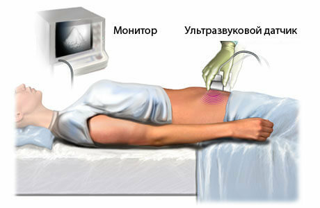
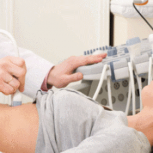
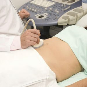
During the study, the patient usually lies on his back.The doctor may ask him to take a deep breath, exhale, turn on his side, sit down.All this is necessary for better visualization of this or that organ.In addition, for better carrying out ultrasonic waves, a special gel is applied to the human body.
Ultrasound examination of the abdominal cavity lasts an average of 15-20 minutes. The patient usually does not feel discomfort, painful sensations during ultrasound. Normally carry ultrasound and children.
All the results obtained during the study, the doctor displays in the protocol.At the end of the protocol, a conclusion is given, the interpretation of which should be handled by a doctor who sent to ultrasound.
Important: can not be diagnosed accurately solely from data obtained during ultrasonic scanning.This study provides guidelines for further diagnostic search( for example, that there is an inflammatory process, that there is some kind of neoplasm, that there are stones in the gallbladder, etc.).Therefore, it is a mistake to believe that ultrasound can diagnose cancer, cirrhosis, viral hepatitis.
Zubkova Olga Sergeevna, medical reviewer, epidemiologist-doctor