The anomaly of Ebstein: causes, symptoms and treatment
 To the rarest congenital heart defects in children without an intracardiac shunt and cyanosis is the anomaly of Epstein. Little is the probability that a child who has such a disease will live to an adult status. The difficulty in diagnosing the Epstein anomaly lies in the different types of defect( there are only 5 of them), which are limited by the number of observations.
Anomaly of Epstein.
This disease is rare. According to statistics, from all children with congenital heart diseases, such a disease occurred in 0,3 - 0,7%, or for 20,000 newborns, only 1 case. Be that as it may, the disease is often a congenital heart disease that affects the tricuspid valve, up to about 40% of cases. There are cases when mothers take medication, with lithium, which contributes to high frequency of malformation. The relatives have a low incidence of disease and the disease is not genetically enough.
Exceptionally poor prognosis in children under 3 days of age who have a natural course of the disease. A slightly different situation in children who survived with the disease the first 3-6 months. Only 70% of sick children are able to survive the first 2 years of life, and 50% usually live up to 13 years.
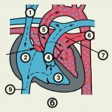
The cause of the disease is the displacement of the tricuspid valve in the right ventricle, which was due to the incorrect attachment of ventricular valves. In this connection, an abnormal right atrioventricular orifice appears. It divides the right ventricle into the distal part and into the proximal( thin walls) part. The distal part becomes a small gastric chamber.
The apical part of the ventricle is small, and the atrioventricular pathological right orifice is of small or normal size. Sometimes the valves can grow together so that they are difficult to differentiate, but in many cases they are blown and deformed along the walls of the right ventricle. The results of the Epstein anomaly show the development of severe tricuspid regurgitation, and in some cases stenosis of the right atrioventricular orifice.
Epstein's anomaly can often be combined with congenital heart defects and other abnormalities: aortic hypoplasia, corrected transposition of the main vessels, an open oval window or an interventricular septal defect, an open arterial duct, and coarctation of the aorta. With Epstein's anomaly, some diseases may occur, most often pericarditis.
In older age, the symptoms of the disease are often: right ventricular failure, shortness of breath, cyanosis, fatigue. Epstein's anomaly is often combined with WPW syndrome. The initial manifestation of the disease is supraventricular tachycardia, which occurred in 25% of cases. A characteristic auscultatory pattern is a clapping prolapse valve that produces multiple clicks.
Signs for electrocardiography:
- syndrome WPW( delta wave),
- right atrial enlargement( prolonged PQ interval and high-amplitude P wave that exceeds the QRS amplitude in the right thoracic leads),
- displacement of the electric axis to the right.
At the expense of X-ray examination, doctors reveal a small right ventricle and an increase in the right atrium.
It's strange, but in many adult sick patients it is very difficult to recognize the defect, although the current surgical correction( implantation of the prosthesis, valve-preserving surgery) and diagnostics could quite extend the life span of the sick person.
Such misdiagnosis can be for two reasons: the variety of clinical options and insufficient information for physicians with a common practice on this defect. There was a case when the patient with an anomaly of Epstein was diagnosed with a defect of the mitral valve. After the patient was delivered in the terminal state due to decompensation of heart failure along a huge circle of blood circulation with jaundice and hepatosplenomegaly. Doctors gave the patient a new diagnosis - cirrhosis of the liver. It was established when Echo KG was conducted.
Due to the identification of other causes, it is necessary to carry out a differential diagnosis with tricuspid regurgitation. The causes can be: myxoma of the right atrium, rheumatic lesion of the tricuspid valve, prolapse of the tricuspid valve as a result of hereditary connective tissue diseases, right ventricular infarction with papillomavirus rupture, infective endocarditis, tricuspid insufficiency formation, carcinoid syndrome.
Itechocardiography can identify the causes of congenital heart disease: the fusion of the septal leaflet to the wall, the displacement of the tricuspid valve into the cavity unit ventricular dilation of the right ventricle and the right atrium, a large amplitude movements of the leaf, the correct attachment to the fibrous ring of the anterior leaflet, tricuspid regurgitation.
its color Doppler investigation can detect pulsation of the renal veins, the reverse current in the lower systolic and superior vena cava, and determine the depth of penetration into the right atrium regurgitant jet. The systolic pressure of the pulmonary artery of the regurgitating jet can be calculated at the maximum rate.
Another method of surgical treatment will be the principles of therapy. Usually therapy venous vasodilators and diuretics amenable to tricuspid insufficiency. Here nitrates sent inside, like patches, as well as cardiac glycosides for atrial fibrillation and ACE.Antiarrhythmic treatment is used in the manifestation of orthodromic tachycardia. Intravenously administered inotropic agent( dobutamine more), if there is severe disfunction and refractory tricuspid insufficiency.
The main purpose of the doctors is to identify as early as possible in the patient Epstein anomaly and pass it to heart surgeon, so he spent a timely surgical treatment.
The results of the surgical treatment of Epstein's anomaly can be either good remotely or directly. Therefore, they are expected in 90% of operated patients.