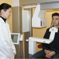
Computer tomography of the jaw

In terms of technical equipment in dentistry, it is worth noting that a significant leap forward is the introduction in the practice of dentists the use of computer tomographs. To a large extent, this tomography of the jaw simplified the work of dentists, thereby increasing the quality of treatment.
What is this computerized tomography
? This jaw study provides maximum diagnostic information in just one scan. As a result of making a panoramic shot, one picture is obtained from which insufficient information is obtained. And the patient has to do a second X-ray examination. As a rule, it is a waste of time, money and health after irradiation. A tomography of the jaw makes the diagnosis safe and accurate, and treatment - more effective. The load in this study is similar to the load when doing a panoramic scan. However, the dentist receives much more information for diagnosis.
Computed tomography of the jaws is of fundamental importance for diagnosis, if necessary, implantation with complete absence of teeth, and with partial absence. With this tomography, you can get reliable and complete information about the height, width and shape of the alveolar process in the zone of the upper jaw and the alveolar part in the area of the lower jaw.
What are the indications for computed tomography of the jaw
Apply this tomography of the jaw in the following cases. This pathology of ENT organs;Trauma and damage to the jaws, facial bones, teeth, skull;Diseases of the maxillary sinuses;Anomalies in development, the position of teeth and jaws;Diseases and injuries of temporomandibular joints;Diagnosis of cysts. Also detection of hidden carious areas;Planning of surgical interventions;Complications due to intracanal treatment;Planning orthodontic, orthopedic, therapeutic treatment;Neoplasms of bones and soft tissues in the maxillofacial area.
What are the advantages of computed tomography of the jaw
Computed tomography is an X-ray imaging, which allows you to obtain a three-dimensional image of the desired zone.
A three-dimensional snapshot increases the informative value of the study and makes it possible to plan the treatment more efficiently. Such diagnostics are needed for reliable analysis of various anatomical structures, including bone structure. This study provides an opportunity to assess exactly the most difficult clinical situation. Computer tomography makes the risks in the operation minimal.
With the implantation of the teeth, tomography makes it possible to obtain an unlimited number of sections, and the thickness and height of the bone tissue while the distance from the nerve of the lower jaw of the future implant is determined with sufficient accuracy. It becomes possible to select the type and size of the implant quite accurately, taking into account the characteristics of the patient.
To detect pathological changes in the early stages and to reveal all the features of the maxillofacial area allows the dentist to clear the image.
A low radial load is applied to the patient during computed tomography. Scanning the jaw is done in just one time, taking up no more than a minute. The results of the research can be written to disk and analyzed on a computer.
With the help of a special program, using a ready shot, the dentist can simulate any situation, in other words, arrange the implants before carrying out the implantation and analyze the restoration features.
Perform tooth implantation with a significant atrophy of the lower jaw. Before carrying out this operation, computed tomography of the lower jaw makes it possible to determine with accuracy the parts of the trigeminal nerve of the jaw( lingual, chin, alveolar).Careful planning of surgery based on the results of computed tomography of the jaw, qualified manipulation to a minimum reduces the risk of nerve damage.
There are practically no contraindications for CT.An exception is pregnancy. With caution, you need to conduct such a study for people with diabetes and serious kidney disease.


