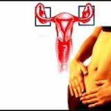
Leiomyoma of the uterus
 uterine leiomyoma is a nodular structure formed by the muscle fibers of the uterine wall with some signs of a benign tumor. The predominance of muscle tissue in the tumor explains its other name - "myoma", and since the uterine leiomyoma also has fibrous tissue, the name "fibromyoma" is often used. All these definitions are a reliable diagnosis, since, in fact, point to one nosology.
The uterine leiomyoma is diagnosed in almost every third gynecological pathology patient at the age of 20-40 years. The term "tumor" with respect to leiomyoma is rather conditional, since the formation of a true tumor is not, but only possesses some of its signs.
Important characteristics of a leiomyoma are:
- good quality: education is not malignant;
- hormone dependence: "behavior" of the tumor is largely determined by estrogens;
- ability to self regression: uterine leiomyoma can completely disappear without any external intervention.
A bit of anatomy .The unique structure of the uterus allows her not only to sustain a developing fetus for a long time, but also to "push" it out at birth to the outside. During pregnancy, it increases significantly, and then, when the child leaves the womb, returns to its original size. Similar changes are provided by the myometrium - a powerful muscle layer within the uterine wall. Myometrium is formed by muscle fibers of several types. They are intertwined, laid by radial layers and twisted into a spiral, forming an extremely strong frame reinforced with elastic fibers and connective tissue. The main function of the myometrium is reduced to contractile movements, during the period of menstruation, they help the contents of the uterus to be evacuated outside, and after the pregnancy is pushed out the fruit.
The uterine wall has two more layers. Outside the myometrium is a perimeter - a dense protective serosa, similar in structure to the peritoneum. The inner uterine layer, the endometrium, is formed by cells of multilayer epithelium, which is constantly updated according to the phases of the cycle. The processes occurring in the endometrium are directly controlled by the hormones of the ovaries.
Thus, the myometrium is the middle layer of the uterine wall. The source of leiomyoma development is its muscular and connective tissue structures. Nodal leiomyoma of the uterus is the presence of a single or several nodes in the myometrium. If the node is not one, the leiomyoma is classified as multiple. Often, all available nodes differ in size and structure, since they have different "ages".
The dimensions of the nodes and their localization largely determine the clinical manifestations of pathology. There are cases when a small leiomyoma node in a patient is diagnosed completely by chance, because it does not manifest clinically and does not affect her health. Such tumors can asymptomatically exist for years, without changing the size and location.
There are no typical clinical manifestations of uterine leiomyoma. Its symptoms are similar to many gynecological ailments, so a reliable diagnosis is possible only after ultrasound scanning. In a small( 2%) part of the examined, the leiomyoma is detected only through diagnostic hysteroscopy.
Leiomyoma therapy is not always performed. Small asymptomatic nodules, especially those entering into menopause of patients who do not tend to increase and grow, can be observed.
To choose the right therapeutic tactics, it is necessary to find out the cause of leiomyoma and to work on it, since the usual removal of nodes inevitably ends with the formation of new ones.
Unfortunately, leiomyomas are able to "return".Relapses are associated with unresolved causes of pathology.
Uterine fibroids: what is it?
Since the most common form of leiomyoma is a node, let's talk about the mechanism of its formation and variants of development.
As already mentioned, the nodal leiomyoma of the uterus is a delimited formation of the myometrium, characterized by the greatest density and ability to grow. If the node does not grow or grows very slowly, it remains "in place" for a long time. When the myomatous node begins to increase, its inevitable shift to other layers of the uterine wall occurs. According to the site localization, the following are allocated:
• Intramural uterine leiomyoma is the nodes located within the boundaries of the myometrium;
• Subserosome leiomyoma of the uterus - these are the nodes of the subperitoneal localization;
• Submucous uterine leiomyoma are submucosal nodes.
Regardless of the final location, any leiomyoma is initially formed in the thickness of the muscle layer. The development of leiomyoma is stage-by-stage. First, near the small vessels, smooth muscle and fibrous fibers begin to expand rapidly - the stage of knot formation begins. He has not yet formed and does not "declare himself" clinically.
Then comes the stage of maturation, it is inherent in the process of active growth of the leiomyoma, when a small "glomerulus" forms in the place of intensive growth of the muscle fiber, it gradually thickens and increases. When a kind of "capsule" is formed around the "glomerulus" from the elements of the surrounding tissues, it becomes like a delimited knot. The most intensive growth of leiomyoma is at this stage of development. As a result, the tumor acquires "adult" traits, is well visualized during examination and can provoke an active clinic.
The stage of "aging" of the leiomyoma occurs against the background of dystrophic processes in its tissues. At this stage, the node no longer increases, in some cases even a decrease is noted.
Leiomyoma in each case has its own peculiarities, it develops, grows and even "ages" not in all patients unequivocally.
Causes of development of uterine leiomyoma
The ability of leiomyoma to appear against the background of prolonged hormonal dysfunction and to undergo a regression in menopause, certainly indicates the hormonal nature of the disease. However, not every hormonal disorder has a patient with a leiomyoma, so they do not speak of reliable causes of its development, but of predisposing factors.
It is believed that the growth of the nodes of the leiomyoma can occur according to the three main pathogenetic variants - central, uterine and ovarian.
1. Variant central
Ovarian hormones affect all processes in the uterus. The ovaries, in turn, are "controlled" by the central structures - the hypothalamus and pituitary gland. Hormones of the pituitary gland( FSH and LH) directly affect folliculogenesis and ovulation processes. Any circumstances leading to a disruption of the function of the brain regions where the "governing" organs are located lead to a disorder of the ovarian function. To such it is possible to carry the expressed psychoemotional and vascular disorders, traumas.
2. Ovarian, "classic" version of
The ovarian function is distorted in case of prolonged inflammatory process( salpingitis, salpingoophoritis), cystic degeneration and similar conditions that change the normal ovarian function. Not only the quantitative secretion of estrogens and progesterone changes, but also their proper correlation. This option is more common than others.
3. The uterine version of
Leiomyoma can appear against the background of normal ovarian function, when estrogens and progesterone are secreted in due rhythm and quantity, but the uterus does not perceive them due to damage to the receptors. This can occur with mechanical damage to the epithelium during scraping or other traumatic procedures.
The function of the ovaries is closely integrated into the endocrine system, so some extragenital ailments( diabetes mellitus, thyroid pathology and the like) can also provoke hormonal dysfunction.
None of the above reasons is not mandatory for the appearance of uterine leiomyoma, they are considered only as possible. It is important to establish why a node appears in one situation, and in another, if there are similar conditions, it does not exist, until it succeeds.
Symptoms and signs of uterine leiomyoma
The localization of the node is of paramount importance with respect to the severity of leiomyoma symptoms, and its magnitude is considered a secondary factor. Interstitially located nodes of small diameter can develop for a long time without a pronounced clinic, but the submucosal, submucous leiomyoma even with small dimensions "makes itself felt" vividly.
Despite the variety of clinical and topographic options, leiomyomies are inherent:
• Menstrual irregularities, often hyperpolymenorrhea.
Menstruation becomes lingering, and blood loss can become significant. At the initial stages of knot formation, menstrual dysfunction is stopped with haemostatic drugs, so patients are not in a hurry to receive. When the nodes become "adults", menstrual dysfunction increases: bleeding is transformed into acyclic, and the amount of blood lost exceeds all permissible norms.
• Pain.
The severity of the pain syndrome is determined by both the magnitude and localization of the leiomyoma. Large leiomyomas do not allow the uterine wall to "shrink" correctly, so pain occurs. The most pronounced are pains with subserous localization of the nodes, and for their intramural location, there are non-bright, aching sensations.
If in the structure of the node there are trophic disorders on the basis of vascular disorders, and in its thickness there are signs of necrosis, the pain syndrome acquires the features of an "acute abdomen".
Irradiation of pain in adjacent areas occurs in situations where the uterus enlarged by leiomyoma compresses nerve fibers that innervate adjacent structures.
• Distortion of normal functioning of neighboring organs.
Dysfunction of the bladder and / or rectum is more pronounced, the larger the size of the uterus. The physiologically located uterus is tilted anteriorly, so urinary disorders are more common.
• Reproductive dysfunction.
Infertility in leiomyomas can be both primary and secondary. To assert that the leiomyoma directly provokes female infertility is incorrect. More often the cause of both pathologies is hormonal dysfunction, which distorts the process of ovulation.
The secondary character of infertility in leiomyoma is more often associated with deformation of the uterine cavity by large nodes or by the "unsuccessful" arrangement of nodes of any size, for example, in the uterine corner.
Uncomfortable is the combination of uterine leiomyoma and pregnancy. More often the presence in the pregnant uterus of the leiomyoma provokes a threat of premature interruption of gestation. Statistically, most pregnancies against the background of leiomyoma have no negative consequences.
What are the uterine leiomyomas?
Above, already listed variants of leiomyom, but we should dwell on each of them in more detail.
Leiomyoma is an education with a predominantly nodular form. A node of a leiomyoma may be the only one, but the variant of multiple nodes is more common, and all of them not only have different topographies, but also differ in "age", when formation processes begin in some, and in others signs of "aging" are already found. Nodes can appear almost simultaneously, and can be formed one after another with different time intervals.
It should be remembered that the "birthplace" of any myomatous node is the muscle layer. Initially, the node that originated there begins to not only increase, but also penetrate into adjacent layers, so as a result, its localization changes.
Intramural leiomyoma of the uterus is a knot of muscular and connective tissue that grows exclusively within the myometrium. Leiomyoma of such localization does not provoke a menstrual cycle, but it affects the character of menstrual bleeding: the knot prevents the muscle from contracting properly and "throwing out" the contents of the uterus outward, so the bleeding increases.
It is considered as the most clinically favorable option, as it rarely provokes serious consequences.
The subserous leiomyoma of the uterus is a knot formed initially in the myometrium, growing toward the outer, serous, envelope. Arranged subperitoneal nodes rarely provoke menstrual dysfunction, but constant aching pains such topography of nodes causes almost always. The subserous node of the leiomyoma becomes in a situation when the knot formed initially on the border with the perimeter is beginning to grow and, accordingly, move towards the peritoneum.
These knots have a thinner base, so when they grow and "move forward" under the serosa, this base stretches out and becomes like a "leg".
Suberosome leiomyomas rarely become large, grow extremely slowly, and in most cases are diagnosed accidentally.
Submucous leiomyoma of the uterus is a node growing toward the uterine cavity( under the mucous membrane).This localization is most clinically and prognostically unfavorable. As the node grows, the knot protrudes into the uterine cavity and deforms it. The uterus perceives the leiomyoma as something alien and tries to get rid of it by intensive cuts, like a generic process when it pushes out the baby. There are pains, especially pronounced during menstruation, and bleeding.
If the uterus shrinks too much, it manages to "push out" the submucous leiomyomia, with pains and dangerous bleeding reminiscent of strong contractions. In such a situation, one speaks of the "birth" of a submucosal node - a condition that is life-threatening.
The described species of leiomyoma are among the most common, among the infrequently occurring forms there is cervical leiomyoma, as well as retroperitoneal and inter-connective.
By nature of node growth, leiomyomas are classified into:
- simple: slow-growing, less well-defined, more often single;
- proliferating: single large nodes or a few small ones, grow rapidly, provoke a bright symptomatology.
Diagnosis of uterine leiomyoma
Based on the patient's complaints, the presence of leiomyoma can only be assumed. Situations when pathology is diagnosed accidentally are not uncommon.
In a bimanual( two-hand) study, you can determine the increase in the size of the uterus against the background of a change in its consistency( it becomes dense).Sometimes it is possible to palpate an uneven uterine contour, deformed by the node. As a rule, the uterus "grows" after leiomyoma, so the dynamics of its magnitude change serves as an important diagnostic criterion.
Dimensions of the uterus in the presence of leiomyoma are evaluated similarly to those in pregnancy - that is, "in weeks".The day of the cycle is chosen and palpation is performed on that day. If the uterus has not grown more than four weeks over the past year, tumor growth is estimated as slow.
When viewed in mirrors, submucosal nodes are sometimes visualized if they "protrude" toward the neck. To clarify what was seen during routine examination, colposcopy is recommended.
The most reliable in the study is the result of ultrasound scanning. It allows:
- to "see" the nodes, determine their number and topography;
- determine the structure and "age" of the leiomyoma;
- assess the type of tumor growth;
- to detect concomitant changes in the endometrium - hyperplastic transformation, inflammatory changes and others.
- to study the condition of the ovaries, determine the nature of folliculogenesis.
Laboratory diagnostics helps to establish the cause of leiomyoma. Swabs and crops are "flora" and oncocytology, blood biochemistry and hormonal examination.
Submucosal leiomyomas are well visualized by metrosalpingography( MSG) or hysteroscopy.
The list of modern diagnostic capabilities is very large, so for each specific situation is selected individually.
Treatment of uterine leiomyoma
There is an erroneous opinion among patients about the inevitability of operative treatment of leiomyoma. There are no universal therapeutic methods for leiomyoma. Meanwhile, not every leiomyoma in general requires treatment. Situations when a randomly detected small asymptomatic nodule of a leiomyoma regresses without medical involvement, are often found, especially among patients entering menopause.
The ability of uterine leiomyoma to regress allows it to be treated conservatively. The decision is made only after studying the features of the pathology, ascertaining its causes and influencing the reproductive capabilities of the patient. Avoid surgery is possible under the following conditions:
- small( up to 3 cm) leiomyoma;
- when the uterus does not exceed the size of a 12-week pregnancy;
- asymptomatic or asymptomatic flow;
- the need to realize reproductive function;
- intramural or subserosal localization in combination with a wide base of nodes;
- if the patient categorically insists on non-surgical treatment( as a rule, it is ultimately unsuccessful).
The conservative technique of leiomyoma treatment should stop the growth of existing nodes, prevent the formation of new ones and eliminate negative consequences.
Removing uterine leiomyoma, whether it is conservative or radical, does not mean a cure. If the cause of the pathology is not eliminated, the nodes will be formed again. Therefore, you should not treat the leiomyoma itself, but its cause. Since almost always at the origins of pathology there is hormonal dysfunction, therapy is directed to its correction.
Hormonotherapy involves a treatment plan that is individual in content and duration. The goal is to eliminate excessive estrogenic effects. With a saved menstrual rhythm, it is achieved with the use of gestagens containing preparations( Dufaston, Utrozestan, Progesterone and analogues).At expressed acyclic bleedings it is expedient to use estrogen - gestagenic agents( Zhanin, Yarina and analogues).
In the treatment it is also possible to use an intrauterine device containing levonorgestrel( gestagen).The hormone is "ejected" into the uterine cavity in portions, simulating the oral route of the drug.
Hormonotherapy is always combined with anti-anemic, sedative, vitamin and improves metabolism.
The duration of therapy is set individually, but it always exceeds the 6-month course. The results of treatment should be monitored by ultrasound scanning, it is performed every three months and the treatment is adjusted according to its results.
uterine leiomyoma: surgery to remove
Surgical treatment of leiomyoma is not carried out without adequate clear indications. More often such are:
- large sizes of the uterus and / or its excessive growth;
- submucous nodes;
- destructive processes in the node and its necrosis;
is a subserous leiomyoma with severe symptoms;
- torsion of the "leg" of the node of the leiomyoma;
- leiomyoma in the cervix;
is a pronounced accompanying hyperplastic process;
- inefficiency of conservative therapy.
To eliminate the leiomyoma, it is not necessary to completely remove the uterus. Modern technologies allow and save the organ, and cut the knot. These include myomectomy - the removal of the leiomyoma by "harvesting" it. Used in situations where it is possible to preserve the entire uterus.
If you leave the entire uterus is not possible, remove only the "affected" area( bottom) together with the leiomyoma - defundation.
In 80% of patients who underwent sparing surgery menstrual function is restored completely, and after a year they are allowed to become pregnant.
In recent years, the technique of uterine artery embolization is gaining popularity. The essence of the technique is quite simple - if you stop eating leiomyoma, it will stop its development, and the nodes will begin to decrease. Since feeding nodes provide uterine arteries, their artificial occlusion( blockage) is made to stop blood flow.
Another innovative method for the removal of leiomyoma is FUS ablation. The focused ultrasonic wave is directed to the node and "evaporates" its cells. Since the technique refers to remote, it is rightly called an "operation without surgery."
Removal of the uterus together with myoma( hysterectomy) is indicated in the absence of the opportunity to eliminate pathology in a more gentle way. The most justified in the climacteric and menopause.
It should be reiterated that it is important not only to remove leiomyoma in surgery. The chosen technique of therapy should exclude the possibility of relapse of the ailment, otherwise there is no sense in the operation. Therefore, if the result of the operation is only the removal of the leiomyoma while preserving the organ, an anti-relapse treatment begins, aimed at eliminating the cause of the leiomyoma.
Prevention of uterine leiomyoma
Reliable causes of leiomyoma are not yet established, but predisposing factors have been studied in sufficient detail. Therefore, preventive measures are reduced to eliminating these factors.
It is well known that leiomyoma is accompanied more often by inflammatory processes( salpingoophoritis, endometritis) and hormonal dysfunction. Timely correction of these disorders reduces the risk of the appearance of leiomyoma.
Often, receiving a conclusion about the presence of leiomyoma, the patient is frightened, especially when the disease is called a "tumor".To correctly treat this pathology, you should know:
- Most leiomyomas are diagnosed accidentally. They are small, asymptomatic, do not tend to increase and are not even subject to serious therapy.
- Cases when small "young" leiomyomas disappeared on their own, are often not considered "miraculous": apparently, the node began to form under the influence of some temporary provoking factor, and after its disappearance, the leiomyoma regressed.
- Most of the uterine leiomyoma is diagnosed at an early stage. Modern diagnostics allows you to detect the process even before the formation of the node, when only a portion of increased density appears in the muscle layer. And this, in turn, allows to conduct a full-fledged examination and begin early therapy.
- As a hormone-dependent tumor, the leiomyi regresses on the background of extinction of the ovarian function in the menopause.