Ultrasound of the heart: preparation, testimony that can be identified
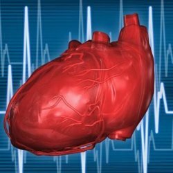
 Heart ultrasound( or echocardiography) is a real-time heart examination using ultrasound. This diagnostic method allows you to assess the status and work of the heart muscle, measure the size of the heart chambers and the thickness of their walls, inspect the valve apparatus, determine the speed and direction of blood flow inside the heart cavities.
Echocardiography can be carried out to patients of any age - from the period of the newborn to the deep old age of .Ultrasound examination of the heart does not harm, but it gives cardiologists the information that is extremely important for revealing cardiac pathologies.
Of course, on the basis of echocardiography alone, an accurate diagnosis can not be made.This study does not allow to assess the state of the coronary arteries and the conduction system of the heart, therefore patients usually have an ECG and coronarography( if there are indications) besides ultrasound.
Contents: What will the heart ultrasound show?Indications for ultrasound of the heart Preparation for ultrasound of the heart How do ultrasound of the heartWhat will show ultrasound of the heart?
Modern ultrasonic devices allow you to explore the heart in various modes: one-dimensional M-mode, two-dimensional, three-dimensional and using a doppler.It is the Doppler that allows evaluation of intracardiac blood flow.The remaining regimes provide information on the movement of the valves and walls of the heart, their condition, size, as well as congenital anatomical features of the organ under investigation.
During the ultrasound of the heart with doppler the doctor necessarily determines the following parameters:
-
 Dimensions of the heart chambers.
 - Thickness of the walls of the heart and intracardiac septums.
- Diameter of the root of the aorta.
- Shock volume( the amount of blood ejected by the left ventricle for one systole).
- Ejection fraction( shows in percentage that part of the blood that the left ventricle during its contraction is able to push into the aorta).
- Blood flow rate through the heart valves in diastole and systole.
Evaluation of this information enables an experienced specialist to suspect( and in some cases, to state):
- Cardiac defects( they can not be detected without an ultrasound of the heart with a doppler).Disturbance of contractility of the myocardium.
- Expansion of cardiac cavities( dilatation).
- Thickening or thinning of the walls of the heart( hypertrophy or hypotrophy).
- Valve valve dysfunction.
- Aneurysms.
- Thrombosis of the heart chambers.
- Violation of the function of the left or right ventricle.
- Pathological changes from the pericardial cavity( eg, the presence of fluid in the pericardium).
Indications for ultrasound of the heart
Indications for an ultrasound of the heart are:
-
 Noises over the heart, found during auscultation.
 - Complaints of chest pain.
- Presence of signs of heart failure( edema, dyspnea, cough, wheezing in the lungs, enlargement of the liver, etc.).
- Dizziness, frequent loss of consciousness, chronic headaches( these symptoms are characteristic for the violation of cerebral circulation - a condition that has a direct connection with heart disease).
- Deviations in the results of the ECG.
- Pathological changes in the heart, identified by radiography.
- Chest trauma.
- Deep vein thrombosis( with this disease, thrombi can penetrate the chambers of the heart, which is fraught with life-threatening consequences for the patient).
- Dynamic monitoring of patients suffering from heart defects, coronary heart disease, cardiomyopathies and other cardiac ailments.
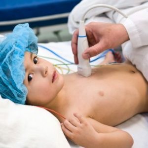
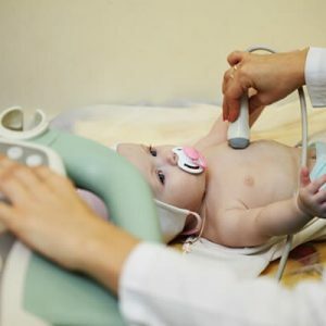
For young children, echocardiography is recommended in the following cases:
- If the pediatrician has detected pathological noises above the baby's chest.
- If even in utero, there have been some deviations in the fetal heart structure.
- If the baby is not sucking on the chest or pacifier, quickly gets tired, badly gaining weight.
- If a child has a nasolabial triangle blue during any physical activity( eg sucking, crying).
- If the relatives of the newborn have or have serious heart defects.
Preparation for ultrasound of the heart
 This study does not require any special preparation.It can be carried out at any time of the day, without being tied to food intake.
For the results of the heart ultrasound to be accurate immediately before the procedure, it is not advisable to take any cardiac medicines( this item should be clarified with your doctor), soothing or otherwise stimulating drugs, drinking coffee, strong tea, drinking alcohol, smoking.It is also recommended to avoid physical exertion and necessarily rest before research, if you had to go fast or climb the stairs.
How to do ultrasound of the heart
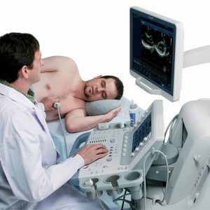
For performing ultrasound of the heart the patient should remove all clothes from the upper body and lie on the couch.The doctor may ask the examinee to turn on one or the other side to install the ultrasound probe on certain points in which it is possible to obtain a representation of the heart in a certain projection.
This examination lasts for an average of 20-30 minutes.All received data together with the ultrasound diagnosis of the doctor are recorded in the protocol.The decoding of this information is subsequently handled by the cardiologist.
Zubkova Olga Sergeevna, medical reviewer, epidemiologist-doctor