Tumors of the chest wall
 Breast wall neoplasms are one of the least researched areas in clinical oncology. Due to the fact that the anatomical structure of the chest is quite complex and it has a list of almost all derivative mechanisms, its tumors are represented by a wide morphological diversity.
Pathogenesis and etiology of
The causes of the appearance of chest tumors, like the tumors themselves, have been little studied to date. The links of the pathogenetics of breast tumors resemble on the basis of their structure and appearance the oncological processes of other locations. A number of tumors can occur as a result of the traumatic injury, and some - as a result of processes preceding the tumor, such as: osteoporosis of the cartilaginous type, cambial promedgerations, fibrous dysplasia, Paget's disease and other diseases. Under the influence of oncological factors, so far little studied, some tumors are able to develop in previously unchanged tissues.
Classification of thoracic wall tumors
In the daily practice of thoracic surgeons, all breast cancers can be attributed to two large groups:
- Tumors in the soft tissues of the walls of the breast;
- Tumors of the skeleton of the walls of the chest.
Both groups are further subdivided into benign( rhabdomyomas, lipomas, osteomas, lipomas, etc.) and malignant( synovial sarcomas, osteoclastomas, liposarcomas, osteosarcoma, etc.).Malignant tumors can be developing, i.e.primary: they develop from tissues or from the rudiments of tissues that are located on the walls;And also secondary, being thus a metastatic tumor of any other localization or which have spread to the chest wall from nearby formations already affected by the blastomatosis process. These formations can act as mediastinal tissues and organs, mammary glands, lungs, etc. Only a qualified conclusion of a professional morphologist can give an idea of the properties and nature of the pathological process.
Diagnosis and clinic of thoracic wall tumors
The presence of a tumor is the most frequent complaint of people with lesions on the chest wall. The first and the only sign of the disease, 66% of all malignant neoplasms is a tumor. Sensation of pain is recorded in about 32% of patients. It should be noted that in most of them the appearance of pain is combined with the appearance of a tumor and in a minority of patients the pains act as the first signal about a tumor preceding the onset of a tumor.
The nature of the pain is dull, aching, especially when touched by a tumor. Painful phenomena are directly related to the rate of tumor growth, and also depend on the localization of the neoplasm. Pains are aggravated when they are in close proximity to joints and when moving. The development of plexitis and neuritis occurs if the tumor is located near large nerve nodes and their plexuses.
In most cases, the intensity of pain syndromes is interrelated with the histogenetic forms of the tumor. Common pain syndrome is more pronounced in malignant tumors, but with sarcomas, especially in soft tissues, the intensity of pain is not strongly pronounced, which does not make this sign dominant in the clinic of the disease.
Different variants of tumor growth rates are identified by their morphological structure and the magnitude of cellular anaplasia. An important role is also played by the individual characteristics of each considered disease. Each specific value for revealing the nature of the pathology process has tumor growth rates.
No more than 25% of patients have skin changes near the tumor with the expansion of veins. A sure sign of a malignant tumor is an increase in skin temperature in the area of the tumor.
Contours, sizes and shape of tumors do not have any characteristic features. Benign formations most often have a rounded appearance with clear boundaries, however, sarcomas also have a similar shape, which greatly complicates the establishment of a correct diagnosis. This can be explained by the fact that the sarcoma has infiltrative growth and is surrounded by something like a capsule formed by the reaction of desmoplasty and the densifying tissues surrounding the tumor.
The size of the tumor can be 2-25 cm in diameter. In most cases, the size of the tumor is from 8 to 12 cm. The size, mainly, depends on the growth rate and the location of the tumor( localization).
Diagnosis of breast wall tumors depends on the patient's clinical examinations and other additional research tools. Of course, the rate of tumor growth, the history of the disease, and also the objective parameters of the tumor are of great importance for the diagnosis of a correct diagnosis.
Benign lesions are subject to differential diagnosis of malignant tumors. In addition, he is subject to such posttraumatic processes as myositis, hematomas, bursitis, etc. Various inflammatory processes are also subject to this diagnosis. A patient with suspected swelling needs a thorough general examination of the systems and organs, using not only radiation, laboratory, but also methods for studying morphology. An important role is played by the level of qualification and experience of the doctor, who determines all further research based on various modern diagnostic methods. Currently, the leading diagnostic method is radiation.
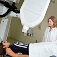
X-ray
The X-ray method of investigation in most cases shows a benign tumor of the soft tissue of the breast wall only due to their local thickening and loss of differentiation. The presence of destruction of the chest bone is a sign of a tumor with a malignant character. More extensive diagnostic capabilities of tumors in soft tissues have CT, ultrasound and MRI.
Morphological studies of the tumor can more accurately determine the diagnosis of a breast tumor, tk. Ray and clinical methods of diagnosing tumors give insufficient information to establish a reliably accurate diagnosis.
Morphological diagnostic data combined with clinical data give a more accurate result. There are various methods of morphological diagnostics, but recently the cyto-morphological method has been used more widely, making it possible to obtain a qualified result relatively quickly and easily about the nature of the cells of the punctate in about 80% of all cases.
The method of trepanobiopsy is more informative - taking tumor tissues with a special needle. This method is less traumatic in comparison with the method of incisional biopsy, however, it is not recognized by all specialists.
In the case of a myxomatous, disintegrating tumor, it is possible to disrupt the abstinence of the intervention, therefore, it is advisable to remove such tumor immediately.
Surgical intervention may be postponed until the final period of tissue treatment only as a last resort. This is possible in case of difficulty in the differential diagnosis method between benign and malignant tumors or a process without a tumor by the results of express biopsy. Such operational delays are undesirable, but permissible in special cases.