Conducting Valsalva varicocele
Valsalva maneuver in varicocele, which is carried out and how?
Valsalva maneuver in varicocele: methods of decoding of the results
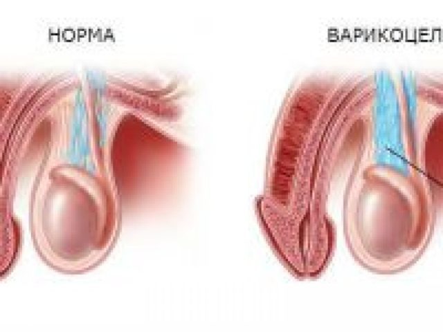
Varicocele - a pathological condition characterized by varicose veins male testes and spermatic cord.
The disease is diagnosed using clinical, laboratory, examination methods and apparatus. Using specific probes also helps to differentiate the disease.
One such method - Valsalva with varicocele. What is this method of diagnosis, it is held, and that shows considered in the article.
Main characteristics of the sample
The method is based on the change in pressure indicators in certain parts of the body to create stress conditions. Such processes are the result of changes in hemodynamics. The diagnostic method is conditional division into 4 phases.
The first and second phase of the process corresponds to the time when the doctor asks diagnosed stretch the abdominal muscles.
Tension causes reflex spasm central and peripheral vessels, resulting in a decrease in specific area entering the body of blood.
Clinically it is accompanied by hypotension (decrease in pressure) and tachycardia (increased heart rate).
The last two phases correspond relaxation process which is accompanied by the inverse processes:
- the amount of blood flowing to the heart, increases;
- increased cardiac output;
- increasing pressure;
- decreased heart rate;
- blood flow in the survey area is restored.
Evaluation of the results depends on the area of the body, which lends itself to the study.
diagnostics Features
The sample has a name well-known specialist anatomist A. M. Valsalva. It has a second name, allows to specify a method of conducting - voltage of Valsalva. Initially, the diagnostic method was created to differentiate and assistance in the field of diseases Upper respiratory tract: inflammation of the auditory analyzer, eliminating pus from the cavity middle ear.
The method is widely used in everyday life. It is used by people when submerged under water, aircraft passengers. Widespread use of the sample found in the field of phlebology, urology, surgery, cardiology.
Often, some of the patient effort and vigilant eye doctor is not enough to properly assess performance. In such cases, use in combination with ultrasound Doppler.
Contraindications
At first glance it seems that the sample is safe enough, in view of the simplicity of its execution. However, there are some contraindications to its performance:
- infectious diseases in the acute phase;
- fever;
- sepsis;
- vein thrombosis in the acute phase;
- myocardial infarction;
- ischemic stroke;
- thromboembolism;
- surgical diseases (appendicitis, peritonitis);
- visual analyzer pathology of the retina.
A source: https://oprostatite.info/urologiya/prochie-zabolevaniya/proba-valsalvy-pri-varikocele
Effective methods of diagnosis varicocele - ultrasound, Valsalva maneuver tests and other
Occurrence varicocele occurs mainly during adolescence, but the disease can occur and after puberty.
Preventing harmful effects of varicose spermatic cord - the loss of fertility and reduce the potency - perhaps, if the disease is time to identify and select effective treatment varicocele.
…
symptomatology
The first stage of varicocele occurs virtually asymptomatic, but the later stages of the disease progression are characteristic symptoms.
Symptoms of varicocele:
- the appearance of pulling the nature of pain (particularly during lifting);
- appearance acinar veins (via the scrotum naked eye physiological changes visible);
- testicular ptosis (veins provokes scrotal swelling);
- decrease in testicle size in (violation of temperature stimulates the abnormal development of the testicles).
Varicose testicular damage also notes reduced amount of sperm and the male hormone - testosterone. In bilateral lesion of testicular hormone flow may stop completely, so men may experience feminization of forms, reducing the vegetation on the body and change in voice tone.
Important. The occurrence of reflux - return throw blood - leads to oxygen starvation eggs and stagnation in the pelvic organs.
About the symptoms of varicocele is told in this video:
Which doctor help?
Get a professional inspection is possible by means of narrow specialists - urologist or andrologist-endocrinologist. After spending palpation and visual examination of the scrotum, the doctor is able to immediately detect the presence of a pathological process, but in controversial situations, it can send the patient to pass a series of tests.
diagnosis of varicocele
Hardware diagnostics provides a comprehensive data on the state of the testes, spermatic cord and appendages in varicocele. Primarily based on the ultrasonic diagnosis of varicocele, but there are other methods.
-
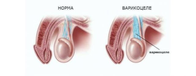
Ultrasound diagnosis of varicocele. The method of non-invasive examinations of the scrotum is used to obtain information about the status of spermatic veins by means of ultrasonic waves.
US-varicocele easily noticeable symptoms in the initial stage where no visual signs of disease.
The thickness of the veins of the spermatic cord is defined in a relaxed and stress state (the man recommended to stretch the abdominal muscles). Preparation for ultrasound a varicocele is not requiredIt is held in a standing position.
You can not only identify the varicocele by ultrasound, but additional diseases of the testes - orchitis, hydrocele - and even cancerous changes.
- Valsalva maneuver with varicocele. Often carried out with any urological examinations and involves the study of the state of the testicle veins on inspiration and expiration. The sample is also carried out during the ultrasound examination, and if the patient has a significant increase in the diameter of the veins in the upright position, the health worker diagnosed varicocele.
-
Phlebography. The method is designed to detect the presence of impaired blood flow in the vein leading to the testicle. The vessel contrast medium is injected, after which radiography is performed, which fixes the rate of the mortar spread on veins. Phlebography also allows to determine the structure spermatic vein and its diameter.
this method It considered obsolete because of his trauma (Requires administering local anesthesia and a catheter), and also the negative influence ray sexual sphere.
-
MRI. The most advanced method for studying the tiniest of structural changes in the sexual sphere.
Magnetic resonance method provides excellent tissue contrast and resolution with which easily defined initial functional transformation.
Final results are recorded on data carriers, allowing the monitor to successfully raise any sites.
passage MRI It recommended for men with suspected varicocele first degree and those with varicose complications.
Given the high cost of MRI services, doctors sent men to study testicular this method only if other methods were uninformative.
Venography does not involve manipulation of the data in the case of the image without the administration of the substance.
reference. The diameter of the testicular vein in healthy men is 4.3 mm, and in patients with varicocele - 6.8 mm or more.
During pregnancy, planning a man who suffers from varicocele, must pass spermogrammuTo detect changes in sperm motility. While the first and second stages of the oppression of the reproductive function is rarely diagnosed in the third stage change in the composition of the ejaculate is almost always observed.
There are other tests in varicocele necessary to study the patient's condition.
-
semen. To obtain reliable data, doctors advise before delivery of sperm to refuse sexual intercourse between two and seven days. See dynamic deterioration can ejaculate by comparing the assays that are dealt in a range between 3 to 12 weeks.
The focus is on the percentage change in the number of live and dead germ cells, as well as the total number of sperm.
-
Marching sample. With the development of varicocele in men can also be diagnosed renal venous insufficiency, but to define it is possible only in this way.
Patient in such a case is dealt blood analysis twice: first - before exercise, the second - after. Changing the content of protein and red blood cells indicates a positive test, and problems with the urinary organs.
-
scrotal thermometry. The presence of reflux and varicose veins of the spermatic cord leads to disruption of testicular temperature (norm - 33-34 degrees). Determination of the temperature changes is carried out with an electronic thermometer.
If a significant increase - more than 36 degrees, the doctor strongly recommends varicocelectomy pass. Alternative - thermography, carried out with the help of remote medical imager.
-
Analysis for testosterone. The material for the study is the blood serum obtained from a man who gave up eating for 8 hours and any medications for 1-2 weeks prior to the study. Donating this analysis recommended by the appearance of sexual disorders: decreased libido, violation of potency, the presence of prostatitis.
Intimate problems associated with sexual activity, often diagnosed with third degree varicocele. The following variations (ug / l) results of analyzes are considered to be within the normal range - 2,8-8,3 (for the male half of the population between 18 and 50 years).
A warning. Long course of the disease can result in pathological conditions associated with a significant reduction of motile sperm or their complete absence - azoospermia and astenospermii.
Identification of varicose veins in a patient in most cases it occurs without the use of laboratory testsBut if necessary to determine the fertility of man and his tendency to kidney failure, the passage of varicocele examination is required necessarily. Regular visits to the urologist - an additional way to notice the dangerous changes and start the necessary treatment.
See also:Prostatitis - what is it and what are they?
A source: https://samec.guru/zabolevaniya/andrologiya/varikotsele/diagnostika-i-analizy.html
Varicocele surgery and indications for its conduct

Varicocele is a fairly common disorder that is diagnosed, usually in their early teens during preventive examinations. Varicocele represents varicose veins testis and spermatic cord.
This disease does not pose a threat to the life of the patient, however, with its intensive development, if you do not carry out the operation on varicocele, it often leads to male infertility. It is believed that a varicocele can cause impotence, but experts reject this theory, claiming that varicoceles has no relation to a decrease in erectile function.
Varicocele surgery is indicated, however, to date in the medical Internet directories and encyclopedias you can find all kinds of methods of treatment of varicocele without surgery.
varicocele disease is often asymptomatic, but sometimes experts note such features as:
- Pain and uncomfortable "pulling" sensation in the scrotum, groin area.
- The increase in the left side of the scrotum. Most often, you will notice when a man should (in most cases it is diagnosed varicocele testicle to the left, the right testicle varicocele occurs in only 2% of men).
- Reduced sperm activity.
The most reliable and effective method to get rid of varicose veins of the spermatic cord and prevention of infertility at the moment It is still surgery varicocele, especially since there are many surgical methods, and we can always choose the least traumatic.
Its useful to note
The main indication for varicocele surgery is the presence of the disease in one of four classes development and the absence of contra-indications and chronic diseases, which may cause complications in the postoperative period.
At this point the patient varicocele different methods of operation can be offered. The choice in this situation it is better to provide the attending physician. Here we look at some of the types of operations on the varicocele.
- Operation Ivanissevicha varicocele. This procedure is generally performed under local anesthesia and is considered the most traumatic. Also, this operation has a high probability of relapse and complications. To date Ivanissevicha surgery for varicocele is done rarely, as is the first method of surgery in varicocele. Plus, this procedure is still available - the cost of such operations is rather low.
- Endoscopic surgery or laparoscopy with varicocele. The procedure is performed under general anesthesia and is today one of the most popular methods of surgical treatment of varicocele. After such an operation, the probability of relapse or complications is practically reduced to zero. The time of endoscopic surgery for varicocele varies from 20 to 40 minutes. As for the post-operative period, it may be noted here that when the patient after laparoscopic varicocele can be ready to be discharged the next day.
- Microsurgical operation with varicocele. To date, this procedure is the most modern and low-impact, however, the cost of such an operation in varicocele is high enough. Microsurgical surgery is performed under local anesthesia and is quite a long time.
operation cost for varicocele may vary depending on the disease and on the method of operation. Thus, the cost of endoscopic surgery of varicocele (laparoscopy) to an average of 35 to 70 thousand rubles, and microsurgical operation will cost from 40 to 90 thousand rubles.
Valsalva maneuver in the diagnosis of varicocele
In the initial stages of the disease it is extremely difficult to diagnose. For such cases, a Valsalva maneuver varicocele. This method of diagnosis is simple and effective and is often used in medical practice with scrotal ultrasonography for varicose veins of the spermatic cord and the testicle.
Valsalva maneuver in varicocele is performed while the patient is in a standing position. At first, inspect and palpate the scrotum in a state where the abdominal muscles are not tense.
Then the patient is asked maximum inflate the abdomen, thus muscles tense and varicocele can detect seal on the affected part of the spermatic cord.
Conducting Valsalva maneuver in tandem with ultrasound at varikotsele helps determine the increase in the diameter of the veins in the affected area.
A source: https://impotencija.net/varikocele/operacija-na-varikocele/
Valsalva maneuver with varicocele
Varicose veins - a problem faced by a huge number of people around the world.
The sooner the disease is diagnosed, the sooner you can start treatment and get rid of the painful problems.
Table of contents:
One such disease diagnostic methods have Valsalva maneuver.
"Remove the sample" on varicocele and varicose veins
Valsalva test in varicocele and varicose help at an early stage to identify the problem with the veins. Plus a diagnosis that the technique is valid even when other methods are powerless.
Valsalva maneuver in varicocele is done in order to determine the pathology of the inguinal vein when viewed women in the prevention and the development of the disease.
When varicose veins, this method helps to identify the disease even when they do not feel any symptoms. The sample helps to establish the state of the valve apparatus of the venous system.
Widely used in the method of vascular surgery and is conducted prior to the appointment ultrasound legs and blood vessels, and sometimes beyond. Thus, when the primary and secondary varices assay enables to assess the presence and degree of increase reflux diameter veins.
The method of characteristics
The diving is widely used a similar technique
This test is named after the famous anatomist - AM Valsalva, and was originally aimed at the treatment of otitis media and removal of pus from the middle ear.
Another name of the method - the voltage of Valsalva, speaks for itself: for diagnosis requires that a person
boosted inhaled, thus covering the nose and mouth. This method is now used much more widely than before.
It is used by divers to a depth, passengers planes, physicians in the diagnosis of varicocele and varicose veins, and many other diseases.
For more specific diagnoses and often require additional studies (eg, ultrasound or electrocardiogram).
After the first examination, the doctor-phlebologist can produce visual diagnostics of the presence of spider veins, and then directed to ultrasound with Valsalva maneuver. Such a study will reveal a complete picture of the state of the vessels and the presence of blood clots.
During the test measured the frequency of contractions of the heart muscle and the pressure in the arteries.
Indications and contraindications for diagnosis
Valsalva test administered to verify or investigate the following:
- condition and operation of the valve apparatus of the venous system of the lower extremities, along with Doppler;
- Study tachycardia with simultaneous electrocardiogram;
- study patency of the auditory tubes;
- Study of heart rate after suffering a myocardial infarction, risk assessment death.
Method can be used independently at time divers dive under water during flight by plane.
Contraindications for this method of diagnosis there are also quite extensive:
- acute infectious diseases;
- fever;
- blood poisoning;
- any disease during the period of exacerbation;
- acute venous thrombosis;
- acute myocardial
- acute stroke;
- surgical diseases in exacerbation;
- thromboembolism;
- problems with the retina of the eye.
Application of the method in phlebology
Method of Vein Valsalva used to study the condition of vessels, the valves and the presence of thrombus.
A source: http://ivanbushman.ru/proba-valsalvy-pri-varikocele/
Diagnostics extension groin vein

Valsalva maneuver in varicocele is used by physicians around the world. This method allows to determine the existence of problems with the veins in the early stages.
Also, this method is used to detect a variety of problems with the cardiovascular system. With a special method doctors can set adverse changes in the absence of visible symptoms.
It is for this reason that experts recommend to immediately visit a medical center with intimate problems.
At various disease symptoms should seek immediate medical care
Characteristics of the inguinal circulatory disorders
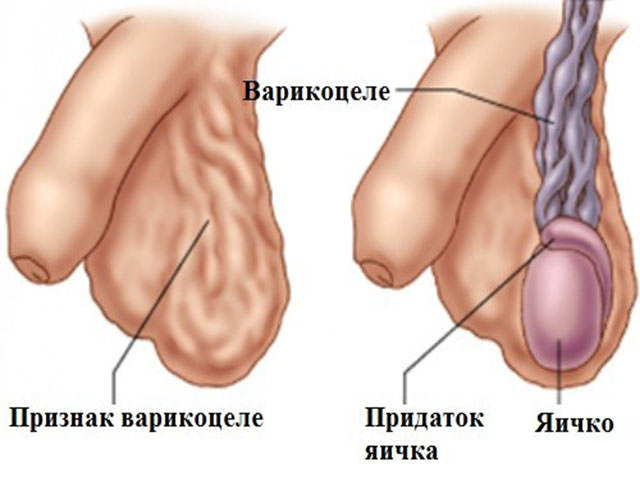
To understand what the sample is needed, you need to understand the features of a varicocele. This pathology affects venous beam carrying Blutwurst liquid to the eggs. Accumulation of veins forms a ball that resembles a bunch of grapes. Thanks to this feature, a tangle called pampiniform plexus. These fibers are responsible for the proper operation of male sex glands.
Through the veins to the organs of blood flows. The blood fluid contains a large number of red bodies. These cells have a biconcave shape. This structure of the red blood cells are due to its function.
The recesses on the surface of the erythrocyte carries oxygen molecule. After contact with the tissues of erythrocyte molecule leaves the surface. Oxygen enters the cell bodies.
When a sufficient amount of the substance tissue able to independently update. Grown old cells are replaced by young cysts.
Varicocele refers to the variety of varicose veins. Varicose veins are characterized by the formation of the stretched pocket on one of the container sections.
The pathological process is influenced by a variety of reasons. Under the influence of adverse factors of elasticity fibers becomes smaller. Blood lingers in this area.
See also:What are the complications of circumcision
The gradual accumulation of blood leads to a strong stretching of the zone.
Because of the intensity of the resulting pocket of blood circulation decreases. When the pressure increases on the affected area. The lack of oxygen leads to disruption of the functioning fed body. Pampiniform plexus carries oxygen to paired gonads. With a shortage of nutrients is a deterioration of spermatogenesis.
The testicles are responsible for producing healthy germ cells. Sperm cells must possess a number of specific properties that contribute to fertilization. Varicocele sperm lose their ability to move.
This causes a massive loss of germ cells. Man becomes infertile. It is for this reason, it is important to determine the presence of varicocele in the early stages.
If the disease becomes severe, there is a risk of death of one of the paired glands.
Doctors are three stages of the disease. The first stage of varicose veins is characterized by the formation of the densified portion at the vessel wall. In this case the patient has no visible symptoms.
Anxiety in men is the second stage of the disease. At this stage, the formation of the pocket. In this case, the patient has the first symptoms of the disease.
Dangerous form of the disease is the third stage. At this stage, the pocket wall much thinner. There is an increase of blood pressure. Damaged tissue may burst. Hemorrhage into the abdominal cavity can lead to death.
Symptoms of varicose lesions
Violation potency indicates a serious problem
In order to timely detect the presence of disease, it is necessary to know the symptoms. In a first step, many men do not pay attention to the signs. This leads to a further worsening of the situation. Doctors recommend pay attention to the following visible symptoms:
- pain in the scrotum;
- swelling of one of the testicles;
- cyanosis of the skin groin;
- erectile dysfunction.
The first visible sign of disease is a pain in the scrotum. The emergence of pain increases with certain activities of the patient. Unpleasant sensations occur when the circulation or active exercise. Also, pain accompanies the ejaculation process. The peculiarity of this symptom is its complete disappearance when you stay in a horizontal position.
When the disease intensity enhancement observed swelling of one side of the scrotum. Often there is swelling on the left side. This localization is characterized Properties pampiniform plexus. In this region, venous beam forms a right angle with the vas deferens. In rare cases, a varicocele is shown on the right side of the scrotal sac.
Also, experts note that due to the reduction of blood flow to particular areas of the veins there is a change of color of the skin. In this zone there is cyanosis. Also palpation of the patient observes portion increased pressure in the abdominal cavity.
The pathological process is accompanied by various disorders and sexual function. Pampiniform tangle feeds testes and vas deferens. During ejaculation, semen passes through the ducts. Against the background of strong blood pressure can decrease the amount of semen released.
Also, problems are identified and during sexual arousal. An erection occurs when filling the cavernous bodies with blood. The liquid is fixed in the cavernous bodies special sphincters. When insufficient blood erection becomes defective. It can also be spontaneous removal of blood from the penis during sexual intercourse.
If the man noticed the presence of one of these signs, it is necessary to consult a specialist. Only physicians can pinpoint the root cause of these symptoms.
Causes of Varicose Veins
Sport activities are beneficial to the male body
Patients often wonder what reasons cause negative changes in the vascular system. The causes of disease are varied. We consider the following negative factors entailing vasodilation groin area:
- genetic predisposition;
- intensive physical activity;
- professional features;
- inflammation of the pelvic organs.
It was found that detected varices in patients due to a genetic predisposition. If the older members of the family suffer from this disease, the younger generation can also get this problem. In this case, doctors are advised to take preventive measures. Only prevention can reduce the likelihood of developing a varicocele.
The risk group includes men, leading an active physical activity. At high physical exertion pressure in the vessels increases. After the end of the exercises, it is normalized.
The frequent expansion and contraction of the lumen of the fiber by stretching fraught tissue. This implies the formation of a stretched portion of the vascular wall.
To reduce the risk of health problems, all exercises should be performed only under the supervision of a sports coach.
Effect on blood circulation and the patient's profession. For many people, work is accompanied by a long stay in one position. This posture is accompanied by a decrease in blood flow rate. Liquid rotates slowly. It is going stagnation in the pelvis. This causes a gradual expansion of the venous beam.
diagnostics Features
When signs of pathology specialist assigns diagnostics. Identify varicocele in several ways. Common diagnosis is Doppler. This study allows you to set force the passage of blood vessels in different areas. When Doppler on the screen can be set to the permeability of veins using a special solution.
Also used electroencephalography. This technique is carried out using a special apparatus. Through separate areas skipped electric wave. In the collision of a wave with an obstacle on the screen image appears. Varikotsele characterized by the formation of one or more nodes on pampiniform beam.
Features of the method
Trial and found application in the field of phlebological. It was found that with a single breath occurs a certain change in the activity of venous sphincters. When deep inspiration liquid fixed in the veins.
Wall easily felt by palpation. This feature is characteristic for the vertical position. In the horizontal position is a sharp inspiratory outflow of blood. The pressure decreases.
Both properties allow experts to carefully examine the condition of the vascular fiber.
After diagnosing a specialist prescribe therapy on an individual basis
Valsalva maneuver was known in the XIX century. He has developed its Italian scientist. Antonio Mario studied the hearing instrument. He observed that membrane retains its shape at the same pressure in the external environment and the auditory tube. The pipe serves as a kind of windows.
It helps air to flow into the middle ear to maintain constant pressure therein. At the same time the pressure drop observed with the swallowing movement. The growth of external pressure on the membrane is observed when moving to a greater depth. To reduce the negative impact on the membrane and avoid its rupture divers forcibly exhale.
It helps to normalize the pressure on both sides of the membrane.
Valsalva has been applied to this sample and in phlebology. With a deep breath veins sphincters are closed. Blood is fixed in the vessel. It allows you to explore the maximum diameter of the venous lumen. When exhalation is studied and the total liquid volume of exhaled air. Using the Valsalva maneuver is possible to accurately detect the presence of pathological areas.
Sample effective for varicose veins peripheral organs. It is also used for the study of the respiratory system and the hearing aid.
during the study
A feature of varicocele is a vascular reaction during inspiration. When there is a strong air delay voltage peritoneal wall. When the voltage pampiniform plexus wall bulge. A specialist can be palpated beam along its entire length. At the outlet walls fall. This helps the doctor to detect even small nodules.
To conduct Valsalva maneuver, a special tube is attached to a pressure gauge. Before the procedure, the patient should take a horizontal position. In the supine position man takes a deep breath. There is a falling off of venous bunch.
The doctor performs palpation. Thereafter, the patient picks it up and squeezes her lips tightly. At the same time there is a gradual exhalation. Device records the pressure output stream. In a healthy person the maximum value should not exceed 40 U / min.
If the reading is higher, there is a pathology.
After the procedure the patient is taking a vertical position. All manipulations are repeated in the same order. According to the results the doctor can draw certain conclusions.
Not all experts are able to accurately determine the existence of changes in the venous beam. To confirm the sample must be accompanied by an advanced method results.
More accurate results are obtained by the joint implementation of the sample with dopplerography. Upon detection of the seal on one of the sections of the vessel doctor uses doplerografii.
It helps to accurately determine the size of the nodules and their location.
It is also necessary to carry out palpation correctly. Probing is carried out over the area of the testicles. When palpation is necessary to determine the state of the ejaculatory ducts and pampiniform plexus. Inspiratory scrutinized scrotum on the affected testicle. duct wall should be smooth and tight. If not, the ducts do not get enough blood fluid.
advantages of techniques
Many of today's experts in the study of varicocele prefer the method of Valsalva. This phenomenon occurs because of several advantages:
- ease of implementation;
- obtain reliable results;
- early diagnosis of the problem.
To carry out the sample does not require the presence of additional devices. The patient must also not comply with any rules. All the action is carried out by means of one small appliance.
See also:What are the symptoms and treated chlamydia in men
When protrusion vascular fiber physician can carefully examine its structure. The presence of small tumors or seal quickly detected. Also palpation can detect changes in the activity of the ejaculatory canals.
With Valsalva maneuver can detect any step varicocele. Negative changes are diagnosed in the early stages of the disease. This gives an advantage over other diagnostic methods.
Any pathological disturbances in the cardiovascular system affect the activity fed bodies. Varicocele negative changes occur in the reproductive system. Early detection of problems can quickly stop it. For this reason it is important checkups every six months.
A source: http://DoktorSos.com/andrologija/varikocele/valsalva-pri-varikocele.html
Applications Valsalva maneuver in both men and women, indications and contraindications
Valsalva maneuver - a technique of diagnostic blood system research work of the body, as well as the heart valve body, respiratory system, and the identification of many pathologies.
This technique is to try to the patient to exhale air flow in a closed mouth organs and nose.
What is a sample reception and Valsalva?
This procedure was invented in the 18th century Italian physician Valsalva. This test was intended originally for removing pus from the eardrum in the ear with otitis.
Valsalva maneuver - a diagnostic technique to study, which is carried out a doctor for diagnosis and detection of disease.
to the contents ↑
Indications and contraindications to the Valsalva maneuver
Valsalva maneuver - this is a test method for the detection of pathologies in such disorders in the body:
- Diagnosing heart palpitations;
- At a fit of myocardial infarction, for determining the risk of death;
- Estimation valve operability vein;
- Diagnosis of varicose veins;
- Diagnosing diseases of the reproductive system - varicocele;
- In determining the functioning of the autonomic system of a person;
- To study the terrain in the ear canal with otitis.
It is not necessary to practice the method of Valsalva in such pathologies:
- Hypertonic disease;
- Palpitations heartbeat - tachycardia;
- When the disease is cardiac organ - cardialgia;
- During the attack suffocation;
- During syncope.
It is prohibited to conduct Valsalva maneuver in the following pathologies:
- In acute myocardial infarction;
- When a brain haemorrhage - stroke;
- In heart failure pathology second or higher degree;
- The disease is pulmonary embolism;
- The pathology of appendicitis;
- In peritonitis;
- When the disease is retinopathy;
- When the state of fever;
- In sepsis;
- When thrombosis of major arteries;
- Clogging of the arteries of the lower extremities;
- Cerebral atherosclerosis;
- Accrual loans blood flow in the brain.
Valsalva maneuver is used for diagnostics in cardiology, but also can detect diseases that develop in the cervical spine - a pathology of the thyroid gland, retrosternal zob.k content ↑
The essence and the mechanism of Valsalva maneuver
The sample according to the method of Valsalva - is a specific method to identify pathologies, which is used by specialists from the field of cardiology and medicine in urology.
Mechanism perform sample:
- The patient is doing through his nose deep breath of air;
- It discharges the air through a special mouthpiece, wherein the very small opening;
- After the procedure deflation, the patient recovers breath.
At the time of the procedure permanently fixed such indicators:
- Fixed heart rate since the start of the procedure, and until its end;
- blood pressure index.
Instead of normal saline exhausting air occurs tension in the muscles of the chest and abdominal muscles.
to the contents ↑
Phase which revealed pathology
- Phase №1 passing method - phase lasting no longer than 3 seconds, which is accompanied by a high air pressure in the chest, as well as in the peritoneum;
-
phase №2 - Voltage phase reveals: hypertensive pathology, increased heart rate - tachycardia, as well as the resistance of the peripheral vascular system.
Heart organ to be shown the maximum sample is filled with blood, the lungs become more transparent. Step second phase lasts not more than seven seconds;
-
phase №3 - a time when the air outlet ends of the body, and a period of relaxation of the respiratory system.
At this stage of testing falls in blood pressure index, heart muscle become less frequent and deeper. The size of the heart and lungs body comes to a normal state;
-
phase №4 - this is the final study period Valsalva maneuver, and the respiratory system relaxation period.
During this period, increases in blood pressure index, manifested cardiac pathology - bradycardia and vasodilation of peripheral vessels sphere. Restored blood flow system, as well as the venous return.
During the Valsalva maneuver occurs reconciliation indicators and cardiogram detected length R-R between the teeth.
Coefficient in cardiography is the distance between the longest and the shortest prong tooth, in a calculated ratio.
If the detected ratio is within the range of 1.30 - 1.70, then it is a borderline condition of the body.
Valsalva maneuver also helps to identify the size of the heart organ, as well as the tone of the heart muscle - miokarda.k content ↑
Valsalva venous pathology
Quite often with varices, patients with other instrumental techniques diagnostic study, also assigned to Valsalva maneuver.
When violations of the venous valves, there is not a proper blood flow in the veins, often there is a process of regurgitation, in which there is over-voltage veins and varicose veins developing pathology.
During the sample period in inhalation, the flow of venous blood becomes weak in phase №2 (voltage), venous blood flow is stopped, and only starts from the third phase (exhalation).
Slow speed in the blood veins, does not timely closed valves of veins. Valsalva maneuver allows to reach the maximum speed in the venous krovotoke.k content ↑
Positive and negative Valsalva maneuver
Positive assay method of diagnosis of Valsalva defines:
- Poor functionality of the venous valves;
- Stagnation of venous blood.
Identification of these factors indicates that the patient has a high risk of venous pathologies.
Not always Valsalva maneuver is as objective as possible.
The problem is that to achieve the maximum description of the information, it is necessary that the abdominal muscles have reached their maximum voltage, but in some patients it does not occur for the following reasons:
- Flabby muscles in the abdominal cavity;
- Obesity;
- Patients who have weakened disease organism;
- The female half of the population.
Such patients carried a modified type of the sample. Its essence is that the sensor is installed on the site of the venous valves. The patient tense the muscles of the abdomen, and the doctor presses on the abdomen in the navel area.
This method reveals venous reflux.
The duration of this procedure - 15 seconds.
to the contents ↑
Diganostika with male pathology - varicocele
Men are at a reception at the doctor urologist, constantly doing Valsalva maneuver, in order to identify the disease varicocele at the initial stage of its development.
With the passage of ultrasound, the sample reveals a deviation from the norm of functionality in the affected areas veins.
If palpation palpable nodules, while this is the first symptom of a varicocele. Also varicocele can be detected by palpation moshonki.k content ↑
Diagnostics for diseases of ENT
Straining for 30 or 40 seconds, the air mass at the maximum of its seizure, there is an increase in eardrums pressure cavity, and open the auditory tube, allowing air to get into the flow middle ear.
In the pathology of purulent otitis media occurs perforation of the eardrum, and the pus comes out of it outside.
When swelling of the auditory tube, when retained permeability therein, at the moment of passage of the sample, the patient hears strong squeak in the patient's ear and feels bubbling in the ear and loud noises.
Reception Valsalva procedure used for the removal of the middle ear exudate caused by an inflammatory protsessom.k content ↑
The use of the sample according to the method of Valsalva in women
The sample according to the method of Valsalva in gynecology (obstetrics) provides for bladder filling and performing this assay for the detection of sphincter of the bladder. A woman should make the most of a deep breath and stretch the abdominal muscles.
This sample allows most accurately detect the pathology of the bladder and urethral sphincter functionality channel.
Using this technique, it is possible to identify the causes of female besplodiya.k content ↑
Functional test on Mueller method
Muller's method allows to calculate the tone of the heart muscle - the myocardium. To perform this method, as well as in the Valsalva procedure should be possible to exhale air from his chest and inhale with a closed nose and mouth.
Strong pulsation occurs vena cava, left-side part body pulsates significantly less than that before test. Delayed maximum breathing is not more than 40 seconds.
to the contents ↑
conclusion
by the method of Valsalva maneuver - this is the best method for the determination of varicose veins and study the state of the lower extremities, as well as identifying the male varicocele disease.
Valsalva maneuver reveals pathology at the initial stage of the occurrence, when there are no obvious symptoms.
A source: https://moyakrov.info/veny/proba-valsalvy