The use of Doppler ultrasound for research vessels of the penis
Surveys for erectile dysfunction

With the development of medicine accurate survey methods for erectile dysfunction becomes more and more. The doctors there is an opportunity to find the cause of the disease faster and assign adequate treatment.
It has long been known for such surveys for erectile dysfunction - clinical analysis of blood and urine laboratory testing of blood sugar, medical examination of the external genitalia, ultrasound of the penis. This basic research, the results of which the doctor prescribes conduct specialized analyzes, taking into account the patient's existing disease.
* * *
baseline survey
Clinical blood test
the composition of the blood study was carried out in each patient appealed.
Blood taken from a patient on an empty stomach, studied in the laboratory for about 10 indicators, consider directly related to the topic of erectile dysfunction.
Hemoglobin in healthy males 18-65 years is between 120-165 units. Decreased hemoglobin indicates anemia (anemia) - deficiency in vitamins and iron, which inevitably leads to a weakening of erection and other functions.
In this case, the body in the shortest possible time to be filled with vitamins, amino acids, minerals and bifidobacteria that boost the immune system and ensure the full functioning of the body.
Increased hemoglobin could indicate the development of benign prostate tumors, impaired kidney function, diabetes mellitus.
The doctor, taking into account the clinical picture of the disease appoint special surveys: a blood test for prostate specific antigen (PSA), ultrasound of the genitourinary system man.
White blood cells from healthy men over 16 years is between 4 - 9 units. Score less than 4 units indicates the inflammatory process in the body. To clarify whether the inflammation is localized in the genitals, the urethra is assigned a tank crop microflora.
* * *
PSA test
In 1970, professor at the University of Arizona, Richard Ablin identified prostate specific antigen [1].
Blood analysis for the presence of PSA can detect prostate cancer and to ensure high quality of existing tumors.
The analysis is performed for all patients older than 45 years, because since 2008 it is part of the all-Russian program of additional medical examination of citizens.
Investigate the blood taken from the vein, it is determined by the total PSA level.
For men of all ages the rate of PSA content - up to 4 ng / ml. PSA content, completely eliminating the presence of tumors in males 40-50 years - no more than 2.5 ng / ml for men older than 50 years - no more than 3.5 ng / ml.
Indicators, slightly above normal may indicate prostate hyperplasia and inflammation, significantly exceeding - for prostate cancer.
To clarify the diagnosis is assigned to the definition of free PSA level, biopsy and cystoscopy.
* * *
Analysis of free testosterone
Determination of free testosterone administered when besides complaints of a history of erectile dysfunction observed infertility, hypogonadism, prostatitis, osteoporosis.
Blood taken from the vein, the laboratory investigated for 6 days.
Norma testosterone in men of all ages: 5,5-42 pg / ml. Norm, taking into account age and calculates the overall picture of the doctor.
Reduced testosterone levels frequently indicate diabetes, depression or hypertension.
* * *
Analysis of blood sugar
Long-term studies of the aging men, conducted in the University of Massachusetts and completed In 1994, confirmed that diabetes - the main cause of erectile dysfunction. It is diagnosed in patients with type I diabetes in 35-75% type II - in 70-89,2% of cases [2].
This analysis is mandatory if there is no other apparent cause of erectile dysfunction.
The blood taken from a finger fasting, the rate of sugar - less than 5.6 mmol / l. Sugar within 5,6-6,1 mmol / l ascertains prediabetes (impaired glucose tolerance), above 6.1 - the development of diabetes.
If enough data of this analysis, analyzed for glycated hemoglobin, which, according to Scientists pediatric department of the University of Pisa (Italy), reveals diabetes in the early stages [3].
* * *
specialized surveys
Conducting research on animals, MD Robert Kloner suggests that about 50% of cases it is impossible to achieve erection in men older than 50 are caused by endothelial dysfunction - the loss of the functionality of the inner layer of blood vessels [4].
* * *
Doppler blood vessels of the penis
Indications for the use of the precision of the method are: the groin injury, Peyronie's disease [5], neoplasms of the prostate gland, structural changes in the vessels.
The method is based on the effect discovered by the German physicist K. Doppler [6], it is possible to observe visually and mathematically calculate the usefulness of sexual organ perfusion using ultrasound. The signal sent by the sensor is reflected from moving blood cells are constantly, the computer detects a frequency signal proportional to an oscillating acceleration or deceleration of blood cells.
Measurements are made twice: the first time a member of the erect medicines, the second time - without an erection.
Settings for normal erection phase:
- delta maximum and minimum blood flow to the average speed -pulsatsionny index: 7-13 cm / s;
- blood resistance index particles (RJ): 1,0;
- peak systolic and end-diastolic velocity 40-50 cm / s and 5.4 cm / s.
No abnormalities of these parameters indicates the nature of psychogenic erectile dysfunction, which is described in detail in the «Structural insights into aberrant cortical morphometry and network organization in psychogenic erectile dysfunction» [7].
* * *
Ultrasound of the penis
Complaint about erectile dysfunction - an occasion for US male organ, because it allows you to see and evaluate the degree of impairment of the structure of the corpora cavernosa, which provide an erection. Ultrasound is prescribed for injuries, fractures, tumors and curvatures of the penis in it.
During the ultrasound the patient lies down, the study body is covered with hypoallergenic gel and movement sensor visualize its structure.
US captures the following parameters:
- Echogenicity denote the "norm". Increased echogenicity indicates cavernous fibrosis, reduced - by inflammation of the corpora cavernosa.
- The structure of the corpora cavernosa is denoted by "uniformity." Heterogeneity indicates cavernous fibrosis.
- Ehogennost cavernous artery walls denoted mark "norm." Increased echogenicity indicates vascular disease caused by diabetes or atherosclerosis.
- The diameter of the cavernous arteries are normal: 0.2-1.4 mm. The larger diameter of the lumen of the arteries speaks to their developmental abnormalities, smaller - about autoimmune, diabetic or atherosclerotic disease.
Ultrasound examination of the leading role in the diagnosis of the causes of erectile dysfunction, it shows a practical report on the inspection 289 patients referred to the clinic with the edge of the Krasnoyarsk hospital 2006-2008 year [8].
* * *
Fallostsintigrafiya
Because ultrasound does not provide a complete picture of the status of microcirculation of blood in the cavernous bodies and always possible to make an accurate diagnosis, to apply this new method of radioisotope fallostsintigrafii.
Before the procedure the patient intravenously administered 1 ml of the solution of inactive labels for erythrocytes. The study was conducted on the emission tomograph, which by means of a gamma camera, moving down from the navel to the proximal third of the hips, a number of pictures (1 per second or 20 per minute). If necessary, a member of the research, erect with the help of pharmaceuticals.
Only your doctor is able to keep track on the obtained images of blood flow in the pathology of arterial and venous segment.
This method of diagnosis determines the real cause of erectile dysfunction with the highest precision!
According to published research findings of Russian scientists microcirculation disturbance in the cavernous bodies were found in 88.8% of the surveyed men. Scientists have also compared the sensitivity and specificity parameters fallostsintigrafii and ultrasound, which is defined as 91% and 94% to 78% and 60%, respectively [9]
* * *
bulbocavernous reflex
This test verified the degree of sensitivity of nerve endings of the penis. During the procedure the doctor squeeze the head of the penis, thus causing reduction of the anal sphincter muscles. In that case, if the function of nerve endings will be broken, the response will be different deceleration or contraction of the muscles is not going to happen.
* * *
Identification Test nocturnal erections
This study confirms or refutes the male erectile function during sleep. Usually in men experiencing sleep about five - six episodes of erection.
The absence of so-called spontaneous erections may indicate a problem with nerve function or blood supply to the penis. To conduct the study used two methods: measurement of the circumference of the penis, as well as the measurement of its rigidity.
In the first method, the member is secured around the three plastic hinges varying degrees of tension. Thus, erectile function is determined based on which of the loop is broken.
In the process of the second method, the loop to be tightened on the circumference at the root of the penis and at the apex. In the event that an erection occurs, the loops to tighten, while the electronic device will register all changes.
* * *
biothesiometer
In carrying out this diagnostic method applied electromagnetic vibration in order to assess, firstly, the sensitivity of the penis, and secondly, its innervation. Thus, reduced sensitivity to these vibrations may indicate nerve damage.
* * *
Introduction of vasoactive drugs
During erection of the dough is caused by injection of a special solution, facilitating the expansion (or increase) in the blood vessels, and blood access to the penis.
* * *
Dynamic infusion kavernozometriya
This test is given to men diagnosed with impotence, in which the doctor suspects a venous leak.
In carrying out this procedure is introduced into the vessels of the penis special dye, wherein the administration is at a certain pressure.
When measuring physiological pressure with which the fluid must flow into the penis to maintain stable erection, the physician can ascertain the extent of venous leakage.
* * *
cavernosography
Used in conjunction with the previous method of diagnosis. This method consists in introducing radiopaque substances directly into the penis. Next is an X-ray of the penis, staying in erection. This establishes a venous leak.
See also:The drug spermaktin: reviews
* * *
A source: http://impotenzia.ru/archives/43
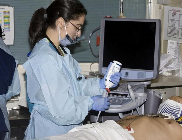
Doppler ultrasound of the penis
Doppler ultrasound of the penis - a modern non-invasive research applied to the study of penile blood flow based on the Doppler effect. It is indicated for erectile dysfunction and impotence of various origins. It is one of the basic techniques used in the process of diagnosis of vascular erectile dysfunction.
Doppler ultrasound of the penis allows you to evaluate the qualitative and quantitative indices of blood flow. For information about the features of the anatomical structure of the penis may be supplemented by other techniques (ultrasound, duplex scanning). The study is completely safe for the patient, has practically no contraindications. It requires no special training.
It can be performed on an outpatient basis.
Doppler ultrasound of the penis (penile Doppler) - ultrasound of the blood vessels and the corpora cavernosa of the penis, which allows to get information about the speed and flow rate.
The method is based on the Doppler effect - changing wave frequency (in this case an ultrasonic) wave when moving the source relative to the receiver. Sensor device for UZDG penile vascular sends waves in the investigated area.
The waves are reflected from the blood corpuscles, advancing through the vessels body arrive at the receiver, and then transformed to a dynamic image on a monitor screen.
Doppler ultrasound of the penis allows you to obtain data on the qualitative and quantitative characteristics of the local blood circulation and blood flow features (turbulent, laminar). The procedure is available, safe and does not use ionizing radiation, so it can be used virtually without restriction in the study of the reproductive system.
Indications and contraindications
The list of indications for penile Doppler ultrasound include erectile dysfunction as a result of disorders of blood circulation in the penis caused by congenital malformations of the arteries and veins, thrombosis and atherosclerosis of vessels of the penis, penile neoplasia, penile structural changes as a result of scarring and bleeding against the backdrop of traumatic injuries and inflammatory processes.
The list of contraindications to Doppler ultrasound of the penis includes the grave condition of the patient, presence of mental and neurological diseases, because of which the patient can remain stationary during the study penile or unable to follow instructions Doctor-diagnostician. A small amount of contraindications, highly informative and safety of penile Doppler ultrasound allowed this method to take a leading position in the list of procedures used in identifying the causes of the violation erection.
The methodology of the
Special training is not required. Before the study should carefully carry out hygiene area of the genitals. Within 1-2 days you need to give up drinking.
Diagnostician must submit to the direction of the procedure, data analysis and other tools of research, doctors' reports and statements of medical history. The patient is placed on his back. Initially, penile Doppler ultrasound is performed at neeregirovannom penis.
Next, a preparation for injection and erection stimulation study vascular state when erect penis. The dynamics of the blood flow is monitored every 5 minutes. The duration of Doppler ultrasound of the penis is 30 to 40 minutes.
The conclusion is usually given immediately to hand, sometimes the patient is asked to wait for 20-30 minutes, or come for a document to a specified time.
If the patient is allergic to injectable or experiencing a pronounced fear of an injection to the penis (under stress are observed autonomic reactions leading to penile vascular spasm and erection insufficiency or absence) of penile ultrasonography performed after taking Viagra and its analogs.
, Manual stimulation (at the time of stimulation doctor comes out of the cabinet so as not to embarrass the patient). If the erection is not weakened by the end of Doppler ultrasound of the penis, the patient can achieve discharge by self-stimulation.
While maintaining an erection for 4 hours or more necessary emergency examination urologist or andrologist.
interpretation of results
Normally, the following parameters are determined during penile ultrasonography: highest systolic the speed of blood flow in the deep arteries - from 40 to 50 cm / sec, diastolic velocity -. 4 to 5 cm / sec.
, Pulsation index (indicator calculated for the difference between the minimum, maximum and mean blood flow velocity) - from 7 to 13, the resistance index - 1. Normal values listed indicators in penile Doppler ultrasound evidence of psychogenic erectile dysfunction version.
Deviations are evidence of venous or arterial disorders.
The cost of penile Doppler ultrasound in Moscow
Doppler ultrasound of the penis - the modern technique, widely used to confirm the vascular genesis of erectile dysfunction. cost of the procedure varies depending on the form of ownership and the clinic's reputation.
The private medical centers on the price of penile Doppler ultrasound in Moscow is often influenced by the presence of the highest category or degree specialist.
Cost method in a specific medical institution determined by the urgency of the study, one stimulation erection (Manual or drug), a view of medicaments for administration to the penis and the need to use Viagra.
A source: http://IllnessNews.ru/yzdg-polovogo-chlena/
Doppler ultrasound of vessels of the penis: indications, technique of
Doppler ultrasound of vessels of the penis: indications, technique of
Doppler ultrasound (Doppler ultrasound) examination of the vessels of the penis and the scrotum is one of the informative and non-hazardous methods of diagnosis which allows teach structure bandwidth vascular walls and in their bloodstream.
Content
Doppler ultrasound is usually done after the ordinary ultrasound, t. E. It is performed using sound waves 2-modes:
To carry out this study used a special ultrasound apparatus operating in the desired mode.
From time to time it may become UZDG other method used when research and inability to perform such diagnostic procedures such as angiography of blood vessels of the penis and scrotum with the introduction of contrast substances. In some cases, the ultrasound scan even superior to their own information content angiography with contrast.
In this article we will acquaint you with the essence, the testimony, the studied parameters, the procedure execution order Doppler ultrasound vessels of the penis. You will also learn about the principles of decoding the acquired results, shortcomings and contraindications of this type of ultrasound scan.
The essence of the technique
This method of diagnosis is based on the Doppler effect.
Used to perform ultrasonography of vessels of the penis and scrotum mode allows you to set a clear diagnosis, and in some Clinical cases eliminates the need for the patient from other more dangerous invasive methods or survey. The principle of its implementation is based on the Doppler effect:
The frequency of the received signal varies directly proportional to the velocity of blood flow. This signal is recorded a computer program that is subject to certain mathematical processing, and receives the necessary special for diagnosis information.
testimony
UZDG vessels of the penis and scrotum can be assigned to customers with subsequent pathologies:
Apart from this, the Doppler scan often cut one in the diagnostic plan in preparation for the surgery on the genitals.
The studied characteristics
After converting the Doppler ultrasound signal allows to obtain follow-up data on the state of the vessels of the penis and scrotum of the patient:
As required Doppler complemented by duplex scanning, which allows you to learn the condition of vessels, the surrounding tissue and the rate of blood flow online.
Rules of preparation and procedure of
For a number of days before UZDG patient should abandon the consumption of alcohol.
See also:Why there are warts on the penis?
When the destination UZDG vessels of the penis and scrotum the doctor explains to the patient the nature and necessity of the study. After that it introduces a special preparation by ordinary rules to the scanning procedure:
The first conversation with the patient's doctor should be able to do with an unhealthy business confidence because UZDG member procedure is performed at outcrop and erection of the penis and endokavernoznoe involves administering drugs that provide erection coming is necessary for more accurate diagnostics.
The patient should be informed and the point that after the end of the scan it will be necessary to achieve the method of self-stimulation ejaculation to address medication-induced erection.
The study day the patient comes into the office of ultrasonic diagnostics, to create a more comfortable test to perform the procedure for around 1 hour. Diagnosis is made on a couch in the supine position.
Before scanning the doctor injects endokavernozno (Vol. e. the penis), one of the drugs, rapidly causing an erection coming. To this mixture may be used:
This measure is undertaken to obtain more informative data UZDG, t. To. Conducted in a relaxed position measuring member can only be speculative or distorted.
Unfortunately, such a method of artificial call desired to scan an erection capable lead to a number of unwanted side reactions:
To prevent these negative effects can be applied other more benign methods to ensure adequate blood flow to the cavernous bodies of the member - receiving pills:
As required perform such a test the doctor, appointing ultrasonography will certainly instruct ailing on the rules of use of the designated product and indicate the time of his admission. Typically Viagra pill should be taken 40-60 minutes before the procedure and to display the erection ultrasound study It carried out the visual and samomanualnaya erotic stimulation, facilitating the flow of blood to the tissues member.
Approximately 20-25 minutes after injecting the product either acts beginning tablet means the corpora cavernosa fill with blood and the doctor begins to scan using a special Ultrasonic sensor. He sets it on the penis and scrotum in sequence every 5 minutes (ie. e. Regardless of erection phase) in over 25 minutes.
After graduating UZDG erection may persist a while. To get rid of her patient is invited to self-stimulation in a separate room. If the excitation of the penis does not go in for 4 hours, the examinee can assist in assigning urgency the necessary measures urologist.
Results UZDG vessels of the penis can get your hands in 30-40 minutes after the procedure or put off the doctor. Some clinics provide research data on electric adresok doctor and / or patient. General state of health after the scan does not change.
the results of decoding principles
Remember! Carry out correct decoding acquired in the process of vascular Doppler ultrasound sex data member can only the attending doctor of the patient. All the characteristics of which are presented in this article are for informational purposes only.
The results of Doppler ultrasound of the penis recorded the following indicators:
Apart from these characteristics in the final result indicates the characteristics that are fixed when the ordinary member of the US (the size of anatomical structures, their echogenicity, etc.). The totality of all data allows to obtain a more complete clinical picture of the disease and to make more effective healing plan.
In assessing the results of the doctor gives special attention to indicators of systolic blood flow rate in the member vessels. This is explained by the fact that it specifically value indicates the presence of stenosis, arteriosclerosis is often observed in the vascular wall.
Other features of the results of Doppler ultrasound blood vessels of the penis also have diagnostic value and are used to calculate the pulsator and resistant index, the value of which may indicate the presence of other diseases:
In the study of ultrasonography results of penile vascular special focuses and evaluation of high performance:
The acquired results allow us to draw conclusions about the presence of stenosis of the arteries. If the scanning process is possible to know the presence of atherosclerotic plaques and blood clots, the doctor may determine their location, structure and surface condition of tissue echogenicity education.
In the study of veins spec can detect signs of varicose, assess the degree of elasticity of the vascular wall and the state of the valves of veins of the penis. To this end, during the scan the doctor makes a Valsalva maneuver, which allows to exclude or confirm the presence of pathological features such as retrograde blood flow.
Contraindications
A relative contraindication to perform ultrasonography of vessels of the penis is an inflammation of tissue member. Apart from this, in some cases a study can not be carried out in the presence of diseases in which means the use of stimulating an erection it is not recommended because of the high risk of complications:
Disadvantages techniques
Lack UZDG penile vessels - the need for reception of a pharmaceutical product, stimulating erection.
The only drawback of such a method and the research vessels of the penis and scrotum blood flow is need for artificial erection call accompanied by injection or oral administration pharmaceutical product. This diagnostic step can be accompanied by mental discomfort and horror of the patient prior to injection or impending occurrence of painful feelings at the injection site.
Such bad causes capable of triggering mechanisms for stress, causing an increase in the tone of the nervous system. As a result, the cavernous artery can spasm and erection can not attack or be insufficient. But this defect can be offset procedure oral administration of agents for erection - Viagra or other drugs with similar effects.
Apart from this, patient should be aware that the information content of penile vascular ultrasonography can assist get rid of the disease and the correct mental attitude to this diagnostic procedure will bring all of its shortcomings to minimum.
To what doctor to address
If you have any problems with the onset of erectile dysfunction, trauma, the appearance of seals or other configurations related to the penis or scrotum, man must turn to an andrologist or urologist.
The doctor, after a series of preparatory research work can schedule a Ultrasound Doppler blood vessels of the penis and other diagnostic procedures (laboratory analysis of seed water, blood, X-rays and so on.
), Allowing to put the correct diagnosis.
Ultrasonic Doppler blood vessels of the penis and scrotum relates to non-hazardous, easily available and informative diagnostic methods and can be administered in many diseases of the genital organs. Such a study is conducted on an outpatient basis and does not ask for special training.
Report of spices on the theme of "vascular Doppler ultrasound in the diagnosis of penile erectile dysfunction":
Ants And A - Doppler ultrasound of vessels of the penis in the diagnosis of erectile dysfunction Watch this video on YouTube
A source: http://vahdoktor.ru/2017/05/13/ultrazvukovaya-dopplerografiya-sosudov-polovogo-chlena-pokazaniya-metodika-provedeniya/
Doppler blood vessels of the penis
Currently, the main method diagnose vascular erectile dysfunction is an ultrasound of the blood vessels of the penis with the determination of blood flow in the vessels (Doppler).
This study is performed on the background andrologom erections induced by introducing into the cavernous bodies of the penis special preparations (known pharmacologically induced erections). This method was proposed by Lue and colleagues in 1985 The study is as follows.
Patient into the penis syringe administered special drugs by injection.
After some time, usually 10-15 minutes, when an erection varying degrees of severity, carried penile ultrasonography, wherein visualized cavernous arteries and evaluate the speed of blood flow in the cavernous arteries (the cavernous arteries - a major artery of the penis blood flow which provides the appearance of erection).
The main indicators of the arterial inflow blood to the penis are:
- value of peak systolic velocity (i.e., the maximum flow rate through the arteries), which should be at least 30 cm / sec
- the rise time of systolic blood flow (ie, how fast the increase in blood flow through the arteries penis), whose values are in excess of 110 ms indicate the presence of arterial violations.
Ultrasound examination of blood flow in the penis against the background of penile erections caused by administration of drugs to diagnose a violation of the venous outflow, ie defeat venoooklyuzivnogo mechanism.
Indicators characterizing venous drainage the blood from the penis are:
- end diastolic velocity in the cavernous arteries - the norm in this figure tends to zero and not more than 5 cm / sec
- resistance index - value it below 0.85 indicates the presence of venoooklyuzivnoy dysfunction.
See also:How to build arm muscles at home for a week
Status venooklyuzivnogo mechanism adopted to assess for normal values of arterial blood flow to the penis (ie, normal values of peak systolic velocity).
The disadvantage of ultrasound blood flow in the penis to cause an erection in the background is the need intracavernous injection of the drug, which may be accompanied by pain at the injection site, and the fear of the patient to prick in penis.
This can lead to launch stress mechanisms are accompanied by an increase in tone of the sympathetic nervous system, followed cavernous artery spasm. The result can be either a lack of erection or incomplete erection of the penis.
Alternatively intracavernous administration of the preparations used reception Viagra (Viagra test) or Levitra (Levitra-test) with the audio-visual (ie watch movies relevant content) and manual (while helping himself with his hands) stimulation.
Other articles on erectile dysfunction:
To contact the author of the site materials "Just the prostate", you can visit the "Contact Us"
A source: https://prosto-prostata.com.ua/menuerection/menupractice/84-dopplerography.html
Doppler ultrasound of the arteries of the penis
Using Doppler ultrasound of the arteries of the penis in the diagnosis of erectile dysfunction has given significant boost to the development of ideas about the state of penile hemodynamics in normal and violations erection.
First Doppler effect has been used in display penile arteries to measure blood pressure [95]. method of determining the value of pulsation of the arteries of the penis to diagnose vascular erectile dysfunction noted Zorgniotti A.W. [271].
High diagnostic value of systolic blood pressure ingredient on the penile arteries and shoulder arteries, as well as relations systolic blood pressure on the arteries of the penis to the systolic blood pressure in the brachial artery, known as penobrahialnogo index (PBI), Was the cause of widespread in the diagnosis of erectile dysfunction these research methods [25, 137, 182].
It noted that PBI a value of less than 0.6-0.75 can talk about the nature of the arterial erectile dysfunction [172. 183, 267].
Using the method of determining the PBI to assess blood flow in the penis has allowed considerable experience which has shown that measurement of blood pressure in the penis and the calculation of the PBI is a reflection of arterial flow only in the dorsal arteries, while as erection depends largely on the blood flow in the deep artery of the penis [162].
Besides, arterial blood flow in the penis evaluation according PBI coincides with the angiography data only in 39% of cases [217], and according to [96] PBI is inaccurate and poorly reproducible blood flow assessment test of the penis.
In this regard, when the ultrasonic Doppler penile arteries is recommended to determine the index of the blood circulation, which is calculated as the ratio of the amplitude with dopplerovskoi curve on the dorsal artery of the penis to the amplitude curve dopplerovskoi brachial artery, as well as the linear blood flow velocity and the nature of the curve changes dopplerovskoi [96. 130].
In our opinion, Doppler ultrasound technique with spectral analysis (UZDG XLA) dopplerovskoi curve could significantly expand the opportunities of evaluation as blood flow through the vessels and the state of the vessels themselves, which explains the use of this technique in our studies.
The highest value in the diagnosis of pathological conditions of blood are indicators characterizing its linear speed as during systole, and during diastole of the heart, the amount of resistance experienced by the blood flow from the vessel wall and the state of blood flow in the lumen (Figure 2).
Fig.2. Ultrasonic dopplerograms penile artery showing the main parameters that characterize the blood flow. (Apparatus for ultrasound Doppler with spectral analysis «Vasoflo - 3» «Sonicaid» company (Great Britain).
Max A - maximum height of the peak Doppler waveform during systole, characterizes the linear blood flow velocity is measured in kHz;
Mach D - mean value of eight consecutive minimum frequency for 20 msec. until the end of diastole, measured in kHz;
Mode - spacing frequency (or speed) in which moves the largest number of blood particles; Mean - average systolic peak frequency;
RP (resistive) - The difference between the maximum frequency of the systolic and diastolic peak, divided by the systolic peak value, i.e.
RP = (A Mach - Max D) / Max A
This figure, known and called "Parselota index"Characterizes the state of the vessel distal to the display space and does not depend on the angle of inclination of the sensor and its frequency;
Pi (pulsation index) - the ratio between the difference (peak to peak) of the maximum frequency values and the time weighted average value of the maximum frequency, i.e.
Pi = F Max (Vpp) / F Max (average)
It is calculated at each cycle and averaged over the selected number of cardiac cycles. This index is informative in the study of peripheral blood flow, especially in the aortoiliac segment, it does not depend on the inclination angle sensor and characterizes the state of peripheral resistance blood flow;
SB - spectral expansion is calculated as the difference between the maximum frequency and average systolic peak frequency, divided by the systolic peak, i.e.
SB = (A Mach - Mean) / Max A
Calculation of this indicator being the percentage, it shows turbulyatsiyu in the bloodstream, ie increased the difference between the maximum and the average frequency of the systolic peak. SB independent of the angle of inclination of the sensor and its frequency.
Since the ultrasonic Doppler penile arteries different authors performed using different apparatus, and the results trudnosopostavimy in its practice, we used data obtained during the examination of the control group of healthy men (28 men), aged 23-67 years old and 37 patients 21-43 years old with functional sex disorders.
At rest, the measured systolic blood pressure and deep dorsal artery of the penis, which ispolzovalimanzhetku width of 3 cm, the supplied diagnostic apparatus, and the index was calculated penobrahialny (PBI). In healthy men the difference between the systolic blood pressure on the dorsal and deep arteries of the penis does not exceed ± 2,5 mm Hg
In patients with functional disorders of erection, this difference was ± 5 mm Hg, which is possible due to the lack of sympathetic tone in this category of men.
PBI patients with functional erectile dysfunction equaled 0.888 ± 0.008 and was not significantly different from PBI healthy males (p> 0.5), which was equal to (0,886 ± 0,007).
The nature of the Doppler curve in healthy men and patients with functional disorders of the same sex in the deep and dorsal arteries of the penis.
However, it should be noted that the ultrasonic location of deep penile arteries is somewhat more difficult than the dorsal artery, although only three people we were not able register blood flow in the left deep artery and one on both the deep arteries of the penis, and only at the initiation of an erection in these arteries was recorded normal bloodstream. blood type on the arteries of the penis refers to the main unchanged and is characterized by the presence on the dopplerograms first antegrade blood phases with significantly high peak frequency retrograde second (reverse) phase flow.
characterized by flow parameters of normal time of rise and fall of the pulse and the ratio between them is, the ratio peak frequency to the duration of the first phase, antegrade and retrograde acceleration notch on the descending knee UZDG complex. Also in this type on dopplerograms blood flow through the arteries of the penis is absent diastolic component.
Based on studies, we believe it is possible to allocate the modified main and collateral types of arterial blood flow disorders.
On dopplerograms izmenennyytip main flow characterized by the absence of the second reverse phase flow, the expansion and deformation UZDG complex due to increasing the pulse fall time or rise time and increasing the pulse fall, decrease in peak frequency UZDG complex, the presence of diastolic component (Fig.3).
Figure H. Bulk modified type of blood flow in the arteries of the penis. (Apparatus for ultrasound Doppler with spectral analysis «Vasoflo - 3» «Sonicaid» company (Great Britain).
Collateral type of flow is characterized by a sharp decrease in the peak frequency, the presence of all patients diastolic component and a sharp increase in the first phase period (pulse rise time, pulse fall time +) (Figure 4).
For the purpose of quantitative evaluation on the arteries of the penis blood flow linear velocity determined by Doppler ultrasound, which is proportional to the maximum complex amplitude (Mach A) dopplerograms resistive parameter (RP) and the pulsation index (Pi), the most accurately characterize the state of the peripheral resistance bloodstream. Statistical processing of the data obtained averages for each group of penile arteries (Table. 10, 11).
Figure 4. Collateral type of blood flow in the arteries of the penis. (Apparatus for ultrasound Doppler with spectral analysis «Vasoflo - 3» «Sonicaid» company (Great Britain).
Table 10. blood flow through the arteries of the penis indicators at rest in healthy men able (M ± m).
indicators Dopplerograms |
penile dorsal artery N = 28 |
Deep artery of the penis N = 25 |
E |
Max A (kHz) |
1,362±0,074 |
1,398±0,078 |
>0.5 |
RP |
0,963±0,005 |
0,953±0,008 |
>0,6 |
Pi |
8,144±0,600 |
13,31±1,766 |
A source: http://paralife.narod.ru/impotenciya/al-sallal/312-dopplerografiya-arterij.htm