Ultrasound of the heart: preparation for the study, how they do it and deciphering the results

Content:
- Preparation and implementation
- How is an ultrasound of the heart done
- Decoding the results
Cardiac ultrasound is by far the most popular initial diagnosis for heart disease.
The main advantages of this type of diagnostics are the authenticity of the readings obtained, a non-contact and simple way of checking and the relative cheapness of the procedure. Most often, the presented diagnostic method is used instead of phonocardiography and radiography. For prevention purposes, it is recommended to undergo an ultrasound scan of the heart every 12 months.
The procedure is called echocardiography, a specialized Doppler attachment is used here. Modern devices allow the diagnostician to evaluate the anatomical structure of the heart muscle and heart valves, and to evaluate the work of the circulatory system.
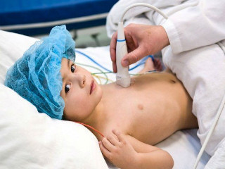
The procedure does not harm the human body, is not painful, and allows you to accurately assess the state of the human heart. The procedure is safe even when carrying a child, it is also performed with newborn babies.
As part of the procedure, a diagnostician examines the structure of the heart muscle and blood vessels, and also analyzes the work of a person's blood flow.
Signs of various disorders of the heart system are too similar to disorders of the respiratory and nervous systems.
However, echocardiography is recommended as soon as the following symptoms appear:
- Accelerated or slowed heart rate, as well as "fading" of the heart.
- Shortness of breath during exertion, cough of a different nature, discomfort and pain in the sternum and upper abdomen, an increase in the size of the liver.
- Frequent dizziness and pain, loss of consciousness.
- Swelling, pallor of the skin, cooling of the hands and feet, increased pressure.
- An increase in temperature, combined with shortness of breath and an increased rhythm.
Due to the overload of the body during training, all people who are professionally involved in sports are periodically subjected to the EchoCG procedure.
In addition, this study is used for the following indications:
- If the doctor detects noise while listening to the chest.
- Changes to ECGcausing the doctor's questions.
- Discovered during the X-ray procedure pathology of the heart muscle or vessels (the size or shape of the organ is changed).
- When a heart attack occurs.
EchoCG is prescribed for juvenile patients with the same manifestations as in adults. This procedure is very important for babies to pass in order to exclude congenital heart defects and other diseases of the heart system.
In addition, when adolescents turn 14 years old, it is necessary to regularly undergo this procedure, since during transition period, when the body is growing and developing intensively, there is a risk of developing cardiac pathologies mechanism.
According to the standards in force on the territory of the Russian Federation, all minors under one year old need undergo ECG and EchoCG procedures during routine medical examination and get a free consultation cardiologist.
During the carrying of a child, a woman is prescribed a procedure with the following indicators:
- If a woman is genetically predisposed to defects of the heart system.
- The previous pregnancy ended in miscarriage.
- With diabetes mellitus.
- When taking antibiotics or drugs for epilepsy in the first and second trimesters.
- In case of illness while carrying rubella or detecting a high level of rubella antibodies.
 Even if the expectant mother has no indications for the EchoCG procedure, it is better to play it safe and go through it, excluding the development of pathologies. The fact is that during pregnancy, the load on the body and on the heart in particular increases significantly. Ultrasound of the heart is used at any age and condition, it has no contraindications and side effects, it is absolutely safe.
Statistics show that in Russia every year 1,300 people die from various heart pathologies. Diagnosis using the procedure in the early stages and the regular ultrasound procedure to study the heart will significantly reduce the indicators.
As such, there are no contraindications for the study.
However, some factors can significantly affect the diagnostic results:
- Large size of mammary glands in female patients.
- Severe curvature of the chest.
- Disease of bronchial asthma and long experience of smoking.
There are several types of ultrasound:
- First - one-dimensional ultrasound examination in M-mode. Instruments show data online and present a top view. During the study, it is possible to calculate the linear dimensions of the heart chambers and the gaps between the valves. It is used to study people of different ages, including newborns. Apparatus were extremely common in the era of the inception of this procedure.
- The second kind - two-dimensional ultrasound. Shows the heart muscle in two dimensions. The doctor is able to calculate the area of the openings of the mitral, aortic and tricuspid heart valves, and the lumen of the great vessels.
- Well, the third one - three-dimensional ultrasound. The most applicable method of examining the heart today. Gives the most complete picture of blood circulation and the anatomical structure of the heart muscle. Today, diagnostic devices provide a real image of an organ in volume online, which has an extremely favorable effect on the speed of diagnosis.
Ultrasound of the heart: preparation for research and methods of conducting
 Preparing for an ultrasound of the heart is not very difficult, therefore it does not cause any discomfort to a person. But first, it's worth talking about the methods of the procedure.
Transthoracic echocardiography - performed through the chest. It is recommended to undergo this ultrasound scan annually, especially for people suffering from cardiovascular diseases, as well as congenital defects.
The procedure does not provide for any special preparation for the ultrasound examination.
You only need to have a sheet with you (it covers the couch) and a towel to wipe the gel off your skin. Sometimes it is not possible to obtain accurate research results. The reason may be an inserted prosthetic valve, too dense muscle tissue and other defects. In this case, they resort to another method of echocardiography.
Transesophageal ultrasound - carry out the manipulation through the esophagus. This method requires preparing the patient for an ultrasound of the heart. It is forbidden to take food and liquids 6 hours before the ultrasound scan. The method is not recommended for patients with esophageal diseases.
Abdominal echocardiography - usually, this method is resorted to when it is necessary to examine the heart of the fetus. Preparation for ultrasound of the heart is necessary only if the gestational age is not long.
The woman is prescribed activated charcoal, a few days before the examination, to clear the intestines of gas. Their presence can negatively affect the objectivity of the results. A procedure is done to identify abnormalities in the fetus, the quality of the heart muscle and other characteristics. The sensor of the device is moved along the anterior abdominal wall of the abdomen.
Stress echocardiogram - the patient is prescribed medication or exercise. The duration of the procedure is 45 minutes, during this time, the cardiac chambers and the changes occurring in them, when exposed to physical activity on the body, are examined.
To obtain a more objective result from an ultrasound of the heart, preparation for the study should include certain actions.
Despite the fact that this manipulation can be carried out without preparatory procedures and be done up to several times a day, it is still worth adhering to some rules before the examination.
These include:
- The need to exclude physically active activities.
- Stop taking sedatives and invigorating agents.
- Try not to eat much.
- Limit the amount of caffeinated drinks you drink.
- If the patient suffers from high blood pressure, he should consult a cardiologist about whether there is a need to reduce the pulse and pressure or not.
To prepare for the study of transesophageal echocardiography, at least a quarter of the day before the procedure, refuse to eat.
How an ultrasound of the heart is done: carrying out various variations of the procedure
 How ultrasound of the heart is done for different types of research, we will figure it out further. To ensure perfect visibility of small structures that appear on the monitor, the illumination level is lowered in the office during the examination.
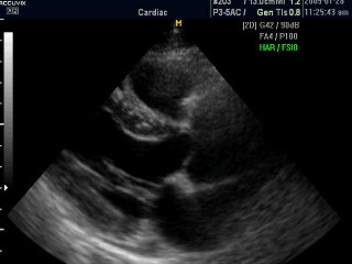
The patient should lie either on his back or, if necessary, on his left side. Typical exploration time is just over 10 minutes. This time is enough for the doctor conducting the diagnosis to carefully examine and measure all the necessary structures. If there are obvious pathologies, the doctor takes a snapshot from the screen.
Echo-KG lasting about 15 minutes is completely painless for patients. To conduct the examination, the patient must be undressed from above, without jewelry. Then the patient lies flat on his back and, with the help of special equipment, an ultrasound of the heart is done. The sensors, which produce images that show the heart and blood vessels, are placed on the chest, which is lubricated with a gel.
In addition to the usual echocardiogram of the heart, there are 2 more methods: stress echo cardiography and transesophageal cardiography. Since ultrasound of the heart is performed only by the most highly qualified specialists in this field, it is possible to undergo examination only in special medical institutions. Below we will characterize each of these methods.
A transesophageal ECG is usually prescribed not in the most trivial cases, for example, if there is a suspicion that the heart valves have been damaged by bacteria, or the valve is artificial, or if there is a likelihood of an atrial septal defect, and sometimes it can be prescribed after a stroke, with atrial fibrillation arrhythmias.
This procedure is carried out as follows: first, the patient's mouth and throat are treated with lidocaine, this is done to reduce the level of discomfort that causes the gag reflex. After that, as the name suggests, an endoscope is inserted through the esophagus into
Stress echocardiography is an examination that is done during stress on the heart muscle. This can make it possible to identify those pathologies that cannot be detected by a conventional ultrasound of the heart.
Obviously, some types of problems appear with a certain level of physical activity, such as movement. For the study, a treadmill or a bicycle ergometer is used, and sensors are installed on the chest that continuously take readings from the work of the heart. This type of ECG does not require any special preparation.
Ultrasound of the heart: what the decoding of the results will show
After the ultrasound of the heart, which will be shown by the decoding of the results of the ultrasound of the heart muscle, the cardiologist or ultrasound specialist will tell the patient.
This is because only a specialist is able to professionally assess the indicators of research and establish a connection with other research methods.
EchoCG can detect defects:
- Mitral valve prolapse.
- Valve defects.
- Abnormalities in the development of the heart muscle.
- LV myocardial underdevelopment.
- Cardiomyopathy.
 Deciphering the results of the diagnostic method should be dealt with by the doctor observing the patient. During the study, the specialist fills in the protocol, which contains the standard indicators of the echocardiogram.
When the doctor receives the data of an ultrasound scan of the heart, deciphering the results will not take him much time. The cardiologist must explain the results to the patient and summarize the resulting picture. It is also extremely important to understand that compliance norms for a child directly depend on the size of the baby's body. When calculating, the doctor uses a special index, which is multiplied by body weight.
As part of an ultrasound scan of the heart, all structural objects of the heart muscle are checked: cavities and valves, the pericardium, the walls of the atria, the pancreas and the left ventricle, blood vessels and muscles.
Defects that the study can show:
- congenital abnormalities (for example, an open window dividing the atria, an open arterial flow, a pathology of the ventricular septum);
- acquired deviations (lack of a mitral valve, stenosis of the aortic valve, valve pathology with endocarditis);
- violations of the muscle layer (myocarditis) - can be seen with an increase in the volume of the chambers, a decrease in the volume of blood coming from the LV, a decrease in the number of contractions, reverse blood flow;
- wateriness in the pericardial sac indicates the presence of exudative pericarditis;
- a heart attack is diagnosed when a dead muscle area is detected due to the lack of contractions;
- a thin and bulging wall indicates an aneurysm;
- ischemia;
- thrombosis and formations of a different nature.
Often, Doppler analysis with ultrasound of the heart is used to compile a complete picture of the disease.
What will this method show? With it, you can conduct a deep analysis of the functioning of the heart muscle and large vessels.
There are several Doppler analyzes:
- Pulse-wave. With the help of this type, an analysis of the speed of blood flow in the vessels and valves is performed, as well as an assessment of the pumping function of the heart muscle.
- Continuous wave. It is used in the study of high blood flow velocity, as well as to measure the pressure in the vessels and parts of the cardio-muscle.
- Color - the study makes it possible to measure and analyze the speed and direction of blood flow through large arteries.
 The territorial location of the medical clinic and the qualifications of the receiving doctors directly affect the prices of the procedure, in particular with Doppler equipment.
An ultrasound of the heart, decoding the results and making a diagnosis will cost from 1,500 to 4,000 rubles. EchoCG through the esophagus will cost a little more - within
It should be noted that the above prices are valid in terms of commercial clinics. State support clinics carry out ultrasound of the heart completely free of charge, but making an appointment is almost always problematic. is carried out in several weeks, but they will immediately tell you what the examination shows, and the doctor will carry out the main decoding of the results at the reception. However, a government agency may not have the additional equipment required for a complete analysis.
In fact, the ultrasound procedure is not that expensive, and sometimes it is much easier to apply for examination at the private medical clinic close to your home or work.
The main advantage of ultrasound research is the absence of harmful effects on the human body, in contrast to X-rays.. The method is absolutely safe for any category of citizens, it is not associated with complications and side effects.
Women who are carrying a child or newborn can also safely carry out the procedure. Today, devices can visualize all components of the heart section with high accuracy and carefully assess the quality of the work of the human heart and blood vessels.
The procedure is available in any private clinic that offers a wide range of services, including the services of a cardiologist. In the clinic, you will be admitted as soon as possible and without annoying, depressing crowds.
The study can be carried out for preventive purposes, and based on the testimony of the observing doctor. The clinic guarantees that all manipulations will be carried out with comfort and convenience for all citizens, both for adults and for young patients.
Thanks to ultrasound studies, doctors manage to save a huge number of lives and avoid dangerous surgeries every year.
Partial use of site materials (no more than 30% of the content of the article) is allowed only with a hyperlink on www.med88.ru, full use of the article (more than 30% of the article content) is possible only with written permission edition.