Deformation of the auricles and otoplasty

A pathology in which there is a violation of the shape or integrity of the external ear is called a deformity of the auricle.Depending on the severity of the condition, it may or may not affect the hearing of a person. Correction is performed by a surgeon specializing in otoplasty, but only after consulting with an otolaryngologist. Table of Contents: General information Reasons Classification Diagnosis Correction of deformities surgically Contraindications Effects of otoplasty
General information
At the heart of the ear shells is a thin elastic cartilaginous tissue that is densely covered with a layer of skin.This fabric is characterized by a large number of bends.Ears are fastened to the bones of the skull with the help of ligaments and muscles.In this zone there is a weak blood supply.
Because of this complex structure, operations to correct deformation of the auricles are considered technically complex.This is also explained by the need to work with a three-dimensional structure, when the expert at best recreates the size, position and structure of the organ, and at worst - the lost auditory functions.
Important! The complexity of carrying out such operations is explained by the importance of the correct shape of the ear buds for the patient.They are responsible for the formation of a harmonious image.In addition, when evaluating a person around, the integrity of perception, in other words, the degree of development of all parts of the body, is of great importance.
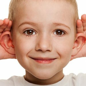
The disease can occur at an early age, giving children a lot of inconvenience.All of them are connected with the ridicule of peers and in the absence of medical care can result in a formed inferiority complex.
Reasons
Defect development can lead to:
-
 Congenital deformities of the auricles.They are reduced to the wrong placement of cartilage, as well as excess or lack of soft tissue.Develop due to the impact of adverse factors on the mother's body during the period of gestation, especially in the first trimester, when the outer ear is formed.To provoke the appearance of pathology, intrauterine infections can also occur.
 - Acquired diseases.Most often appear due to damage to the integrity of the ear as a result of injury, neglect of safety rules at work.To provoke the appearance of a defect may also be an operative intervention, a thermal or chemical burn, an inflammatory process.
Please note! A keloid scar that is, in fact, a growth of the skin along the entire length of the auricle as a result of inflammation or trauma, is also the cause of deformation.In some cases, lead to it can even puncture the ear lobe.
Inflammatory processes in the auricle often result in a thickening of the cartilage.In medical circles, pathology is called the "boxer's ear."Another option for the development of a defect is the appearance of neoplasms in the parotid zone.The most terrible form of the defect is the complete absence of the auricle as a result of trauma.
Classification
Among the congenital defects of the auricles, physicians distinguish:
-
 Macro is a pathological condition in which there is a disproportionate increase in hearing due to an excess of cartilaginous tissue;
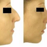
 - Lopoustness - location of the ears not parallel to the temporal bone, but at an angle of 30 to 90 degrees;
- "coagulation" of the auricles - when the cartilaginous tissue bends inward or downward;
- pathology of the lobe - increase in size, bifurcation, growth or complete absence;
- curl defects - the appearance of a protrusion on it, which is called the tubercle of Darwin, the sharpening of the upper pole of the shell in a complex with a smoothed curl( "satire ear"), excessive flattening( "macaque ear");
- microtia - it lies in the underdevelopment of the auricles.
Please note! In microtia, the ears may be ingrown, too small or flat.Pathology is often accompanied by underdevelopment of the middle ear, as well as anomalies in the development of a certain side of the face.If there is a bilateral microtia, the patient becomes disabled as a result of hearing loss, speech.
There are 3 degrees of underdevelopment of the auricle:
-
 The first - is characterized by the presence of separate parts of the body, although in a reduced size.
 - The second - is characterized by the presence of a roller instead of a curl, which is located in place of the auricle.
- Third - diagnosed with complete absence of auricle.In place of the latter may be a formless tubercle.
Diagnosis
Problems with the auricle are visible to the naked eye.Meanwhile, to assess the situation, doctors can assign additional research, the essence of which is to identify the true causes of deformation and the degree of its complexity.Most often such methods are computed tomography and MRI.
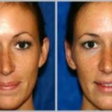
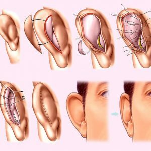
Correction of deformities surgically
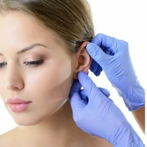 Otorhinolaryng surgery is one of the areas of plastic surgery that corrects defects in the auricles. During the operation, a specialist tries not only to accurately reproduce all parts of the outer ear while maintaining its size, structure and position, but also to return the patient to a lost hearing.The success of the procedure is in the correctness of the choice of technique.
Important! In childhood, elimination of defects in the auricles is not performed.The optimal age is 7 - 9 years.Previously, it is difficult to achieve satisfactory results due to the features of growth and development of the body, because at this time it is still being formed.An exception to this rule are situations in which the slightest delay is fraught with loss of hearing or the development of complications.Acquired deformations due to the transferred injuries are eliminated not earlier than 18 months after the event.
The defect removal operation is performed in several stages, between each of which an interval of 2 to 4 months is maintained.This technique is due to the complexity of the relief of the outer ear, lack of parotid tissue and increased scarring of the operated area.The basis of the new auricles is created from the costal cartilage.
Before performing otoplasty, the specialist informs the patient about the risks associated with it, as well as the possible consequences of a prolonged rehabilitation period.After that, images are created in different angles, on the basis of which, using the Gonzalles-Wola technique, missing elements of the auricle are drawn.
Please note! In case of severe deformations, the patient is operated under general anesthesia, and with mild deformations, under local anesthesia.
At the initial stage of otoplasty, a subcutaneous pocket is formed on the site of the missing element of the auricle, which is subsequently filled with a cartilaginous tissue.When it takes root( it usually takes up to six months), it is detached with the skin, and in its place the ear is formed with the help of a skin graft.As a rule, the course of otoplasty with all stages is extended for up to 12 months.
The operation to eliminate the defects of the "ears of wrestlers", which are distinguished by cartilages broken during the competition, is reduced to recreating their anatomical shape. This cartilage plate is cut from the back of the ear, after which the specialist restores the integrity of the cartilage and fixes them in the desired position by using special rollers.The latter is left for a period of up to 7 days, and then taken off.
Despite the fact that the seams are removed for 10 to 12 days, the fixing bandage, which protects the body from injury, is recommended to wear for at least 14 days.
Important! In some cases, the auricle is held in the correct position with the help of treads or elastic rings.
Contraindications to otoplasty
The operation to correct defects of the auricle is not carried out if:
-
 internal diseases;
 - bleeding disorders due to medication;
- of ongoing infectious processes;
- malignant or benign neoplasm;
- of ear inflammation;
- of diabetes;
- hypertension.
It is worth noting that when healing wounds after otoplasty, ear tissue can be deformed again.The reason for this is a keloid scar.That is why, if there is a high risk of its formation, the patient may be refused surgery.
Effects of otoplasty
Despite the complexity of the operation, it lasts from half an hour to two hours and usually ends successfully.Complications are fixed no more often than in 0.5% of cases. The most common of these:
- suppuration;
- appearance of gross scars;
- cutting of joints.
All these manifestations can negatively affect the appearance of the auricles, so the plastic surgeon necessarily informs the patient about them. The most dangerous complication is necrosis of cartilage.In order to exclude the risk of its development, operated after otoplasty is left under medical supervision for another 2 to 3 days.
The right choice of a specialist and the procedure for conducting an operation is the key to its success.It is justified in 90% of cases.In the rest, due to asymmetry or complications, the patient returns to the operating table repeatedly.
Deformation of the auricles is a serious pathology, which at best spoils the appearance and provokes the development of an inferiority complex, and at worst - leads to hearing loss.Meanwhile, the diagnosis is not a verdict.The defect is successfully corrected with the help of one of the directions of plastic surgery - otoplasty.
Совинская Елена, medical reviewer