
Keratitis: causes, symptoms and treatment, photos, consequences
Keratitis is the general name for a number of diseases of the visual analyzer, a distinctive characteristic of which is inflammation of the cornea of the eye.
Due to the accumulation of infiltrate, opacity occurs, which, in the case of a purulent infection, is accompanied by necrosis of the corneal tissue.
Pathology can arise as an independent disease, or become a consequence of already existing disorders in the body.
Keratitis of the eye leads to a decrease in visual acuity, glaucoma and even loss of an eyeball. The sooner medical care is provided to the patient, the more chances to avoid undesirable consequences.
This article will help you understand what keratitis is, describe its symptoms and treatment.
Content
- The causes of keratitis
- Provoking factors for the development of the disease
- Keratitis symptoms
- Classification of keratitis
- Congenital keratitis
- Diagnostics
- Keratitis treatment
- Drug treatment
- Surgery
- Treatment of keratitis with folk remedies
- Keratitis prevention
- Forecast
- Related Videos
The causes of keratitis
Keratitis has a variety of etiologies. But in most cases (about 70%), the causative agents of the disease are viruses of a simple and herpes zoster (shingles). Other reasons include:
- infections:
- viral (causative agents are: adenoviruses, varicella-zoster virus, measles);
- bacterial. Causes purulent keratitis of the eye (pathogens: streptococci, staphylococci, Pseudomonas aeruginosa and Escherichia coli, causative agents of tuberculosis, diphtheria, syphilis);
- fungal (candida, aspergillus, fusarium mushrooms);
- chlamydial infection (chlamydia);
- parasitic (amoeba, leishmania, worms).
- Injuries. The development of the disease is provoked by damage to the cornea. These can be microdamages due to contact lenses, eye damage with sharp objects, chemical burns, operations on the visual analyzer;
- allergic reactions (allergic conjunctivitis, allergic rhinitis);
- excess ultraviolet radiation (photokeratitis);
- rheumatoid arthritis, periarteritis nodosa, Sjogren's syndrome.
Provoking factors for the development of the disease
The factors that favor the development of pathology include:
- avitaminosis;
- decreased immunity;
- disruption of the trigeminal nerve;
- diabetes;
- Availability gout history.
Keratitis symptoms
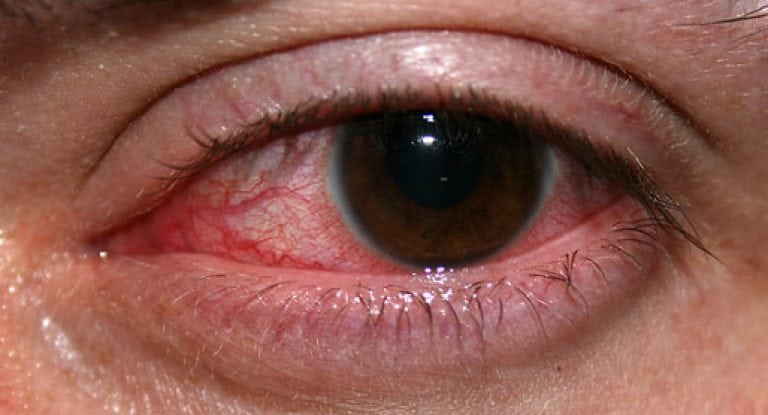
The main and common symptom for all keratitis is the corneal syndrome. They are characterized by:
- photophobia (photophobia);
- excessive tear production;
- reflex eyelid closure (blepharospasm).
Corneal syndrome occurs due to the pressure of the infiltrate on the nerve endings. The accumulation of leukocytes, plasma cells and histiocytes leads to a decrease in the transparency of the cornea, a violation of its shape and opacity.
Against this background, sensation of a foreign body in the eye, discomfort and pain. The cornea of the eye becomes rough and reddens (see. photo above). Patients report deterioration of vision.
Read also:Retinal macular degeneration (macular degeneration)
Progressive keratitis of the eye leads to flaking and sloughing of the corneal epithelium, and then the appearance of superficial ulcers. Without proper therapy, these disorders turn into exudate ulcers. Even after recovery, the tissues are not restored, and a thorn forms in this place.
Very often, keratitis occurs in parallel with conjunctivitis and inflammation of the choroid (keratouveitis) and sclera (keratoscleritis). In cases where purulent inflammation of most of the membranes of the visual analyzer is observed, the likelihood of necrosis of the organ as a whole increases.
It should be noted that the severity of symptoms and the rate of development of the inflammatory process in a person directly depend on the state of health and individual characteristics.
Classification of keratitis
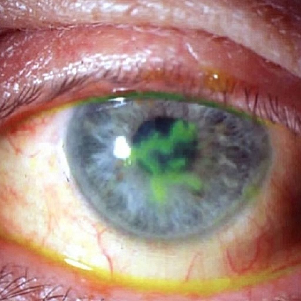
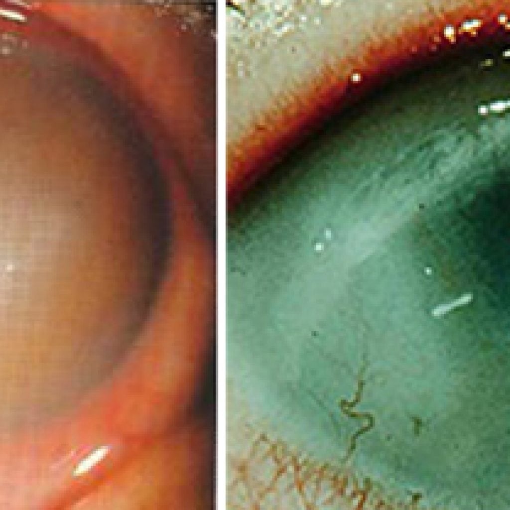
Keratitis is classified according to several criteria.
-
Due to the occurrence:
- bacterial keratitis (congenital parenchymal keratitis is separately isolated, the development of which is caused by the pathogen syphilis);
- viral keratitis;
- allergic;
- fungal keratitis;
- caused by protozoa, most often amoebas;
- photokeratitis;
- traumatic keratitis;
- neuroparalytic keratitis, occurs due to damage to the trigeminal nerve;
- unexplained etiology (idiopathic keratitis).
-
Depending on the nature of the cause that caused the inflammation of the cornea of the eye, keratitis is divided into two groups:
- endogenous, or developed due to internal reasons (vitamin deficiency, rheumatism, allergic reactions, etc.);
- exogenous, or developed due to external factors (infection, injury).
-
By the degree of damage:
- superficial (inflammation is localized in the upper layer);
- deep (all layers of the cornea are affected).
-
By the way of accumulation of inflammatory infiltrate:
- central (at the pupil);
- paracentral (opposite the iris);
- peripheral (at the limbus).
Congenital keratitis
Parenchymal keratitis is an extreme stage of congenital syphilis. It manifests itself even after several generations. People are especially susceptible to it up to 20 years.
Pathology is characterized by cyclicity, bilateral lesions, reddening of the choroid and the absence of corneal manifestations.
Diagnostics
Corneal syndrome is the first sign that will help an ophthalmologist diagnose inflammation of the cornea of the eye in a patient.
For further research, the specialist instills anesthetic drops in the patient. This will help relieve the spasm and open the eyelids.
The next step will be taking anamnesis, general examination and checking visual acuity.
To clarify the diagnosis, ophthalmologists resort to such additional research methods as:
- biomicroscopy (study of the structures of the visual analyzer by means of a slit lamp);
- ophthalmoscopy (allows you to assess the changes that have arisen as a result of the inflammatory process);
- staining of corneal tissue with fluorescein for better visualization of structural changes;
- Ultrasound.
Read also:Strabismus
After the diagnosis is made, in order to prescribe an individual therapy, the doctor prescribes additional tests. The goal is to find out the causes of the disease. The patient hands in:
- allergy tests;
- rheumatic tests;
- blood for PCR (for the detection of the herpes virus) and for antibodies to the causative agents of syphilis;
- smear for bacteriological analysis.
Based on the research results, the doctor prescribes treatment. Often, keratitis is treated by specialists of several directions. For example, in cases where a patient is diagnosed with parenchymal keratitis, a venereologist is connected to therapy, if the source of the disease is a tuberculosis pathogen - a phthisiatrician.
Keratitis treatment
Treatment of keratitis of the eye is mandatory in a hospital and lasts five days. Continues at home if necessary. If a patient has keratouveitis in parallel with keratitis, the symptoms of the disease can persist for up to several months.
Drug treatment
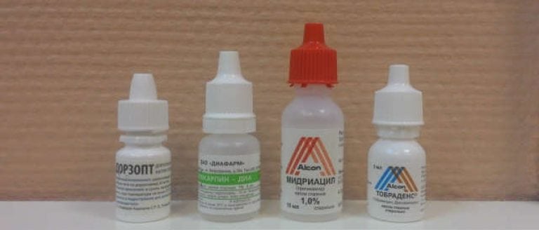
To achieve a speedy recovery, local and systemic therapy is used.
Systemic therapy necessarily implies:
- taking drugs designed to destroy pathogens. With purulent inflammation, antibiotics are prescribed (Ciprofloxacin, Ceftriaxone, Moxifloxacin). With the viral nature of keratitis - Acyclovir. Fungal keratitis is treated with Voriconazole, Fluconazole, Itraconazole;
- detoxification activities (droppers with saline or Rheosorbilact, drinking plenty of fluids);
- the appointment of antihistamines to reduce puffiness (Zyrtec, Zodak, Desloratadin).
Additionally, the patient is prescribed aloe extract to improve the trophism of the affected tissues and multivitamin complexes.
Keratitis of any nature requires local treatment. For this purpose, apply:
- eye drops containing antibiotics (Tobrex, Floxal);
- eye drops with an antiseptic (Oftamirin, Okomistin, Albucid), which will not only adversely affect pathogenic microorganisms, but also prevent the addition of a secondary infection;
- drops of Okoferon, if a virus is the cause of the disease;
- Zovirax (ointment), if the causative agent of keratitis is the chickenpox or herpes virus;
- drops with corticoids (Maxidex, Sofradex, Tobradex), which prevent damage to the deep layers of the cornea;
- ointments or gels that accelerate the regeneration processes (Lakropos, Korneregel, Vidisik).
Medical treatment of eye keratitis, as a rule, is complemented by physiotherapy (phonophoresis, electrophoresis, magnetotherapy).
Parenchymal keratitis requires specific treatment. First step - preparation - taking iodine preparations, then intramuscular injection of Bioquinol.
Second phase - penicillin treatment for 14 days. Together with the antibiotic, mydriatics (Atropine, Irifrin, Cyclomed), yellow mercury ointment, dionin solution and corticosteroid drugs are locally prescribed.
Surgery
The indication for surgery is the presence of ulcers on the cornea of the eye. To eliminate tissue defects, modern methods of microsurgery are used: laser coagulation and cryoapplication. If a scar has formed, keratoplasty is performed.
Read also:Keratoconjunctivitis
With advanced forms of the disease, when the inflammatory process cannot be stopped by conservative methods, they resort to removing the eyeball.
Treatment of keratitis with folk remedies
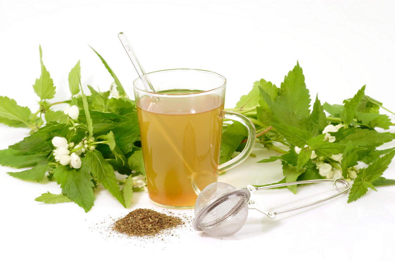
Traditional medicine suggests treating diseases of the cornea of the eye with the help of compresses, lotions, steam baths.
- To prepare the mixture you need to boil potatoes "in their uniforms" (1 piece), knead them, add one egg yolk. Then add boiled milk until gruel is formed. The resulting mixture on a cotton cloth is applied to the closed eye for 20 minutes. The procedure is repeated three times a day.
- Flush the inflamed organ it is possible with a solution of honey at the rate of 1 tsp. 200 ml of boiled water.
- Relieve pain and reduce photophobia sea buckthorn oil helps. It is recommended to bury it every hour, 1 drop in the first days of the disease, then every 3-4 hours.
- Eyebright decoction erect is taken orally. Prepare it like this: 3 tbsp. dry herbs are poured with boiling water and insisted for 6 hours, then passed through cheesecloth. The finished solution is stored at a temperature not exceeding 10 degrees. Take 100 ml 2-3 times a day. The course is 30 days.
It is important to remember that you should not risk your health and self-medicate! Any therapy should be carried out only under the supervision of a doctor!
Keratitis prevention
You can prevent inflammation of the cornea of the eye if you are attentive to your health and follow simple rules:
- observe eye hygiene;
- choose the right contact lenses and take care of them;
- avoid ingress of foreign bodies and injuries;
- promptly treat any diseases, and in particular, the visual analyzer;
- eat balanced.
Forecast
The outcome of the disease mainly depends on the nature of tissue damage, the location of the infiltrate and the complications that have arisen.
Therapy at the initial stages of pathology shows good results - the infiltrate dissolves without consequences or there are slight opacities.
If the treatment was not provided on time, the outcome of the disease can be:
- thorn;
- glaucoma;
- optic nerve atrophy;
- atrophy of the eyeball;
- loss of visual function.