Dacryocystitis: what is it, causes, symptoms, treatment, prognosis
Content
- Description of the disease
- ICD 10 classification
- Anatomical structure of the lacrimal canal
- Dacryocystitis reasons
- Dacryocystitis symptoms
- Diagnostics
- Dacryocystitis treatment
- Antibiotic therapy
- Infusion therapy
- Massage (recommended for children)
- Dacryocystorhinostomy
- Physiotherapy
- Treatment with folk remedies
- Prognosis and complications
- Prevention of dacryocystitis
Description of the disease
Dacryocystitis Is an infection or inflammation of the lacrimal sac, usually accompanied by a blockage in the nasopharyngeal duct. Dacryocystitis can be acute or chronic and congenital or acquired. May cause pain, redness and discomfort.
The disease is more susceptible to women than men. The lacrimal canal of women is narrower, this anatomical feature is a provoking factor. Dacryocystitis occurs between the ages of 30 and 60.
ICD 10 classification
Assigning a code according to ICD 10 makes it possible to differentiate the disease of the lacrimal apparatus for statistical and dynamic research in medicine of the incidence of the population. Dacryocystitis belongs to the H04.3 group "Acute and unspecified inflammation of the lacrimal ducts." It includes:
- Phlegmatic. Canal obstruction, narrowing of its ducts leads to the accumulation of fluid. Inflammation begins when there are foci of infection in the sinuses or mucous membranes of the nose. An abscess develops, fluid accumulates in the cavity.
- Subacute (unspecified). It occurs in the presence of provoking factors that lead to an exacerbation of acute dacryocystitis. It can be an allergenic factor, traumatic and other forms after acute dacryocystitis.
Dacryocystitis of newborns is included in the P39 class, which includes infectious diseases in the perinatal period. Dacryocystitis of newborns has code P39.1. Its formation takes place both in the prenatal period and after birth for the first time 4 weeks.
A separate group includes chronic dacryocystitis, which has code Н04.4. It occurs when the nasolacrimal ducts are narrowed during pathological processes in nearby mucous membranes (conjunctivitis, sinusitis and other inflammatory diseases).
Anatomical structure of the lacrimal canal
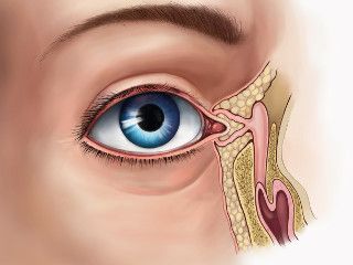
The tear performs antibacterial and moisturizing functions of the cornea and conjunctiva of the eye, it also nourishes and cleanses the eyes. Covering the cornea with a thin layer, it provides normal biophysics of the visual apparatus (refraction and reflection of light).
According to its composition, the tear is 98% transparent, consisting of water, slightly alkaline liquid, which consists of protein, lysozyme, fats, mucus, sugar, Na, K, Cl, urea. During the day during wakefulness, 0.5 - 1 ml of tear fluid is produced, and during crying or ingestion of a foreign body, about 2 teaspoons.
The lacrimal gland belongs to the accessory organs of the eye. It belongs to the group of lacrimal organs, which also include the lacrimal ducts, small lacrimal glands.
The lacrimal gland is a complex tubular serous gland. It consists of parts:
- Orbital (orbital) - in the fossa of the gland, in which there are 3-5 excretory ducts. Large-caliber ducts are covered with two-layer epithelium, and smaller-caliber - single-layer. The excretory tubules of the upper eyelid have ducts between the lobules of the eyelid.
- The secular is located under the upper fornix of the conjunctiva. Has 3 - 9 tubules and tubules flowing into the orbital.
Read also:Photophobia (photophobia)
It is supplied with blood by the orbital artery, and the lacrimal vein carries out the outflow of blood. It is innervated by trigeminal, facial, sympathetic nerves. The secretion of parasympathetic nerve nodes is regulated.
Sometimes in newborns, the nasolacrimal canal is closed by a membrane that dissolves in the first weeks of life. After 2-3 months, it is replaced by cellular tissue.
Dacryocystitis reasons
Main reasons:
- sinusitis (form ethmoiditis);
- atresia of the nasolacrimal ducts (their absence) - congenital pathology;
- rhinitis;
- polyps in the nasal cavity;
- inflammation of the conjunctiva;
- impaired patency of the Gasner valve (congenital);
- neonatal infectious processes;
- infections (tuberculosis, syphilis, intestinal, viral);
- helminthic invasions;
- immunodeficiencies;
- candidiasis of internal organs;
- anatomical anomalies of the structure of the facial part;
- ECC syndrome;
- diabetes;
- injury to the lacrimal gland;
- allergy;
- dustiness.
The main cause of the inflammatory process is the accumulation of microorganisms in the ducts of the lacrimal gland and a violation of the outflow of fluid. Often, when sowing tear fluid, the following are found:
- Staphylococcus aureus;
- Pseudomonas aeruginosa;
- Epstein-Bar virus;
- pale treponema;
- mushrooms.
The development of dacryocystitis can be provoked by the internal hematogenous spread of infectious agents.
Dacryocystitis symptoms
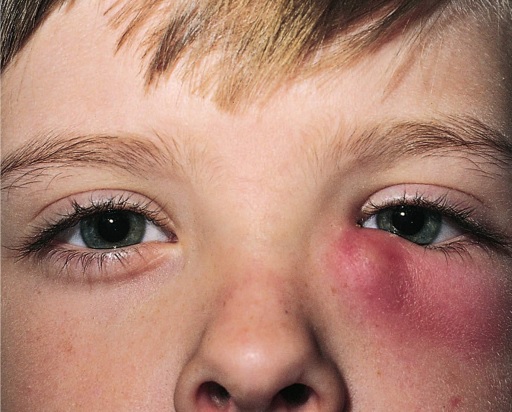
Dacryocystitis has specific symptoms. The acute form is characterized by:
- pain;
- redness;
- lacrimation;
- swelling;
- pus;
- hyperemia;
- fever;
- weakness;
- narrowing of the palpebral fissure.
Soreness of the inner corner of the eye and irradiation of pain into the sinuses, their spread to the conjunctiva and cornea creates the risk of complications and the spread of inflammation in the sinuses.
Chronic dacryocystitis is manifested by persistent lacrimation and bean-like protrusion.
Redness of the mucous membrane, swelling and thinning of the eyelid skin, enlargement of the gland are specific symptoms of the clinic of the chronic form of the disease.
Dacryocystitis of newborns is expressed by lacrimation, purulent secretion, swelling of the eyelids. The anatomical structure of the nasolacrimal canal is wide cavities, allowing the risk of spreading to nearby sinuses and to other parts of the visual apparatus.
Diagnostics
Clinical manifestations of the disease when contacting a specialist requires some differentiating diagnostic procedures. The specialists they turn to are an ophthalmologist, an otorhinolaryngologist, an infectious disease specialist, and a dermovenerologist. When contacting a doctor, the primary diagnosis consists of:
- collection of anamnesis of the disease (when it began, how long and other points last);
- palpation;
- directions for instrumental and clinical research.
In clinical studies, leukocytosis is detected in a general blood test. Research is also conducted:
- blood sugar;
- Australian antigen;
- Analysis of urine;
- ELISA for syphilis.
Read also:Barley on the eye, what is it, causes, symptoms and how to treat
Instrumental research:
- Biomicroscopy of the eye - a slit lamp examination of the visible parts of the eye with magnification. The procedure is widely used in the diagnosis of eye diseases.
- Vesta color test. A tampon is inserted into the nasal cavity, and a colloidal solution is instilled into the eyes. With an average patency of the lacrimal canals, the tampon is stained after 8-10 minutes, this time is average. If more time has passed, then this is a diagnostic sign of obstruction of the lacrimal canals.
- Radiography. X-rays with the use of a contrast agent allow diagnosing foreign bodies and seals. X-rays are taken together with the sinuses. Children under three years of age do not have sinus x-rays.
- Dye disappearance test. Fluorescein is buried and after a while the height of the lacrimal meniscus is measured.
- Echography. Diagnoses expansion, stagnation of the lacrimal canal.
- Sounding.
- Bacteriological culture of the contents of the lacrimal canal.
This is a standard program in the diagnosis of eye infections.
Dacryocystitis treatment
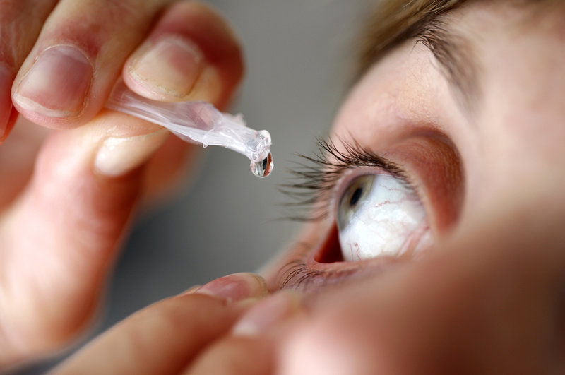
The tactics of treating dacryocystitis depends on the severity and is prescribed only by an ophthalmologist, with control of the dynamics of the disease. The options are described below.
Antibiotic therapy
Drops in the conjunctival sac are applied 1 drop 5-6 times a day for 7-10 days:
- gentamicin (0.3%);
- sulfacetamide (10-20%);
- polymyxin;
- chloramphenicol (0.25%).
Eye ointments are placed in the conjunctival sac 1 time per day for 7 to 10 days:
- tetracycline 1%;
- tobramycin 0.3%;
- erythromycin 1%.
Intramuscular injections, which are calculated by weight and the frequency is determined individually:
- gentamicin;
- ampicillin.
The drugs are used in combination with bacteriostatic and bactericidal action on the body.
Infusion therapy
Within 1-3 days, glucose-salt solutions are used to relieve the intoxication symptom. Vitamins (ascorbic acid), hormones (prednisolone, dexamethasone) and other substances that are used for symptomatology are added to the solutions.
Massage (recommended for children)
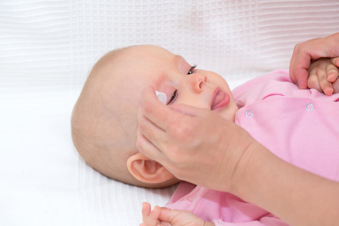
Contraindications for massage is a purulent-inflammatory process (risk of infection of surrounding tissues). Before the procedure, hands are washed and treated with an antiseptic, gloves are put on. Rinse the child's eyes with furatsilin solution. The multiplicity of massage performance for newborns and adults is different, so for adults - 3-5 times, and for newborns up to 7 times a day. Course 10 days.
Execution technique: with warm, warm hands, light movements from the beginning of the eyebrows in circular movements go down the wings of the nose to its tip.
Dacryocystorhinostomy
Dacryocystorhinostomy is a surgical operation performed to restore the patency of the lacrimal canal by reconstructing the anastomosis between it and the nasal cavity.
Distinguish between balloon, laser, endoscopic. Endoscopic surgery or probing of the nasolacrimal canal is performed to clear the canal with a probe and avoid complications in the future.
Read also:Redness of the eyes: causes and treatment
For newborns, it is performed under general anesthesia. Anesthesia is designed for the period of the operation (sparing mode). A probe made of soft materials is used that easily accepts bends and does not damage the canal walls.
Physiotherapy
UHF, UHF, carried out in complex therapy with antibiotics at an early stage, reduces the risk of developing suppuration.
Manipulations complicating the course of the disease are contraindicated in the treatment. These include:
- wearing contact lenses;
- blindfolds;
- conduct corneal studies (eye ultrasound, tonometry and other manipulations).
The result of the treatment depends on the accuracy of the manipulations and the qualifications of the specialist, as well as the conditions of the clinic in which the treatment is being carried out.
Treatment with folk remedies
Alternative medicine in ophthalmology is powerful. But the use of traditional medicine methods requires consultation with a specialist. Use infusions (inward and outward), washings. The treatment uses:
- chamomile (rubbing the inflamed areas with a cotton pad);
- Kalanchoe leaf (juice as eye drops);
- eyebright (inward and outward);
- collection (sage leaf, eucalyptus, oregano, mint, dill) is used as a lotion.
Prognosis and complications
With high-quality and short-term treatment, the prognosis is favorable. Complications can be:
- Phlegmon (abscess lacrimal sac): spread of suppuration to nearby tissues causes swelling. Complicated by the development of sinusitis and corneal damage.
- Corneal ulcer: destruction of corneal tissue during infectious - the spread of infection outside the lacrimal sac. In case of non-infectious disease, insufficient moisture in the cornea leads to dry eye syndrome. This is a concomitant factor in dacryocystitis.
- Thrombophlebitis of the orbit of the eye: the spread and settling on the walls of the veins of microorganisms from the foci of infection leads to blockage of the veins and the appearance of their pattern under the skin, difficulty in the movement of the eyeballs.
- Endophthalmitis: develops with perforation (penetration) of a purulent process into the internal structures of the eye.
The complication develops both locally and spreading throughout the body. Such complications include - sepsis, meningitis, which can lead to disability and death of a person.
Prevention of dacryocystitis
The prevention of the development of dacryocystitis has several rules that apply to any disease. This is maintaining personal hygiene, avoiding traumatic situations and using protective equipment. The use of high-quality cosmetics, caring products, as well as the treatment of colds and infectious diseases.
Dacryocystitis is a disease that can be disguised as ENT diseases. Complications of the disease can lead to blindness. Timely treatment ensures the preservation of vision. Recovery depends on the tactics of treatment and the method of the specialist.