Syringomyelia: what is it, causes, symptoms, treatment, prognosis
Content
- What is Syringomyelia?
- Syringomyelia reasons
- Forms of syringomyelia
- Syringomyelia symptoms
- Affected populations
- Diagnostics
- Syringomyelia treatment
- Forecast
- Complications
What is Syringomyelia?
Syringomyelia Is a condition in which a fluid-filled cyst forms inside the spinal cord called a fistula. Over time, the fistula can enlarge or lengthen the damaged spinal cord and compress and damage the nerve fibers that carry information to the brain and from the brain to the rest of the body.
Usually, a kind of body fluid known as cerebrospinal fluid (CSF) surrounds and protects the brain and spinal cord. Cerebrospinal fluid also fills the ventricles, which are interconnected cavities within the center of the brain, which extend to a small central duct that runs along the spinal brain. When a person has syringomyelia, this fluid accumulates in the tissue of the spinal cord, widening the central duct and forming a cyst (fistula). Typically, a cyst occurs when the normal flow of cerebrospinal fluid around the spinal cord or brain stem is disrupted. When the fistula affects the brainstem, the disorder is called syringobulbia.
Syringomyelia reasons
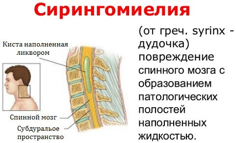
Syringomyelia can have several possible causes, but most cases are associated with Arnold-Chiari syndrome, a structural defect in which brain tissue passes through a hole in the base of the skull (foramen magnum) to the spinal canal and obstructs the flow of cerebrospinal fluid.
Injuries, spinal cord tumors, and damage caused by inflammation around the spinal cord can also cause syringomyelia. In other cases, the cause is unknown (idiopathic).
Forms of syringomyelia
There are two main forms of syringomyelia.
- Congenital syringomyelia. In most cases, the cause is an Arnold-Chiari syndrome (anomaly), leading to the formation of a fistula, most often in the cervical (neck) spine. Symptoms usually begin between the ages of 25 and 40. People with congenital syringomyelia often suffer from hydrocephalus, an accumulation of excess cerebrospinal fluid in the brain with enlargement of the cerebral ventricles. Exercise or coughing can force cerebrospinal fluid into the ventricles, causing a headache or loss of consciousness (fainting). In addition, the person may have a disorder called arachnoiditis, an inflammation of the arachnoid, one of the three membranes surrounding the spinal cord.
- Acquired syringomyelia (also known as primary spinal syringomyelia or non-infectious). The causes of acquired syringomyelia include spinal cord injury, meningitis (inflammation of the lining of the brain and spinal cord, usually caused by infection), arachnoiditis, a tumor in the spinal cord, and bleeding inside the spinal cord.
Syringomyelia symptoms

The specific symptoms and severity of syringomyelia can vary greatly from person to person. Some patients do not have any noticeable symptoms (asymptomatic course); others may have various, progressive symptoms, causing significant disability. It is important to note the highly variable nature of syringomyelia and to realize that sick people may have different sets of symptoms and different rates of progression (or no progression of the disease at all).
The disease usually progresses slowly, but a rapid onset may occur. Common symptoms include pain in the neck and shoulders. The pain can also affect the hands and can cause burning and tingling sensations. Some people who are affected also experience numbness or numbness, especially to heat and cold. Muscle weakness and wasting can also develop, especially in the arms and ultimately the shoulders. Syringomyelia often affects the upper (cervical and thoracic) parts of the spinal cord. Affected people may initially notice a loss of pain and temperature in the fingers, hands, and upper chest. In the early stages, the sensation is present. The loss of sensation can spread to the shoulders and back.
Those affected may also develop pain and stiffness (spasticity) in the legs and incoherent movement (ataxia), which at the end of the illness affects the ability to walk. In severe cases, paralysis of the arms or legs may occur. Some people experience muscle contractions, such as small involuntary muscle contractions or "twitching" (fasciculations).
Can develop skeletal abnormalities, including three-plane spinal deformity (scoliosis). In some children, scoliosis may be the only symptom. Some people develop Charcot's joints, which cause chronic progressive degeneration of the joint due to damage to the nerves that supply the joint. Charcot's joints are initially visible as swelling and redness of the affected area. Without treatment, deformity of the affected joints may occur.
Some people develop symptoms associated with damage to the autonomic nervous system, the part of the underlying nervous system that controls involuntary functions. Such symptoms include:
- loss of bowel and bladder control;
- increased sweating (hyperhidrosis);
- fluctuations in blood pressure.
Horner's syndrome, a rare condition that occurs when one of the nerves supplying the eyes and face is damaged, can occur. Horner's syndrome usually affects one side of the face and is characterized by:
- sagging eyelid;
- narrowing of the opening between the eyelids;
- a decrease in the size of the pupil;
- reduced sweating on the affected side of the face.
People with syringomyelia associated with Arnold-Chiari malformation may also have a condition called hydrocephalus, in which there is an abnormal accumulation of cerebrospinal fluid in the brain. In infancy, hydrocephalus causes a variety of symptoms, including:
- an abnormally enlarged head;
- vomiting;
- headache;
- drowsiness;
- irritability;
- convulsions;
- deviation of the eyes.
Affected populations
Syringomyelia is more common in young people between the ages of 20 and 40, but it can also develop in young children or the elderly. Some reports suggest that syringomyelia is slightly more common in men than in women. In one study, the incidence in the United States is 8.4 per 100,000 people. On the territory of Russia, the disease is spread unevenly - from 0.3 to 7.3% of the total neurological morbidity.
Diagnostics
The diagnosis of syringomyelia is based on the identification of characteristic symptoms, a detailed medical history, careful clinical evaluation, and various specialized studies. In some cases, the disease is discovered by chance when a person is examined for another reason.
A specialized imaging test called magnetic resonance imaging (MRI) is used to diagnose syringomyelia. MRI uses a magnetic field and radio waves to produce cross-sectional images of individual organs and tissues in the body, such as the brain and spinal cord. An MRI can reveal a fistula or other condition associated with a disease, such as an intraspinal tumor or an Arnold-Chiari malformation.
Syringomyelia treatment
Syringomyelia treatment focuses on the specific symptoms that each person has. Treatment may require the coordinated efforts of a team of specialists. Pediatricians, neurologists, neurosurgeons, surgeons, eye specialists (ophthalmologists) and others healthcare providers may need systematic and comprehensive treatment planning sick children.
Specific therapeutic procedures and interventions can vary depending on numerous factors, such as:
- disease progression;
- the presence or absence of certain symptoms;
- root cause;
- the effect of symptoms on quality of life;
- the person's age and general health.
Decisions regarding the use of specific drug regimens and / or other treatments must be made physicians and other members of the healthcare team, in careful consultation with the patient based on their characteristics case; careful discussion of potential benefits and risks, including possible side effects and long-term consequences; patient preferences; and other relevant factors.
Some asymptomatic patients with the disorder may not need treatment but should be monitored regularly to see if the disorder progresses.
Common therapeutic interventions include:
- pain relievers (analgesics);
- physical therapy and decreased activity, especially in the case of weightlifting.
The goal of treating syringomyelia is to restore the correct outflow of cerebrospinal fluid and eliminate the pressure exerted by the cyst on the spinal cord.
Initial treatment usually focuses on the underlying cause of syringomyelia.
For the most common cause of spinal cord fistula, Arnold-Chiari syndrome, there is no specific, agreed-upon therapy or treatment regimen. Neurosurgeons and other physicians disagree on the best approach to treating this malformation. Different neurosurgeons may recommend different surgical procedures or treatment regimens.
Like syringomyelia, people with Chiari malformation who are asymptomatic are usually not treated but monitored to make sure the disease does not progress. If you have mild or nonspecific symptoms, such as neck pain or headaches, doctors may recommend conservative treatment.
Symptomatic malformations are most often treated with surgery. There are no specific criteria or objective research to determine when to have surgery or what procedures are best. The most common surgery is known as decompression of the posterior fossa. With this procedure, the surgeon creates space by removing small pieces of bone at the back of the skull, thereby enlarging the foramen magnum. This relieves pressure and decreases the compression of the brainstem.
If a fistula in the spinal cord caused by Chiari malformation triggers the hydrocephalus, the surgeon will usually treat it first. The most common surgical treatment is a shunt that connects the dilated ventricles of the brain to another body cavity. The most common choice is the abdominal cavity. After the hydrocephalus is treated, the issue of malformation is resolved.
Surgery can be used to treat other conditions that cause syringomyelia, including surgery to remove the tumor.
In some cases, a tiny tube called a stent can be placed in the cyst. The stent allows fluid inside the cyst to drain into the area outside the spine. The procedure can stop the disorder from progressing and relieve some of the symptoms, such as pain and headaches. However, stenting can be associated with significant side effects, including spinal cord injury or infection, bleeding (hemorrhage), and blockage.
PTSD syringomyelia can be difficult to treat. The operation in this case is recommended for people with neurological impairment and / or overwhelming pain. Surgery is aimed at expanding the area around the spinal cord at the site of injury and reducing the volume of fluid. A shunt insertion can also be used to treat PTSD syringomyelia. Shunting for PTSD carries risks, including further damage to the spinal cord, and may require replacement if the shunt becomes clogged or deformed. Many doctors consider shunts to be the last resort for patients with post-traumatic syringomyelia.
Surgery can often improve symptoms and stabilize the disorder in many patients. However, after successful treatment, syringomyelia may recur, which requires additional operations.
After surgery, the doctor examines the cyst with an MRI to make sure the cyst is stabilizing or shrinking.
Forecast
The prognosis depends on the underlying cause, the magnitude of the neurological dysfunction, and the location and expansion of the fistula.
Patients with moderate to severe neurological deficits do much worse than patients with mild neurological deficits.
The natural history of syringomyelia is still not fully understood. Although earlier studies suggested that 20% of patients die at an average age of 47 years, mortality rates in modern patients as a result of surgery and better treatment of complications associated with significant paresis, such how pulmonary embolismprobably lower.
Complications
Myelopathy is the most serious consequence of syringomyelia. Below are seven classifications of myelopathy disability according to the Modified Nurik classification.
- 0 degree - no root signs / symptoms.
- I degree - root signs / symptoms are observed; there is no evidence of a fistula present.
- II degree - signs of damage to the umbilical cord; normal gait.
- III degree - mild gait problems; the patient is able to work.
- IV degree - gait disturbance interferes with working capacity.
- V degree - the patient is able to move only with help.
- VI degree - the patient is bedridden.
Complications of myelopathy include:
- recurrent pneumonia;
- paraplegia or quadriplegia;
- bedsores;
- dysfunction of the intestines and urination.