Chordoma: what is it, causes, symptoms, treatment, prognosis
Content
- What is a chordoma?
- Signs and symptoms
- Causes
- Affected populations
- Diagnostics
- Symptomatic Disorders
- Standard treatments
- Forecast
What is a chordoma?
Chordoma Is a rare tumor that develops in the bones of the skull and spine. These tumors arise from the remnants of the notochord, a flexible, rod-shaped structure that provides support for the developing embryo. During fetal development, the notochord is replaced by the bones of the spine. Notochord cells that persist in the spine can cause chordoma.
Chordomas are slow-growing tumors that cause destruction of the surrounding bone and eventually spread to the surrounding soft tissue. Sometimes the chordoma spreads through the bloodstream to other organs (metastasizes), such as the lungs, lymph nodes, liver, or other bones. Although chordoma can develop at any time in life, it is most common in older people.
Symptoms associated with chordoma depend on the size and location of the tumor. Almost all cases of the disease occur randomly for no apparent reason. In extremely rare cases, chordomas can develop in several members of the same family as a result of certain genetic risk factors (familial chordoma).
Chordomas can be classified as a type of sarcoma. Sarcoma is a general term for a group of cancers that affect bones or connective tissue - the tissues that connect, support, and surround various structures and organs in the body. Although chordomas are primary bone tumors, they are sometimes classified as central nervous system tumors when they occur near the base of the skull.
Signs and symptoms
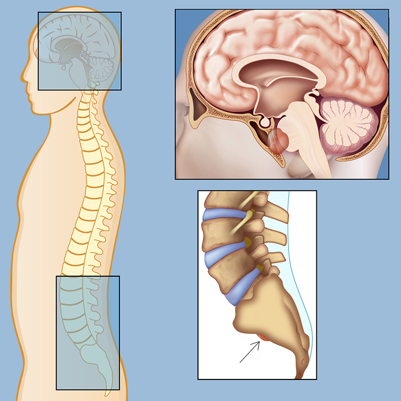
Chordoma can develop anywhere along the spine from the base of the skull to the coccyx (lower spine).
Symptoms vary from person to person and depend in part on the location and size of the tumor. Chordomas located in the lower part of the spine may be associated with pain in the lower back, in the legs, weakness and numbness in the lower back or legs, and also disorders that affect the bladder and bowels, including loss of bladder control (urinary incontinence) and / or loss of control of intestines. In some cases, swelling may be felt in the lower back.
Chordomas of the base of the skull (cranial chordomas) may be associated with double vision (diplopia), headaches and / or facial pain. Paralysis of certain facial nerves can also occur, resulting in difficulty swallowing, speech and voice impairments, and abnormal eye movements.
In some cases, an intracranial chordoma can block the outflow of cerebrospinal fluid (CSF), causing CSF to build up in the skull and put pressure on the brain (hydrocephalus). Hydrocephalus can cause several symptoms that vary with age. In infants, it can cause a bulge in the soft spots on the skull, an increase in head circumference, and a roll of the eyes downward. In older children, the condition can cause nausea, vomiting, drowsiness, double vision, rapid eye movements, and problems with balance. In adults, the condition can cause headaches, personality changes, and problems with focusing the eyes.
Chordomas in the area immediately below the skull (cervical spine) can cause neck pain, hoarseness, difficulty swallowing (dysphagia) and, less commonly, bleeding from the voice box (bleeding from larynx).
Causes
The underlying causes of chordoma are unknown. Most cases occur spontaneously and are not caused by hereditary genetic changes. The prevailing theory is that acquired genetic abnormalities or mutations result in cancerous growth of the remnants of the notochord. These genetic abnormalities can occur spontaneously for unknown reasons or, less commonly, be inherited.
Both nonfamilial and familial chordomas have been linked to the gene Tlocated on the long arm of chromosome 6 (6q27). This gene creates (encodes) a protein known as transcription factor T or Brachyury homologue. This protein is important in notochord development and is highly expressed in chordoma cells.
Most people with sporadic chordoma have one nucleotide polymorphism (SNP) in the gene T. SNPs are the most common genetic variations and are often found in human DNA. Most SNPs do not affect human health. SNP in the T gene was identified in more than 80% of patients with sporadic chordoma (compared with about 50% of people without chordoma), and, therefore, it is believed that it predisposes to the development of chordomas. However, since SNP is common in the general population, and most people who have it do not develop chordoma, it is believed that additional factors are needed for chordoma to develop.
Scientists have also learned that many familial chordomas are caused by a specific chromosomal abnormality known as duplication, in which instead of two copies of a gene T there are three copies. This additional copy T-geneappears to be associated with a strong genetic predisposition to the development of chordoma.
Abnormalities on chromosome 7 have been studied as a potential cause of familial and nonfamilial chordoma. Multiple additional complex chromosomal abnormalities (including chromosomes 1p, 3, 4, 9p, 9q, 10, and 13) have also been identified in some tumors. Whether these various abnormalities in chordoma development play into certain cases is unknown. Scientists need further research to identify the complex mechanisms responsible for chordoma development.
Affected populations
Chordomas can affect people of all ages, including young children, but they are most commonly diagnosed in people between the ages of 40 and 75 (the median age at diagnosis is 55). Collectively, chordomas affect men more often than women, in a ratio of about 2: 1. However, tumors of the base of the skull have an equal gender distribution (1: 1). In children, tumors of the base of the skull are more common. Chordomas account for about 1-4% of all malignant bone tumors and about 20% of primary spinal tumors. The incidence of chordoma is estimated at about 1 in 1,000,000 people. According to some reports, these tumors are more common in people of European descent.
Diagnostics
Chordoma symptoms are not specific. Therefore, the diagnosis is based on characteristic radiological pathological findings.
Plain X-rays or specialized imaging techniques can be used to diagnose chordoma. Such specialized imaging techniques may include computed tomography (CT) and magnetic resonance imaging (MRI). During computed tomography, a special machine uses X-rays to create cross-sectional images of the body. MRI uses magnetic fields and radio waves to produce cross-sectional images. These imaging techniques are used to detect the presence of a tumor and evaluate the size, location, and local expansion of the tumor, which helps surgeons plan any surgical procedure.
A biopsy is needed to confirm the diagnosis of chordoma. During the procedure, the doctor inserts a needle through the skin and into the tumor to obtain a small sample of cells. In some cases, a surgical procedure is required to obtain sufficient tissue for examination. A pathologist examines a tissue sample under a microscope to determine the specific type of tumor present.
Symptomatic Disorders
Symptoms of the following disorders may be similar to those of chordoma. Comparisons can be useful for differential diagnosis:
- Chondrosarcoma Is a general term for a type bone cancerthat arises from cartilage cells. Cartilage is a specialized tissue that buffers or cushions joints. Most of the skeleton of the embryo is made up of cartilage, which slowly turns into bone. Chondrosarcomas usually affect the arms, legs, and pelvis, but can affect any area containing cartilage, including the base of the skull and spine. Most cases are diagnosed in adults between the ages of 20-60. Chondrosarcomas are malignant and can spread to other parts of the body.
Standard treatments
Treatment usually requires the coordinated efforts of a team of specialists. Doctors who specialize in the diagnosis and treatment of cancer (oncologists), doctors who specialize in the use of ionizing radiation for the treatment of cancer (oncologists-radiologists), neurosurgeons, doctors specializing in diagnosis and treatment musculoskeletal system (orthopedic surgeons), as well as other healthcare professionals should systematically and comprehensively plan treatment the affected person.
Specific therapeutic procedures and interventions can vary depending on many factors, such as stage of the disease, size and the location of the tumor, the specific subtype of the tumor, the presence or absence of certain symptoms, and age and general physical health the patient. Decisions regarding the use of certain drug regimens and / or other treatments must be made by physicians and other members of the healthcare team with the patient. Thorough discussion of the potential benefits and risks of specific treatments, including possible side effects, helps the patient to make an informed decision about the preferred therapy.
Chordoma treatment usually involves surgery to remove as much of the tumor as possible while maintaining neurologic function and quality of life. Because these tumors are located near the brain or spinal cord, surgical removal may be difficult and the surgeon may not be able to remove the entire tumor despite the many operations. Removal of the entire tumor by surgery in one segment (complete resection in a single block) is only possible for approximately 50% of the sacral chords. With chordomas of the spine and base of the skull, this percentage is even lower. Chordomas of the base of the skull can rarely be removed by block resection, and often require other advanced neurosurgical techniques.
Radiation therapy in combination with surgery is often used to treat chordoma and reduce the risk of recurrence. Unfortunately, chordomas are generally resistant to radiation therapy, and high doses of radiation are often required.
Chordoma can recur despite successful treatment with surgery and radiation. Relapse is common and may require additional surgery and / or radiation therapy.
It is highly recommended that you go to a specialized cancer center with doctors who are experienced in diagnosing, treating and managing chordoma patients.
Forecast
The prognosis of chordomas usually depends on the success of the surgery to remove the tumor. Although chordomas are usually slow-growing tumors, they are locally aggressive and tend to invade adjacent tissues and organs and have multiple local recurrences (i.e. come back). Localized recurrences lead to tissue destruction and are usually the cause of death. Spread to distant sites of the body (metastases) has been observed, but is rare.
One study found that about 67% of patients with base of skull chordomas were alive after 5 years, and 58% of patients the tumor did not worsen (progression-free survival) compared to the time of staging diagnosis; about 57% of the patients were alive after 10 years, and about 44% of the tumor did not worsen. The prognosis is better if more tumor is removed, the patient undergoes radiation therapy, and if there is no nasal or pharyngeal invasion. A large study confirmed that both the 5-year progression-free survival and overall survival of the cranial chordomas are better with complete tumor resection.
Knowing the completeness of tumor resection helps predict patient outcome in terms of duration time when the patient will not have tumor recurrence, and when determining the need for radiation therapy.
In general, chordomas may recur at 3.8 years for radically resected tumors, 2.1 years for subtotal resection followed by radiation therapy and 8 months with subtotal excision without radiation therapy. Due to the high relapse rate, frequent follow-up is required because when a relapse is detected early, it is easier to treat.
Survival among children undergoing surgery was significantly higher than among adults, and overall survival was longer.