Hypophosphatasia: what is it, causes, symptoms, treatment, prognosis
Content
- What is hypophosphatasia?
- Signs and symptoms
- Causes
- Affected populations
- Symptomatic disorders
- Diagnostics
- Standard treatments
- Forecast
What is hypophosphatasia?
Hypophosphatasia (HFF) is a rare genetic disorder characterized by abnormal development of bones and teeth. These disorders are due to defective mineralization, the process by which bones and teeth absorb minerals such as calcium and phosphorus. These minerals are essential for the proper hardness and strength of teeth and bones. Impaired mineralization leads to the fact that the bones become soft and prone to fracture and deformation. Impaired tooth mineralization can lead to premature tooth loss. Specific symptoms can vary greatly from person to person, sometimes even among members of the same family.
There are six main clinical forms of GFF, which range from the extremely severe form, which can cause stillbirth, to a form associated only with premature loss of milk teeth, but no bone violations. Hypophosphatase is caused by mutations in the tissues of the nonspecific alkaline phosphatase gene (gene
ALPL). This gene is also known as the gene TNSALP. Such mutations lead to low activity of the tissue nonspecific enzyme alkaline phosphatase. Depending on the specific form, HPF can be inherited in an autosomal recessive or autosomal dominant manner.Signs and symptoms
Hypophosphatasia is a highly variable disorder. Six main clinical forms were identified, based mainly on the age of onset of symptoms and diagnosis. They are known as:
- Perinatal;
- Infantile form;
- Juvenile form (severe or mild);
- Adult;
- Odontohypophosphatasia.
Typically, the severity of these different forms of HPP correlates with residual alkaline phosphatase activity in the body, with less enzyme activity causing more severe disease. Because hypophosphatasia has a wide range of severity, it is important to note that those affected individuals may be missing all the symptoms described below and that each individual case is indeed unique. Some children develop severe complications early in life; others have mild disease that may improve with age. Parents should talk with their doctor and their child's healthcare team about specific symptoms and general prognosis.
Perinatal GFF associated with deep inactivity of alkaline phosphatase and markedly disturbed mineralization. Consequently, the skeleton cannot form properly in the womb. Specific skeletal malformations can vary, but short, curved arms and legs and underdeveloped ribs are common. Some pregnancies end in stillbirth. In other cases, affected newborns survive for several days but die of respiratory failure due to chest deformities and underdeveloped lungs.
Prenatal benign HFF associated with drooping limbs at birth. Deformities of the skeleton can be detected by ultrasound examination before birth. The skeletal malformations associated with this form of the disorder improve after birth, ultimately eventually resembles signs and symptoms in individuals who have infantile hypophosphatasia or odontohypophosphatasia.
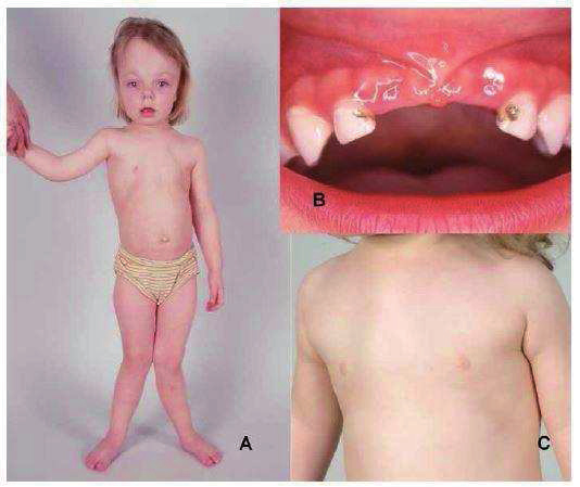
At infantile form of GFF There may not be any noticeable abnormalities at birth, but symptoms can appear at any time during the first six months. The initial symptom may be an inability to gain weight and grow at the expected rate for one's age and gender. Some affected children have early fusion of the skull bones later (craniosynostosis), which can lead to a disproportionately wide head (brachycephaly). Craniosynostosis can be associated with increased pressure of the fluid that surrounds the brain (cerebrospinal fluid), a condition known as "intracranial hypertension." This complication can cause headaches, swelling of the optic nerve head (papilloedema), and bulging of the eyes (proptosis). Affected babies have soft, weakened bones, which leads to rickets. Rickets Is a general term for bone diseases that occur during growth with softening of the bone and characteristic deformities of the legs when bending from growth plate abnormalities. Wrists and ankles may be enlarged. Affected children may also have chest and rib deformities, as well as rib fractures, predisposing them to pneumonia. Various degrees may develop pulmonary insufficiency and labored breathingpotentially progressing to life-threatening respiratory failure. Episodes of fever and bone pain may occur. In some children, muscle tone decreases (hypotension), so the child becomes “flexible”, which is associated with increased blood calcium levels (hypercalcemia). Hypercalcemia can cause vomiting, constipation, weakness, and poor nutrition. Increased excretion of calcium can damage the kidneys. In rare cases, seizures may occur. Sometimes there is a spontaneous improvement in mineralization in early childhood. Short stature and skeletal deformities can persist throughout life.
Juvenile GFF varies greatly in children, and severe and mild forms should be considered. Sick children may occasionally have craniosynostosis and show signs of intracranial hypertension. Skeletal malformations that resemble rickets can appear between the ages of 2 and 3 years. Pain and fractures of bones and joints can occur. Typically, one or more deciduous teeth fall out earlier than usual. Some children are weak and have delays in walking, and when they learn to walk, they may have a distinct, winding gait. Spontaneous remission of bone symptoms has been noted in young adulthood, but such symptoms may recur in middle or late adulthood.
Adult GFF characterized by a wide range of symptoms. Affected people have osteomalacia, a softening of bones in adults. Some childhood patients have a history of rickets or premature loss of deciduous teeth. Individuals with adult HFF may suffer from fractures, especially stress fractures of the foot or pseudo-hip fractures. Repeated fractures can lead to chronic pain and weakness. Spinal fractures seem to be less common, but they do occur as well. Bone pain is a common complication. Some affected adults develop joint inflammation and pain near or around certain joints due to accumulation of calcium crystals (periarthritis calcific) or a condition called chondrocalcinosis, characterized by the accumulation of calcium crystals in the cartilage of the joints, sometimes damaging the joint. Others have sudden, severe joint pain. Affected adults may experience tooth loss during adulthood.
Odontohypophosphatasia is characterized by premature loss of milk teeth in childhood or tooth loss in adulthood. Dental problems are a single feature found that does not show up along with the characteristic bone symptoms of other forms of HFF.
Causes
Hypophosphatasia is caused by mutations in the gene for nonspecific alkaline phosphatase (TNSALP), also called the genome ALPL. This is the only gene involved in GFF. Genes provide instructions for making proteins that play critical roles in many bodily functions. When a gene mutation occurs, the protein product may be defective, ineffective, or missing. Depending on the function of a particular protein, one or more body systems can be impaired.
With HFF, mutations in the gene ALPL can be inherited in an autosomal recessive or autosomal dominant manner. Perinatal and childhood forms of GFF are inherited in an autosomal recessive manner. The childhood form can be autosomal recessive or autosomal dominant. The adult form and odontohypophosphatasia are usually autosomal dominant disorders, but rarely can be inherited as an autosomal recessive trait.
Most genetic diseases are determined by the status of two copies of a gene, one from the father and the other from the mother. Dominant genetic disorders occur when only one copy of an abnormal gene is needed to cause a specific disease. The abnormal gene can be inherited from either parent, or it can be the result of a new mutation (gene change) in the affected person. The risk of passing the abnormal gene from the affected parent to the offspring is 50% with every pregnancy. The risk is the same for men and women.
Recessive genetic disorders occur when a person inherits two copies of an abnormal gene for the same trait, one from each parent. If a person inherits one normal gene and one gene for the disease, the person will be a carrier of the disease, but usually asymptomatic. The risk that two carrier parents will both pass on the abnormal gene and therefore infect the baby is 25% with each pregnancy. The risk of giving birth to a child who will be a carrier of the disease, like the parents, is 50% with each pregnancy. The probability for a child to receive normal genes from both parents is 25%. The risk is the same for men and women.
Gene ALPL creates (encodes) an enzyme known as tissue nonspecific alkaline phosphatase or TNSALP. Enzymes are specialized proteins that break down other chemicals in the body. TNSALP is essential for the proper development and health of bones and teeth and is expressed in the liver and kidneys as well as in bones. Mutations in ALPL gene leads to insufficient levels of functional TNSALP, which, in turn, leads to the accumulation certain chemicals in the body, including phosphoethanolamine, pyridoxal-5'-phosphate, and inorganic pyrophosphate. Inorganic pyrophosphate helps regulate skeletal mineralization. Elevated levels of inorganic pyrophosphate can indirectly lead to increased levels of calcium in the body and insufficient bone calcification. Typically, TNSALP enzyme activity correlates with the severity of hypophosphatasia, usually with less residual enzyme activity in more severe disease.
Individuals with an extremely rare form of HPF called pseudohypophosphatasia have had normal alkaline phosphatase levels as determined by routine clinical laboratory tests research.
Affected populations
GFF affects men and women in equal amounts. In Canada, it is estimated that severe HFF is of concern in about 1 in 100,000 live births. The overall incidence and prevalence of all forms of HPF is unknown. Mild cases may go undiagnosed or misdiagnosed, making it difficult to determine the true frequency of HFF in the general population. HFF occurs most frequently in the Mennonite population in Canada, is relatively common in Japan, and relatively rare in blacks.
Symptomatic disorders
Signs and symptoms of the following disorders may be similar to those of hypophosphatasia. Comparisons can be useful for differential diagnosis.
- Osteogenesis imperfecta (BUT) is a group of rare diseases that affect connective tissue and are characterized by fragile bones that break easily. As with GFF, specific symptoms and physical signs vary greatly from person to person. The severity of OI can also vary widely among individuals in the same household. Osteogenesis imperfecta can be mild or severe. Four main types of OI have been identified. OI type I is the most common and mildest form. OI type II is the most severe. Most forms of OI are inherited in an autosomal dominant manner.
- Rickets (vitamin D deficiency), a disorder that often manifests itself in infancy or childhood is the result of inadequate intake vitamin Dcaused by poor diet, lack of sunlight, or malabsorption syndromes, in which the intestines cannot adequately absorb nutrients, including vitamin D from food. Vitamin D is required for the absorption of calcium and phosphorus from food in the body, which in turn affects how calcium is deposited in the bones. Thus, an adequate amount of vitamin D is essential for proper bone development and growth. The main symptoms of rickets include soft bones, slow growth, and weakness. This disorder is more common than HFF.
- X-linked hypophosphatemia is a rare hereditary disease characterized by impaired phosphate transport and altered activation of vitamin D in the kidney. In addition, phosphate can be poorly absorbed from the intestines. Low phosphate levels in the blood (hypophosphatemia) as a result of these disorders can lead to rickets or osteomalacia. In children, growth rates may be slower than normal, often resulting in short stature. X-linked hypophosphatemia is caused by mutations in PHEX a gene located on the X chromosome. It is the most common hereditary form of hypophosphatemia (incidence of about 1/25 000 births). However, autosomal dominant and recessive forms of hypophosphatemia are less common.
Diagnostics
Diagnosis of hypophosphatasia is based on the identification of characteristic signs and symptoms, a detailed medical history, a thorough clinical evaluation and a variety of laboratory tests, including routine x-ray and biochemical examinations. Diagnosing HFF correctly is easy for doctors who are familiar with or have experience with this disorder. However, most physicians have little or no knowledge of GFF. Consequently, affected individuals and families can face a frustrating delay in diagnosis. Gene mutation analysis is also available in commercial laboratories today. ALPL.
Standard treatments
Currently, the pathogenetic therapy of GFF has been developed. Asphotase Alpha (“Strensiq”, trade name) is a human recombinant tissue-specific chimeric Fc-deca-aspartate alkaline phosphatase glycoprotein.
Other treatments for hypophosphatasia target specific symptoms and complications that may differ from person to person. Treatment may require the coordinated efforts of a team of specialists. Pediatricians, orthopedic surgeons, dentists, pain specialists, and other health care providers may need systematic and comprehensive treatment planning.
Non-steroidal anti-inflammatory drugs (NSAIDs) can be used to treat bone and joint pain. NSAIDs require caution and monitoring when used because they can cause side effects (e.g. they can damage the stomach and kidneys), especially if they are administered in excessive doses and for prolonged time. If craniosynostosis is causing intracranial pressure, surgery may be needed to relieve the pressure.
Vitamin B6 can help control specific seizures in severely affected children. Severely affected children who have elevated blood calcium levels may be treated with dietary calcium restriction, hydration, certain diuretics and possibly calcitonin, but hypercalcemia is often difficult to control as it occurs in severely affected patients.
Regular dental treatment in the early stages is recommended. In some cases, physiotherapy and occupational therapy may be recommended.
Adults with recurrent fractures of the long bones can be treated with orthopedic internal fixation, also known as osteosynthesis. During this procedure, the orthopedic surgeon places a metal rod through the central opening of the bone to make it more stable and stronger.
Affected people should avoid bisphosphonates, a class of drugs used to treat other bone conditions such as osteoporosis. These drugs can worsen hypophosphatasia or cause symptoms in people with undiagnosed HPF. Examples of bisphosphonate drugs include alendronate, ibandronate, pamidronate, risedronate.
Forecast
The most severe form is perinatal hypophosphatasia, which is considered fatal in most cases. The infantile form is fatal in about 30% of patients. Life expectancy studies have not been conducted for the juvenile form. Individuals with an adult form and odontohypophosphatasia have a normal life expectancy.