Leishmaniasis: what is it, causes, symptoms, treatment, prognosis
Content
- What is leishmaniasis?
- Signs and symptoms
- Causes and risk factors
- Affected populations
- Diagnostics
- Standard treatments
- Prophylaxis
- Forecast
What is leishmaniasis?
Leishmaniasis is an infectious disease caused by protozoan parasites of the genus Leishmania. Protozoa are microscopic unicellular organisms. The parasites that cause the disease are transmitted to humans through the bites of certain types of infected sand flies. In humans, these parasites cause three main forms of infection: cutaneous leishmaniasis, mucosal leishmaniasis, and visceral leishmaniasis. In each of these forms, infection ranges from no symptoms (asymptomatic infection) to serious, even life-threatening complications. Only a small percentage of people infected with parasites develop the disease.
Leishmaniasis is broadly classified based on its location in the western or eastern hemispheres. In the Western Hemisphere, the disease is known as New World leishmaniasis and occurs in parts of Mexico, Central America, and South America. In the Eastern Hemisphere, the disease is known as Old World leishmaniasis and occurs in parts of Asia, in The Middle East, southern Europe (especially the Mediterranean region), North Africa and tropical regions Africa. New World and Old World leishmaniasis are caused by different types
Leishmania.Signs and symptoms
Leishmaniasis can affect people in different ways. Some have a silent infection and have no signs or symptoms. Others develop mild to moderate illness, but some develop severe infections that can cause permanent damage and potentially life-threatening complications.
Acute leishmaniasis.

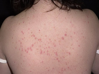
This is the most common form of leishmaniasis. Symptoms may begin within weeks or months after being bitten by an infected sand fly. Affected people may develop one or more sores (skin lesions), especially on exposed areas of the body such as the face, ears, arms, and legs. Lesions are formed at the site of the bite.
Lesions can be papules (bumps) or nodules (hard, raised bumps), plaques (flattened, bulging lesions), or ulcers (open, eroded areas). Skin lesions can change in size, get smaller, but often enlarge and do not heal.
Wounds may be wet and oozing fluid (such as pus), or they may be dry, crusty, and usually painless. Patients may develop lesions that are confined to one area of the body and that may slowly heal on their own over 6-18 months. The lesions, however, usually leave noticeable scars. Sometimes people also have swelling of nearby lymph nodes (lymphadenopathy).
— Diffuse cutaneous leishmaniasis.
This very rare form is characterized by an initial lesion of the skin that spreads to various parts of the body. People often have poorly functioning immune systems, making them susceptible to widespread widespread skin lesion, predisposes to poor response to treatment and allows infection to last endlessly. Affected people may have multiple plaques, ulcers, and nodules all over their body. Diffuse cutaneous leishmaniasis progresses slowly but is a chronic condition that usually recurs after treatment, even if treatment appears to have been effective initially.
- Recurrent leishmaniasis.
This term is used to define the recurrence of a skin lesion years after the initial lesion has healed. Recurrent leishmaniasis often develops on the face, especially on the cheeks, with a new sore or papule forming over or near the scar of the old lesion. Sometimes this lesion can gradually increase.
Leishmaniasis of the mucous membrane.
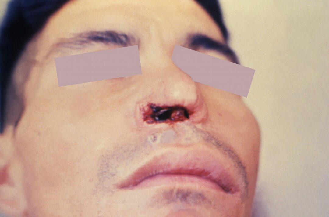
Parasites can spread from the initial skin lesion through the bloodstream to other distant sites, such as the mucous membranes of the nose, mouth, and throat. Individuals with mucosal leishmaniasis usually have skin lesions that heal on their own. to yourself or during treatment, often leading to damage to the mucous membrane after several years, and sometimes decades.
The first signs of illness may be persistent nasal congestion or nosebleeds. Over time, inflammation and partial or complete destruction of the mucous membranes of the mouth, nose and throat may develop. If left untreated, mucosal leishmaniasis can lead to disfigurement and scarring of the nose and mouth. This damage can result in nasal congestion and bleeding. Complications can be difficult to treat and gradually worsen.
Mucosal leishmaniasis can develop in people who were not initially treated for cutaneous leishmaniasis. Known risk factors for mucosal disease include: being infected with certain types of parasites common in South America; numerous, large, long-term lesions of the scalp or neck; suppressed immune system; or an infection acquired in Bolivia.
Visceral leishmaniasis.
This form of leishmaniasis, usually the most clinically severe, also develops when certain types of parasites leave the skin, enter the bloodstream, and reach internal organs, including spleen, liver and bone marrow. Clinical signs range from asymptomatic infection to mild illness that resolves on its own to severe, life-threatening infection. If symptoms develop and full illness is left untreated, visceral leishmaniasis is usually fatal.
Affected persons may develop repeated bouts of fever, weakness, unintentional weight loss, or even severe wasting (cachexia), significant enlargement of the spleen and liver and pancytopenia (low levels of the three main types of blood cells (red blood cells, white blood cells, and platelets)). A low red blood cell count is called anemic and can cause fatigue, pale skin, shortness of breath, dizziness, and a fast or irregular heartbeat. Affected people often get worse over the course of weeks or months.
Visceral leishmaniasis in India is also called kala azar. Kala azar usually refers to severe, chronic cases of the disease. Many additional names can be used for visceral leishmaniasis, including Dum-dum fever, Burdwan fever, Sirkari's disease, and Sahib's disease.
— Post-kalaazarcutaneous leishmaniasis.
This form of infection occurs in people who have or have had visceral leishmaniasis. It is most common in Africa and India. In Africa (eg Sudan), the disease is present or occurs soon after the diagnosis and treatment of visceral leishmaniasis. People may develop an increased rash on the face, buttocks, arms, and legs. These lesions heal on their own over time or after the visceral infection has been treated.
In India, post-calaazar cutaneous leishmaniasis develops several years after treatment for visceral leishmaniasis. People often develop multiple, flat, discolored patches of skin (macula). Ultimately, these lesions develop into plaques or nodules on the face and trunk. In India, this form requires intensive treatment.
- Coinfection with HIV with leishmaniasis.
Some people contract both the Human Immunodeficiency Virus (HIV) and leishmaniasis. These people are more likely to develop severe, life-threatening complications and death. They can develop complications not usually seen in healthy people with leishmaniasis, including damage to the gastrointestinal tract and other atypical areas. Due to HIV-related immunodeficiency, treatment can be ineffective and relapse rates are high even after apparently effective treatment.
Causes and risk factors
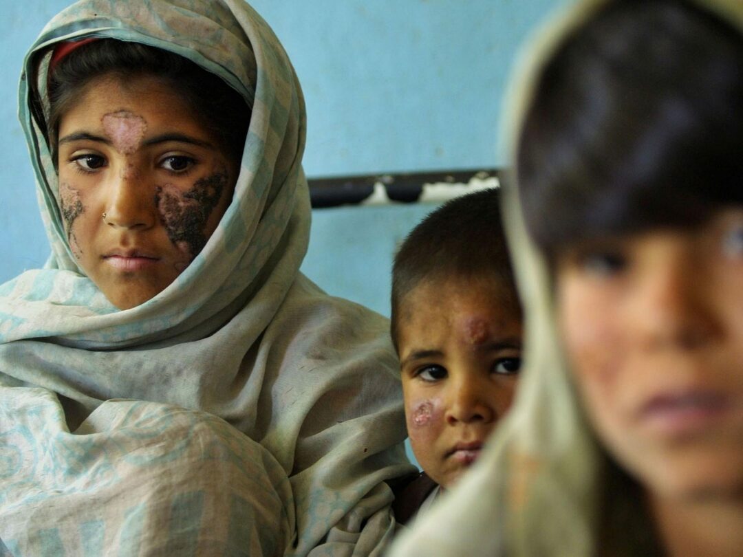
Leishmania infections are caused by 20 different types of parasites Leishmania. Parasites are microscopic organisms that live in another organism (called a host, such as a human) and survive by taking in nutrients from the host. Leishmania transmitted to humans or animals through the bite of an infected sand fly. Sand flies become infected by biting and sucking the blood of infected people or animals, especially dogs. Sand flies are very small and do not make noise. Sometimes their bites can be painful; most often, the bites are painless and go unnoticed. Sand flies are most active from dusk to dawn.
Sand flies can bite and infect anyone of any age in areas where leishmaniasis is found. The disease is much more common in rural areas than in cities. There are several risk factors for leishmaniasis, including socioeconomic status (the disease is common in some of the poorest areas on earth), malnutrition and poor housing, and there is some evidence of genetic predisposition. Additional environmental risk factors include deforestation, mining, construction of buildings, alteration or creation of new irrigation schemes and other aspects of urbanization that can lead to increased exposure to sand flies and, as a result, to infestation leishmaniasis. Migration patterns (the movement of large groups of people susceptible to leishmaniasis) can lead to an increase in the number of infected people.
Affected populations
Leishmaniasis is rare in Russia. Most people from Russia who become infected do it on trains or live in regions where the disease is common.
The exact number of people who develop leishmaniasis worldwide each year is unknown. Cutaneous leishmaniasis is estimated to have between 700,000 and 1.2 million new cases each year worldwide. For visceral leishmaniasis, this number ranges from 200,000 to 400,000. More than 90% of people who develop visceral leishmaniasis live in poor rural areas of Bangladesh, Ethiopia, India, Nepal, South Sudan and Brazil. Cutaneous leishmaniasis is much more common, especially in South and Central America, the Middle East, and Central Asia.
Diagnostics
Diagnosis of leishmaniasis is based on characteristic symptoms and signs, a detailed medical history, careful clinical evaluation, and a variety of specialized tests. A detailed medical history includes information about whether the person has traveled to areas where the disease is common. For example, cutaneous leishmaniasis should always be considered for non-healing or progressive skin lesions in a person who has traveled or lived in an area where leishmaniasis is found.
Clinical testing and examination.
Doctors take samples of infected tissue for examination. They may take biopsies or scrapers from skin lesions if cutaneous leishmaniasis is suspected, or from bone marrow (material found in the bones of the body) if visceral leishmaniasis is suspected. These samples are sent to laboratories where they are examined in various ways for the presence of parasites. Leishmania.
To detect antibodies against parasites Leishmania blood tests may be done. Antibodies are specialized proteins created by the immune system in response to foreign or invading pathogens. Doctors readily find antibodies against parasites Leishmania in the blood of people with visceral leishmaniasis. However, sometimes the test can be negative even if the person has a medical condition. Antibody blood tests are not often helpful for people with cutaneous or mucosal forms of leishmaniasis.
Standard treatments
Leishmaniasis is a curable and treatable disease. Specific procedures and therapeutic interventions can vary depending on numerous factors such as the form of the disease (cutaneous, mucous, visceral); the presence or absence of certain symptoms; the geographic location where the person was infected; specific types Leishmania participating in the process; the person's age and general health.
Decisions regarding the use of specific drug regimens and / or other treatments should accepted by physicians with experience in the treatment of leishmaniasis, in consultation with the patient, based on the specifics of his case. The potential benefits and risks of treatment (including side effects) should be carefully discussed, patient preference, the possibility of skin infection scarring and other appropriate questions. For example, patients should be clearly informed that initial treatment may be extended, that more than one course of treatment may be required, and given a tendency to relapse in all forms of this infection, that after treatment, additional observation is necessary for 6-12 months before considering that a person is successful healed.
According to the Centers for Disease Control and Prevention (CDC), people with cutaneous leishmaniasis may or may not need a special treatment, while all symptomatic, clinically significant cases of visceral leishmaniasis and mucosal leishmaniasis require treatment. Specific people or groups such as young children, pregnant women, women who are breastfeeding, people with a functioning immune system or people with other medical conditions may need different medications or different regimens dosage.
Cutaneous leishmaniasis can heal on its own, although it can take a long time to heal and often leaves scars. In special cases, treatment of small, uncomplicated lesions may include the application of warm or cold lotions to the wounds. to kill parasites, or an antibiotic known as paromomycin can be applied as an ointment directly to wounds.
For people with cutaneous leishmaniasis who have multiple large skin lesions, a weakened immune system, or those infected with Leishmania species that may potentially cause mucosal leishmaniasis, treatment may include various medications such as liposomal amphotericin B, miltefosine, or stibogluconate sodium.
The best treatment for mucosal leishmaniasis is not known. Treatment for this condition includes liposomal amphotericin B, miltefosine, and sodium stibogluconate. Surgery may be necessary for people with severe complications of the mucous membranes of the mouth and nose (orofacial surgery).
For people with leishmaniasis and HIV, treating HIV infection with antiretroviral therapy can improve response antileishmaniasis treatment, avoid or delay leishmaniasis recurrence and improve overall survival.
Prophylaxis
Prevention is the most effective treatment for leishmaniasis. In areas where leishmaniasis is known to be present, steps should be taken to avoid exposure to sand flies, especially from dusk to dawn, using an insect repellent containing DEET and clothing that covers the arms and legs outside. Travelers should stay in well-protected areas, especially at night when sandflies are most active. You should also use insect nets, a nets around your bed, and wear insecticide-treated clothing.
Forecast
Cure rates are high with appropriate medications, mainly when treatment is started before it affects the immune system. Cutaneous leishmaniasis can lead to disfigurement.
Death is usually caused by complications (such as other infections) and not by the disease itself. Death often occurs within 2 years.