Melorheostosis: what is it, signs and symptoms, treatment, prognosis
Content
- What is melorheostosis?
- Signs and symptoms
- Causes
- Affected populations
- Symptomatic disorders
- Diagnostics
- Standard treatments
- Forecast
What is melorheostosis?
Meloreostosis(Leri's disease, rhizomonomelo rheostosis) Is a rare and progressive disease characterized by thickening or expansion (hyperostosis) of the outer layers of the bone (cortical bone). Melorheostosis affects both growth and development of bones and soft tissues. This disorder is benign (noncancerous) but often results in severe functional impairment; chronic pain; joint contractures and / or stiffness of muscles, tendons or ligaments; and deformities of the limbs, hands, or feet.
Signs and symptoms
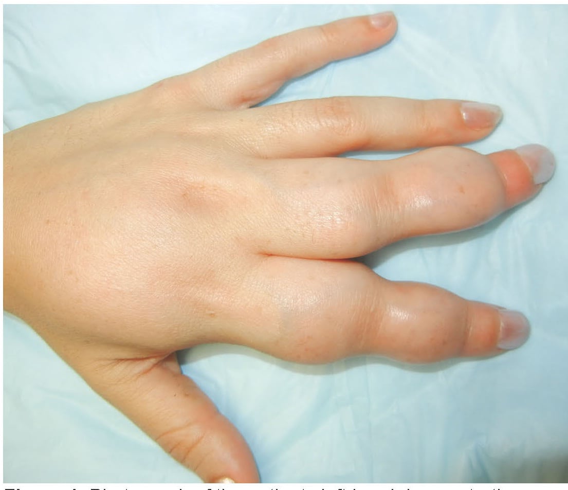
Signs and symptoms of melorheostosis include irregular bone growth, including thickening of the cortex and a candle wax pattern on x-rays; unequal limb length; soft tissue abnormalities, including shortening of tendons and ligaments, absence or abnormalities of muscles, subcutaneous calcification, joint inflammation, and contractures leading to deformity or immobilization joints; limited range of motion; pain and stiffness;
edema extremities and vascular abnormalities. Less commonly, severe bone lesions can compress surrounding nerves.Melorheostosis usually affects one specific segment of the appendicular skeleton (arms and legs). It is usually limited to one side of the body (rarely bilateral) and within a limb bounded by the medial or lateral side of the bones. The disease can also affect the axial skeleton: the pelvis, sternum, ribs, and, less commonly, the spine and skull. Symptoms may gradually get worse over time.
In children, the disorder usually causes uneven growth of the limbs, deformities, or contractures (limitation of passive movement in the joint). In adults, symptoms of pain, joint stiffness, and more noticeable progressive deformities are more common.
The age at diagnosis usually depends on the severity of the onset and symptoms, and varies widely between children and adults. Melorheostosis is usually seen in early childhood and may even appear in the first days of a child's life. In 50 percent of patients with melorheostosis, symptoms only develop by age 20.
Causes
Mutations in the MAP2K1 gene are estimated to cause about half of all cases of melorheostosis. Gene MAP2K1 contains instructions for making a protein called MEK1 protein kinase. This protein is active in many types of cells, including bone cells. It is part of a signaling pathway called RAS / MAPK. This signaling pathway helps control the growth and division (proliferation) of cells, a process through which cells mature to perform specific functions (differentiation) and cell movement (migration). RAS / MAPK signaling is critical for normal development, including bone formation.
V MAP2K1 the gene mutations that cause melorheostosis are somatic, which means that they occur throughout human life and are present only in some cells, in this case, bone cells in a certain area of the body. The mutations result in a version of MEK1 protein kinase that is overactive, which increases RAS / MAPK signaling in bone. The increased signaling disrupts the regulation of bone cell proliferation, allowing new bone to grow abnormally. Research shows that enhancing RAS / MAPK signaling also stimulates excess bone remodeling, a normal process in which old bone breaks down and new bone is created, to replace it. These changes in bone growth and replacement underlie the bone abnormalities characteristic of melorheostosis.
In cases of disorder without an identified mutation in the gene MAP2K1, the cause of the disease is usually unknown. Research suggests that somatic mutations in other genes, especially genes associated with the RAS / MAPK signaling pathway, can also cause the disorder.
Affected populations
The estimated incidence of melorheostosis is 1 in 1,000,000. Both sexes suffer in the same way, with about 400 cases reported.
Symptomatic disorders
Symptoms of the following disorders may be similar to those of melorheostosis. Comparison can be useful for differential diagnosis:
- Osteopoikilosis Is a rare and benign bone disease characterized by many rounded or oval areas of increased bone density at the ends of long bones. They are usually present on both sides (bilateral).
- Buschke-Ollendorff syndrome is a rare hereditary connective tissue disorder characterized by a combination of skin growths (called connective tissue nevi) with osteopoikilosis.
- Stripedosteopathy - a benign bone disease characterized by longitudinal "stripes" of increased density in the affected bones.
- Linear scleroderma manifests itself as a strip-like thickening of the skin on the arms and legs. It usually first appears in early childhood and is characterized by the inability of one limb to grow as quickly as the other.
- Desmoidfibroids usually develop in the fibrous (connective) tissue of the body that forms tendons and ligaments, usually in the arms, legs, or midsection, and in the head and neck. Tumors can be invasive to surrounding tissues and difficult to control.
- Hemangiomas appear in early childhood and are neoplasms consisting of small blood vessels.
Diagnostics
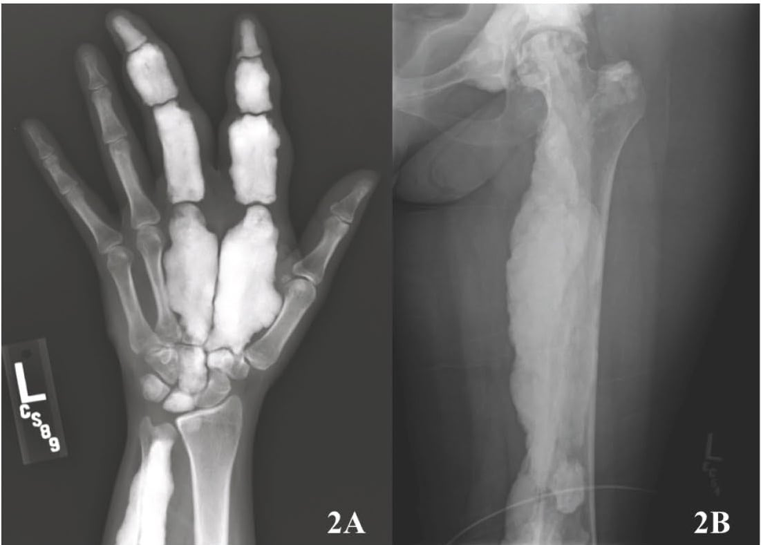
Diagnosis is mainly based on conventional radiography. On radiographs, melorheostosis is presented as linear lesions of increased bone density along the major axis (trunks or diaphysis) of tubular bones. The lesions increase the thickness of the cortex and can spread to the outer surface of the bones, creating the appearance of "wax dripping from the candle." In polyostatic lesions, linear lesions are usually continuous along the same side of the affected limb and "jump" over the joints. Calcification of soft tissues and even ectopic ossification may occur.
Magnetic resonance imaging is used to study soft tissue lesions. Radioisotope bone scans can help distinguish melorheostosis from other bone lesions. The biopsy shows varying degrees of bone marrow fibrosis, along with markedly irregular bone with mixed areas of lamellar and tissue bone. In the soft tissue masses, a mixture of osteocartilagenic, fibrovascular and adipose tissues is observed.
Standard treatments
Treatment is currently limited and is mainly aimed at relieving symptoms. None of the treatment options have been found to be completely effective, and what may be beneficial to one person may be ineffective or even harmful to another. Treatment options can include surgery, physical and occupational therapy, hydrotherapy, and drugs to alter the bone remodeling process.
Surgical treatment options include limb and tendon lengthening, fasciotomy, capsulotomy, osteotomies, excision of fibrous tissue and / or hyperostosis, contralateral epiphysiodesis, arthrodesis, callotasis and amputation. Surgical treatment is best done after skeletal maturity, but deformities can recur.
Pain management can be challenging. Pain medications may include non-steroidal anti-inflammatory drugs (NSAIDs), steroids, or rarely opioid analgesics. These medications are sometimes useful in the early stages of chronic disease progression, but may be less effective for severe lesions.
Forecast
Melorheostosis is not life threatening, but it can greatly affect quality of life due to chronic pain that can worsen or reappear even after surgery.