Necrotizing enterocolitis: what is it, symptoms, treatment, prognosis
Content
- What is necrotizing enterocolitis?
- Signs and symptoms
- Causes and risk factors
- Affected populations
- Symptomatic disorders
- Diagnostics
- Standard treatments
- Forecast
What is necrotizing enterocolitis?
Necrotizing enterocolitis, abbreviated NECK, Is a devastating disease that affects the intestines of the newborn. The disorder usually occurs in premature babiesborn less than 37 weeks old and is characterized by severe inflammation of the baby's small or large intestine, which can progress to tissue death (necrosis).
NEC occurs in about 1 in 1000 live births. NEC can occur in term infants, but is much more common in very preterm infants, especially infants with very low birth weight - incidence ranges from 3% in infants weighing at birth 1251 to 1500 grams (2 lb 12.13 oz to 3 lb 4.91 oz) up to 11% for infants born less than 750 grams (1 lb 10.46 ounces).
Necrotizing enterocolitis usually occurs when a newborn is several weeks old and is receiving enteral nutrition. Babies initially complain of vomiting.
bloating, bloody stool, long pauses in breathing and decreased activity. This can lead to intestinal necrosis and perforation.Medical treatment includes cessation of enteral nutrition (delivery of nutritious food directly to the stomach), broad-spectrum antibiotics, and supportive care. Surgery is indicated if there are signs of bowel perforation and necrosis. This serious gastrointestinal disease is associated with significant morbidity (complications associated with the disease) and death. Despite treatment, about 15% of babies who develop NEC die, and some surviving babies suffer from multiple complications such as short bowel syndrome, stunted growth and long-term nerve disorders development.
The exact mechanism of this disease, although not fully understood, is considered multifactorial and associated with premature bowel development, abnormal colonization of intestinal microorganisms, and inflammation intestines. NEC remains the leading cause of morbidity and mortality in the neonatal intensive care unit, despite significant advances in the care of preterm infants.
Signs and symptoms
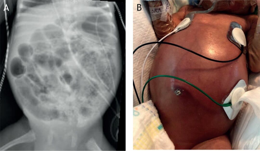
The onset of NEC usually occurs within the first few weeks after birth when breastfeeding is initiated, and age of onset is inversely related to gestational age at birth. At an early stage of the disease, newborns may show signs of food intolerance in the form of vomiting, an increase gastric aspirate, bile (green) gastric aspirate, or abdominal murmur reduction with bloating and soreness.
Read also:Enteropathic acrodermatitis
Coarse or occult blood may be present in the stool, indicating damage to the mucous membrane. Many of these signs are nonspecific and can occur with other diseases. The progression of necrotizing enterocolitis leads to systemic signs such as lethargy, long pauses in breathing, called apnea, temperature instability and poor perfusion (pumping fluid through an organ or tissue). Ultimately, this can lead to respiratory failure and cardiovascular collapse, requiring mechanical ventilation and vasopressors. Perceptible mass and erythema (abnormal redness of the skin due to capillary blockage, as in inflammation) of the abdominal wall indicate a more progressive disease process.
Causes and risk factors
After years of research and clinical observation, the etiology and pathogenesis of NEC remains elusive. Several key risk factors have consistently been identified as important prerequisites for the initiation of intestinal damage leading to NEC. These include prematurity, formula feeding, abnormal microbial colonization of the intestine, and ischemia (when blood vessels in the intestine become narrowed or blocked, which reduces blood flow).
Prematurity remains the main important risk factor associated with necrotizing enterocolitis. Immaturity of the intestinal epithelial cell barrier and immune system appears to contribute to pathogenesis. Before birth, the fetus has a sterile intestinal environment, and after birth, it quickly becomes colonized with bacteria. Incorrect colonization with a predominance of gram-negative bacteria can lead to disruption of normal intestinal epithelium, bacterial translocation and induce an excessive inflammatory response. The distinctive histological findings seen in NEC are inflammation and coagulation necrosis (tissue death pattern). Ischemia is another important pathophysiological factor in the development of necrotizing enterocolitis. Reduced oxygen supply to intestinal cells can lead to cellular damage and necrosis.
Affected populations
NEC affects 5 to 10% of preterm infants weighing less than 1500 g. Among the risk factors identified for NEC, prematurity and birth weight remain inversely proportional to the risk of NEC. Term babies who develop NEC usually have special risk factors, such as congenital heart defects, sepsis and low blood pressure (hypotension).
Read also:Duodenal ulcer
Symptomatic disorders
Many other gastrointestinal disorders can mimic necrotizing enterocolitis, so it is important to consider and treat an alternative etiology when evaluating a patient. Sepsis can cause intestinal obstruction with bloating and food intolerances. Premature babies, especially those taking indomethacin or steroids, can also have spontaneous bowel perforation (SPP). SPK is characterized by isolated perforation of the distal small intestine.
Other causes of severe bloating in newborns include bowel obstruction due to Hirschsprung's disease, atresia of the small intestine, meconium ileus and volvulus malrotation. NEC can also be mistaken for allergic enterocolitis secondary to cow's milk protein allergy.
Diagnostics
NEC is diagnosed clinically and radiographically. Once clinical suspicion arises, an abdominal x-ray is taken as an initial assessment. This is repeated serially depending on the severity and clinical course to assess the progression of the disease.
Characteristic signs of NEC on abdominal radiographs include intestinal pneumatosis (air in the wall intestines), pathological permanent dilated loops, thickened intestinal wall, pneumoperitoneum and gases of portal veins. Pneumoperitoneum, defined as free air in the abdomen, is a surgical emergency that indicates bowel perforation and usually requires intervention.
Abdominal ultrasound can also be used to assess the presence of free fluid in the abdomen or formation abscess.
Additional laboratory tests to assess the severity of NEC include blood culture, coagulation studies, and a manual differential CBC to assess leukocytosis with bandemic. neutropenia, anemic and thrombocytopenia. Blood gases are tested serially to assess the severity acidosis and the need for respiratory support or to aid in fluid drainage.
Standard treatments
Treatment for necrotizing enterocolitis depends on the clinical stage. If stage I NEC is suspected, initial treatment consists of intestinal rest with cessation of enteral nutrition, nasogastric decompression, blood culture, and broad-spectrum antibiotics. When oral feeding is stopped, intravenous parenteral nutrition is started. Careful observation with serial examinations and radiographs is necessary. The surgeon is consulted after confirmation of stage II or III NEC. Supportive care includes respiratory support, inotropic (cardiac) support, fluid resuscitation, and correction of acid-base imbalance. Patients with NEC may develop disseminated intravascular coagulation (DIC, a condition that prevents normal blood clotting) due to consumption of clotting factors and require transfusion of food blood. The main indication for surgery in NEC is a perforated or necrotic intestine. Other indications include clinical deterioration and severe abdominal distension causing the syndrome abdominal compartment (organ dysfunction or failure due to a strong increase in pressure in the abdominal cavity). Usually, two surgical approaches are used, depending on the clinical picture, laparotomy with resection (removal) of intestinal necrosis or primary peritoneal drainage (a procedure for introducing a Penrose drainage into the space in the abdominal cavity that contains the intestines, stomach, and liver).
Read also:Pancreas: Symptoms and Causes of Disease, Medication and Diet
Prevention of NEC has the greatest potential to reduce adverse outcomes associated with NEC. It is now clearly shown that breast milk protects against NEC when compared to formula feeding. Establishing a standardized feeding protocol with objective criteria for refusing to feed also reduces the risk of necrotizing enterocolitis. Probiotics may prevent NEC by restoring gut microbial flora, but more research is still needed on optimal dosage and duration of treatment.
Forecast
The currently available therapeutic and surgical methods of treatment have improved the prognosis in children of the first year of life with necrotizing enterocolitis. About 70–80% of these newborns survive. The most common long-term complication is narrowing (stricture) of the small intestine. Strictures occur in 10–36% of children in the first year of life who survived the first episode of necrotizing enterocolitis. Strictures usually cause symptoms for several weeks to several months after an episode of necrotizing enterocolitis. In some cases, surgical treatment of strictures is required.