Sirenomelia (mermaid syndrome): what is it, symptoms, photos, treatment, prognosis
Content
- What is Mermaid Syndrome?
- Signs and symptoms
- Causes and risk factors
- Affected populations
- Symptomatic disorders
- Diagnostics
- Standard treatments
- Forecast
What is Mermaid Syndrome?
Sirenomeliaalso known as mermaid syndrome, Is an extremely rare congenital developmental disorder characterized by anomalies of the lower spine and lower extremities. Sick babies are born with partial or complete fusion of the legs (see. photo below). Additional malformations may also occur, including genitourinary abnormalities, gastrointestinal diseases, abnormalities of the lumbosacral spine and pelvis, as well as the absence or underdevelopment (agenesis) of one or both kidneys.
Sick infants may have one foot, both feet missing, which may look outward. The tailbone is usually absent, the sacrum is also partially or completely absent. Additional conditions may occur with mermaid syndrome, including an imperforate anus, spina bifida and malformations of the heart. Sirenomelia is often fatal during the neonatal period.
Some sources in the medical literature classify sirenomelia as the most severe form of caudal regression syndrome, a complex developmental disorder. More recently, however, many researchers have pointed out that mermaid syndrome is a similar but distinct disease.
Signs and symptoms
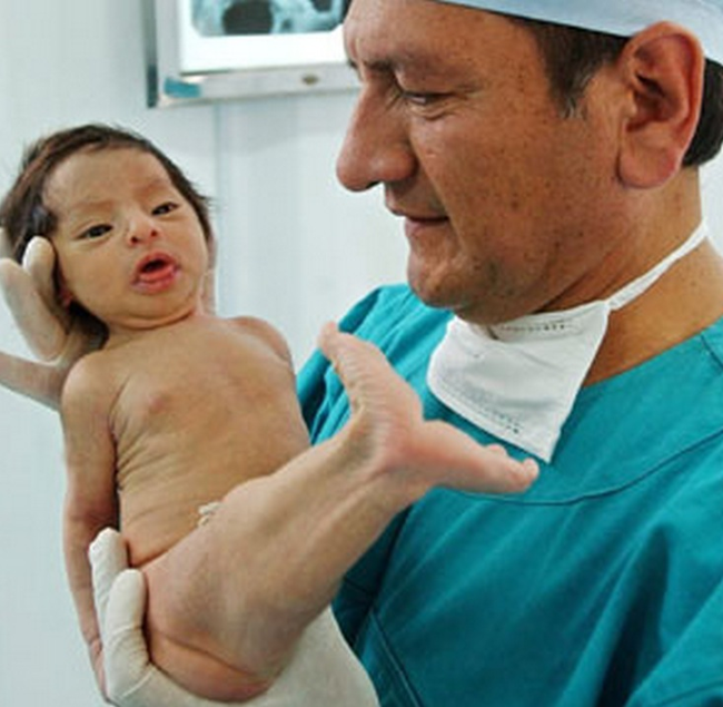
There is a wide range of physical malformations that can potentially occur with sirenomelia, and the specific symptoms can vary greatly from one person to the next. Sirenomelia is associated with serious life-threatening complications and is often fatal in the early years of life. However, a few cases have reported survival after infancy into later childhood or adolescence.
A characteristic sign of sirenomelia is partial or complete fusion of the legs. The severity varies greatly. Affected infants may have only one femur (long bone of the thigh) or two femurs within one skin shaft. Affected infants may have one foot, legs without a foot, or have both feet turned so that the back of the feet is pointing forward.
Affected infants also have a variety of urogenital abnormalities, including the absence of one or both kidneys (renal agenesis), cystic kidney anomaly, absence of the bladder, narrowing of the urethra (atresia urethra). In addition, patients develop a non-perforated anus - a condition in which a thin coating that blocks the anus, or passageway that usually connects the anus and the lower part of the large intestine (rectum), cannot develop.
Babies with sirenomelia can also have pathologies affecting the sacral and lumbar spine. Some patients have an abnormal curvature of the spine from front to back (lordosis). Patients may have no external genitals. It was also reported that spleen and / or gallbladder.
Read also:Angelman syndrome in children - causes and diagnosis. Karyotype, symptoms and treatment of Angelman syndrome
Defects in the abdominal wall also occur, such as protrusion of a portion of the intestine through an opening near the navel (hernia of the umbilical cord / omphalocele). Some sick children with mermaid syndrome may have meningomyelocele, a condition in which the membranes, covering the spine, and in some cases the spinal cord itself, protrude through the vertebral defect pillar. Congenital heart defects and respiratory complications such as severe lung underdevelopment (lung hypoplasia) can also be associated with sirenomelia.
Causes and risk factors
The exact cause of sirenomelia is unknown. Researchers believe that environmental and genetic factors play a role in the development of the disorder. Most cases of mermaid syndrome occur randomly for no apparent reason (sporadically), indicating environmental factors or a new mutation. Most likely, sirenomelia is multifactorial, which means that the disease is caused by several different factors. In addition, different genetic factors can contribute to the development of the disease in different people (genetic heterogeneity).
Environmental factors that play a role in the development of sirenomelia are unknown. Some patients have a genetic predisposition to the development of the disease. A person who is genetically predisposed to the disease carries the gene (s) for the disease, but it may not show up if the disease does not start or is not "activated" under certain circumstances, for example, due to certain environmental factors Wednesday. Researchers believe that environmental or genetic factors are teratogenic in the developing fetus. Teratogenic effects are the effects of certain physical, chemical and biological agents that interfere with the development of the embryo or fetus.
In some patients, sirenomelia is considered the result of disorders in the early development of the circulatory system (impaired development of the vascular system) inside the embryo. In some patients, one large artery emerging from the upper abdominal cavity was found, without two common arteries, which usually extend from the lower part of the aorta and carry blood to the posterior tail (caudal) end embryo. The only artery present (called a "stealing" vessel because it essentially draws blood from the lower part of the embryo) deflects the blood flow that normally circulates from the aorta to the lower parts of the embryo and to placenta. Thus, the stealing vessel redirects blood flow to the placenta, never reaching the tail (caudal) end of the embryo. As a result of this redirected blood flow, the marginal vessel also removes nutrients from the blood-deprived part of the embryo. The arteries in this caudal region are underdeveloped, and the tissues that depend on them for nutritional substances, cannot develop, are deformed or stop growing at some unfinished stages. In patients with sirenomelia, the bud of the lower limb of the embryo cannot divide into two legs. The underlying cause of these disorders is unknown.
Read also:Patau syndrome: what is it, symptoms, methods of diagnosis and treatment, prognosis
Affected populations
Sirenomelia affects men more often than women in a 2.7: 1 ratio. The exact incidence is unknown, but it is estimated that mermaid syndrome occurs in about 1 in 60,000 to 100,000 births. Sirenomelia is more common in one of the identical (monozygotic) twins than in fraternal (dizygotic) twins.
Symptomatic disorders
Symptoms of the following conditions can mimic the symptoms of sirenomelia. Comparisons can be useful for differential diagnosis.
Caudal regression syndrome Is a broad term for a rare complex disease characterized by abnormal development of the lower (caudal) end of the spine. The spine is made up of many small bones (vertebrae) that together form the vertebral column. The vertebral column is usually divided into three segments: the cervical spine, which consists of the vertebrae just below the skull; the thoracic spine, consisting of the vertebrae in the thoracic region; and the lumbar spine, which consists of the lumbar vertebrae. A triangular bony structure called the sacrum connects the lumbar spine to the pelvis. The sacrum consists of five vertebrae fused together. At the end of the sacrum is the coccygeal bone (coccyx).
Infants with caudal regression syndrome can potentially experience a wide range of abnormalities, including abnormal development (agenesis) of the sacrum and coccyx and abnormalities of the lumbar spine. In some cases, more serious malformations may occur.
Lower spine abnormalities cause many additional complications, including joint contractures, clubfoot, and a tear or injury to the end of the spinal cord, which can potentially cause urinary incontinence. Additional abnormalities of the gastrointestinal tract, kidneys, heart, respiratory system, upper limbs, and upper spine may also occur. The exact cause of caudal regression syndrome is unknown. It is assumed that both environmental and genetic factors play a role in the development of the disease.
VACTERL Association Is a non-random group of birth defects that affects multiple organ systems. The term VACTERL is an abbreviation, where each letter represents the first letter of one of the most common findings seen in sick children:
- V (eng. Vertebral anomalies) - spinal anomalies (70%),
- A (eng. Anal atresia) - anus atresia (55%),
- C (eng. Cardiovascular anomalies) - septal defects and other heart defects (75%),
- TE (eng. Tracheo-esophageal fistula) - tracheoesophageal fistula with esophageal atresia (70%),
- R (eng. Renal defects) - kidney anomalies (50%) - agenesis, dysplasia, hydronephrosis; the only umbilical artery.
- L (eng. Limb defects) - defects of the radius - hypoplasia of 1 finger or radius, preaxial polydactyly and syndactyly (70%);
Read also:Shereshevsky-Turner syndrome
In addition to the aforementioned features, patients also suffer from other less frequent anomalies, including deficits in height and inability to gain weight and grow at the expected rate (inability to development). Other low-frequency signs include facial asymmetry (hemifacial microsomia), malformations of the outer ear, defects in lung lobe formation, bowel malrotation, and genital anomalies. VATER / VACTERL functions are more common in twins. In some cases, the abbreviation VATER is used. Some researchers add (S) to the abbreviation VACTERL or VATER to denote one umbilical artery instead of the usual two. Mental functioning and intelligence are usually not impaired; developmental delay / mental retardation should indicate an alternative diagnosis. The exact cause of the VACTERL association is unknown. Most cases occur by chance, for no apparent reason (sporadic).
Diagnostics
The diagnosis of sirenomelia can be made prenatally, most often in the second trimester, using fetal ultrasound (fetal ultrasound). An ultrasound scan is a scan that uses high-frequency sound waves to produce an image of a developing fetus. Fetal ultrasound can detect some of the defects associated with sirenomelia.
Standard treatments
Treatment may require the coordinated efforts of a team of specialists. Pediatricians, surgeons, cardiologists, orthopedists, orthopedic surgeons, kidney specialists (nephrologists) and others health care professionals may need to systematically and comprehensively plan the treatment of the victim child.
Surgery to separate the leg joints was successful. In preparation for the operation, tissue dilators in the form of a balloon are inserted under the skin. When they are filled with saline over a period of time, the balls expand, causing the skin to stretch and grow. The excess skin is then used to cover the legs when they separate. Despite treatment, mermaid syndrome is usually fatal to the newborn.
Forecast
Sirenomelia is a rare and fatal congenital anomaly. In many pregnancies with a lilac-melon fetus, a spontaneous miscarriage occurs. Between one third and one half of babies are stillborn, and almost all, with the exception of a few, die in the neonatal period.