Deep vein thrombosis: what is it, symptoms, causes, treatment, prognosis
Content
- What is deep vein thrombosis?
- Signs and symptoms
- Causes and risk factors
- Epidemiology
- Diagnostics
- Standard treatments
- Forecast
- Prophylaxis
What is deep vein thrombosis?
Deep vein thrombosis (DVT) Is the formation of a blood clot in a deep vein, most often in the legs or pelvis. Symptoms may include pain, edema, redness and dilated veins in the affected area, but some do not have DVT symptoms. The most common life threatening DVT is the chance that a clot (or multiple clots) detaches from the vein (embolizes), passes through the right side of the heart, and gets stuck in the arteries that supply blood lungs. It is called pulmonary embolism (TELA). Both DVT and PE are considered part of the same disease process called venous thromboembolism (VTE). VTE can only proceed as DVT, like PE with DVT or PE without DVT. The most common long-term complication is post-thrombotic syndrome, which can cause pain, swelling, a feeling of heaviness, itching and, in severe cases, ulcers. In addition, recurrence of VTE occurs in about 30% of cases within 10 ten years after the initial VTE.
The mechanism of formation of a clot (thrombus) usually involves some combination of a decrease in blood flow velocity, an increased tendency to clot, and damage to the blood vessel wall. Risk factors include recent surgery, advanced age, active cancer, obesity, personal and family history of VTE, trauma, lack of movement, pregnancy and postpartum, and antiphospholipid syndrome. VTE has a strong genetic component, which accounts for approximately 50 to 60% of the variability in the frequency of VTE. Genetic factors include blood group other than 1, antithrombin, protein C and S deficiencies, and factor V Leiden and prothrombin G20210A mutations. In total, dozens of genetic risk factors have been identified.
People with suspected deep vein thrombosis can be assessed using a clinical Wells score. D-dimer testing can also be used to rule out a diagnosis or to signal the need for further testing. The diagnosis is most often confirmed by ultrasound examination of the suspected veins. About 4-10% of DVTs affect the hands. About 5–11% of people will develop VTE during their lifetime, with VTE becoming more common with age. Compared to people age 40 and under, people age 65 and over are about 15 times more at risk. However, evidence has historically been dominated by European and North American populations, and individuals of Asian and Latino descent have a lower risk of VTE than whites or blacks.
The use of blood thinners (anticoagulants) is standard treatment, and typical medications include rivaroxaban, apixaban, and warfarin. The initiation of warfarin requires the addition of a non-oral anticoagulant, often in the form of heparin injections. Prevention of VTE in the general population includes preventing obesity and maintaining an active lifestyle. Preventive measures after low-risk surgery include early and frequent walking. Risky surgery is usually prevented with a blood thinner or aspirin in combination with intermittent pneumatic compression.
Signs and symptoms
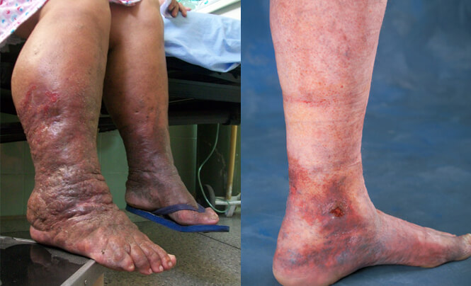
The signs and symptoms of deep vein thrombosis, although highly variable, include soreness, edema, burning, enlargement of superficial veins, redness or discoloration, and cyanosis with fever. However, some with DVT are asymptomatic. Signs and symptoms themselves are not sensitive enough or specific for a diagnosis, but when considered in combination with pre-test likelihood, they can help determine the likelihood DVT. In the most suspected cases, DVT is ruled out after examination, and symptoms are more likely to occur for other reasons, such as rupture Baker cysts, cellulite (inflammation), hematoma, lymphedema, and chronic venous insufficiency. Other likely causes of symptoms include tumors, venous or arterial aneurysms, and connective tissue disorders.
Causes and risk factors
Three main factors that can contribute to the development of deep vein thrombosis:
- damage to the inner lining of the vein;
- predisposition to blood clotting;
- slowing down blood flow.
- Injury of a vein.
Damage to a vein can occur during surgery, an injury to an arm or leg, injection of irritants, inflammation, or certain conditions such as thromboangiitis obliterans. They can also be damaged by a blood clot, making it more likely that a second clot will form.
- A tendency to increased blood clotting.
In some diseases, such as cancer and some hereditary blood clotting disorders, there is an increased ability of the blood to clot. Certain drugs, including oral contraceptives, drugs containing estrogen, or substances estrogen-like (such as tamoxifen and raloxifene) may also lead to increased clotting blood. Smoking is also a risk factor. Sometimes blood clots are more likely to form after childbirth or surgery. In the elderly dehydration of the body usually leads to increased readiness of the blood to clot and, therefore, may contribute to the development of DVT.
Read also:Cyclic neutropenia
- Slowdown of blood flow.
During prolonged adherence to bed rest and in other cases when a person is forced to remain immobile (for example, after leg injury or stroke), blood flow in the veins slows down because the calf muscles do not contract and do not push blood towards heart. For example, deep vein thrombosis can develop in people who have had myocardial infarction or other serious illness (heart failure, chronic obstructive pulmonary disease [COPD] or stroke) and lie in a hospital bed for several days without sufficient movement, or in people with paralysis of the legs and lower body (with paraplegia). DVT can develop after major surgery, particularly on the pelvic organs, hip, or knee. Thrombosis can develop even in healthy people who have been in a sitting position for a long time, for example, during long trips or flying in an airplane, but in such a situation, thrombosis is extremely rare and usually develops in people with other risk factors.
Epidemiology
Deep vein thrombosis and thromboembolism remain a common cause of morbidity and mortality in bedridden or hospitalized patients, as well as in generally healthy people. The exact incidence of DVT is unknown because most studies are limited by the inherent bias of clinical diagnosis. The existing data, which probably underestimates the true incidence of DVT, suggests that about 80 cases per 100,000 population occur annually. About 1 in 20 people develop DVT during their lifetime.
Diagnostics
Assessment of clinical likelihood using Wells scale (cm. Special column in table below) to determine whether a potential DVT is “likely” or “unlikely” is usually the first step in the diagnostic process. The scale is used when deep vein thrombosis of the first lower limb is suspected (without any symptoms TELA) in primary health care and outpatient settings, including the emergency department help. The numerical result (possible score from –2 to 8) is most often grouped in the “unlikely” or “likely” categories. A Wells score of two or more means DVT is considered “probable” (about 28% probability), while while those with a lower score are considered “unlikely” to develop DVT (about 6%). In those who are unlikely to have DVT, the diagnosis is ruled out due to a negative D-dimer blood test. In people with probable DVT, ultrasound is the standard imaging used to confirm or exclude a diagnosis. Imaging is also necessary for inpatients with suspected DVT and those who were initially classified as unlikely DVT but tested positive for D-dimer.
| Clinical data | Points |
|---|---|
| Paralysis, partial paralysis, or orthopedic plaster cast on the lower limb in the recent past | +1 point |
| Patient lying down (more than 3 days) or major surgery in the last 4 weeks | +1 point |
| Local soreness of the deep veins of the lower extremities | +1 point |
| Swelling of the entire leg | +1 point |
| Swelling of the back of the lower leg, the difference with the other leg is 3 cm (the measurement is taken 10 cm below the tuberosity of the tibia) | +1 point |
| Swelling with preservation of a trace on the skin when pressing on the sore leg | +1 point |
| Collateral non-varicose superficial veins | +1 point |
| Acute cancer or cancer treatment in the past 6 months | +1 point |
| An alternative diagnosis, more likely than deep vein thrombosis (Baker's cyst, cellulitis, damage muscles, superficial vein thrombosis, postphlebitic syndrome, inguinal lymphadenopathy, external compression veins) | −2 points |
Although Wells' score is the predominant and best-studied rule for the clinical prognosis of deep vein thrombosis, it has drawbacks. The Wells Scale requires a subjective assessment of the likelihood of an alternative diagnosis and performs less well in the elderly and those with a history of DVT.
Standard treatments
In deep vein thrombosis, the primary goal of the physician is to prevent pulmonary embolism. Initially, hospitalization may be required, but thanks to modern advances in science and technology, most patients with deep vein thrombosis can be treated at home. Bed rest is not required unless it relieves symptoms. Patients do not have to restrict their physical activity. Physical activity does not increase the risk that the blood clot will come off and cause pulmonary embolism.
Treatment usually includes:
- anticoagulants (most common) g
- thrombolytics
- in rare cases, a clot filter (umbrella filter).
Read also:Increased blood eosinophils in an adult
- Anticoagulants.
All patients with deep vein thrombosis receive anticoagulants. Usually doctors prescribe a low molecular weight heparin (such as enoxaparin, dalteparin, or tinzaparin) or fondparinux, which is injected under the skin (subcutaneously), in parallel with warfarin, taken inside. The injectable works immediately, and the full effect of warfarin appears after a few days. After warfarin takes effect, patients stop taking the injectable drug. In some patients (people with cancer or people with recurrent blood clotting problems despite treatment with oral anticoagulants) doctors use only an injectable drug and do not prescribe warfarin.
The duration of treatment with these drugs (warfarin or injectable drug) varies depending on the degree of risk. People who have DVT for a specific short-term cause (such as surgery or the drug they stopped taking) usually continue anticoagulant therapy for 3 to 6 months. Unless a specific cause is identified, patients usually take warfarin for at least 6 months. Warfarin should be taken indefinitely if there is an inherently persistent cause (for example, blood clotting disorder), or if the patient has two or more episodes of deep vein thrombosis.
The use of warfarin increases the risk of bleeding, both internal and external. To minimize the risk, people receiving warfarin should have periodic blood tests to assess the blood's anticoagulant ability. Doctors use the blood test result to adjust the dose of warfarin. Blood tests are usually done once or twice a week for 1 or 2 months and then every 4-6 weeks. Many different drugs and foods affect the metabolism of warfarin in the body. Certain drugs and food increase its cleavage, leading to ineffectiveness of the dose of warfarin and increasing the risk of recurrent clot formation. Other drugs and food slow down cleavage of warfarin, leading to an increase in the effectiveness of the warfarin dose and, therefore, an increase in the likelihood of bleeding. Some patients are also more sensitive to warfarin and may need warfarin sensitivity testing to adjust levels.
New drugs, given by mouth, have been developed that can be used as an alternative to warfarin. These drugs, called direct oral anticoagulants (PACA), include rivaroxaban, apixaban, edoxaban, and dabigatran etexilate. These drugs have a faster anticoagulant effect than warfarin and are as effective as warfarin for treating blood clots. With these new drugs, patients do not need frequent blood tests to adjust the dose, as with warfarin, but the risk of bleeding is still higher.
The most common complication of anticoagulants is excessive bleeding, which can be life-threatening. Risk factors for excessive bleeding include:
- age 65 or older;
- recent heart attack, stroke, or bleeding in the digestive tract;
- diabetes;
- renal failure.
To eliminate the anticoagulant effect in patients receiving warfarin, doctors may prescribe vitamin K, plasma transfusion (containing clotting factors) or prothrombin concentrate complex. Doctors may prescribe protamine to partially reverse the anticoagulant effect in patients receiving low molecular weight heparin.
Newer direct oral anticoagulants (PACA) (apixaban, dabigatran, edoxaban, and rivaroxaban) that are taken by mouth tend to cause less episodes of severe bleeding than warfarin, but antidotes are now widely available for these drugs (in the event of excessive bleeding) absent. Doctors are investigating additional antidotes.
- A filter that captures blood clots.
In very rare cases, with intolerance to anticoagulants, development of serious side effects or ineffectiveness in preventing formation additional blood clots inside a large vein between the heart and the area affected by deep vein thrombosis, a filter (umbrella filter) can be placed. Typically, the filter is placed in the inferior vena cava, which returns blood to the heart from the lower body. A filter can trap emboli, preventing them from entering the lungs, but unlike anticoagulants, filters do not prevent new clots from forming. Filters are usually intended for patients in whom anticoagulant treatment is impossible or ineffective.
- Thrombolytics.
Doctors are considering using intravenous drugs such as alteplase to dissolve blood clots. These drugs (also called thrombolytics or fibrinolytics) may be prescribed if less than 48 hours have passed since the clot formed. After 48 hours, scar tissue begins to develop in the blood clot, making it less likely to dissolve.
Doctors sometimes use thrombolytics in combination with mechanical removal techniques in individuals with large clots in the thigh. In these cases, doctors may place a small, flexible tube (catheter) into the blocked vein, remove as many clots as possible with an instrument and inject a diluent through the catheter thrombus.
Read also:Fisher-Evans Syndrome
- Treatment of complications.
If pulmonary embolism develops, treatment usually includes oxygen (usually given through a mask or nasal cannula), analgesics to relieve pain, and anticoagulants. If pulmonary embolism is life-threatening, thrombolytics are prescribed or surgery to remove the emboli is performed.
Veins never fully recover from deep vein thrombosis. Valvular vein repair surgery is under experimental development. With the development of chronic venous insufficiency, it is advisable to wear elastic compression stockings below the knee.
With the development of painful skin ulcers (trophic ulcers) Correctly applied compression bandages can have a good effect. With careful application of these bandages once or twice a week, the ulcer will almost always heal as the blood flow in the veins improves. The sores can become infected, and each time the dressing is changed, there may be pus and foul-smelling discharge on the dressing. Pus and discharge are washed off with soap and water. Skin creams, balms and medicines applied to the skin, regardless of the type, are ineffective.
After the blood flow in the veins improves, the ulcer heals on its own. Wearing an elastic stocking every day after the ulcer has healed helps prevent recurrence. The stocking is changed as soon as it stretches and ceases to tightly encompass the leg. If possible, the patient should purchase seven stockings or pairs of stockings (if both legs are affected), one for each day of the week, so that the stockings remain effective for a longer period.
In rare cases, non-healing ulcers require skin grafts. After the transplant, elastic stockings should be worn to prevent recurrence of the ulcer.
Forecast
Deep vein thrombosis is most commonly a condition of old age that occurs in nursing homes, hospitals, and active cancers. This is associated with a 30-day mortality rate of about 6%, with pulmonary embolism accounting for most of these deaths. Proximal DVT is often associated with PE, in contrast to distal DVT, which is rarely, if ever, associated with PE. About 56% of patients with proximal DVT also have PE, although chest CT is not required simply because of DVT. If proximal DVT is left untreated, about half of people will develop symptomatic PE in the next 3 months.
Another common complication of proximal DVT and the most common chronic complication is post-thrombotic syndrome, in which patients have chronic venous symptoms. Symptoms may include pain, itching, swelling, paresthesia, a feeling of heaviness and, in severe cases, leg ulcers. After proximal deep vein thrombosis, about 20-50% of people develop the syndrome, and 5-10% have severe symptoms. Postthrombotic syndrome can also be a complication of distal DVT, although to a lesser extent than proximal DVT.
Recurrent DVT is another possible consequence. Within 10 years after primary VTE, about 30% of patients will have a relapse. Recurrence of VTE in patients with prior DVT is more likely to recur as DVT than PE. Cancer and unprovoked DVT are significant risk factors for recurrence. After an initial proximal unprovoked DVT with and without PE, 16-17% of patients will have a recurrence of VTE within 2 years after completing the course of anticoagulants. Recurrence of VTE with distal DVT is less common than with proximal DVT. In deep vein thrombosis of the upper extremities, the recurrence of VTE is about 2–4% per year. After surgery, the recurrence rate of provoked proximal DVT or PE is only 0.7%.
Prophylaxis
To prevent blood clots in the general population, it is recommended that you exercise your legs and walk while sitting for hours, be active, and maintain a healthy body weight. Walking increases blood flow through the veins of the legs. Overweight can be changed, unlike most risk factors, and interventions or lifestyle changes that help overweight or obese people lose weight reduce risk of DVT. Statins have been investigated for primary prevention, and the JUPITER study (from the English. Justification for the Use of Statins in Prevention: an Intervention Trial Evaluating Rosuvastatin), in which rosuvastatin was used has provided some preliminary evidence efficiency. Of all the statins studied, rosuvastatin was found to be the only one that could reduce the risk of VTE. However, the amount required for treatment to prevent one primary VTE is about 2000, which limits its applicability.