Biliary atresia: what is it, causes, symptoms, treatment, prognosis
Content
- What is biliary atresia?
- Signs and symptoms
- Causes and risk factors
- Epidemiology
- Histopathology
- Diagnostics
- Treatment
- Forecast
- Complications
What is biliary atresia?
Biliary atresia (biliary atresia) Is an obstructive cholangiopathy of unknown etiology, affecting both intrahepatic and extrahepatic bile ducts. It manifests itself in the neonatal period as persistent jaundice, clay-colored stools and hepatomegaly. The condition is fatal if left untreated, and the survival rate at 3 years is less than 10%.
The earliest mention of this condition dates back to 1817 by Dr. John Burns, who described it as an incurable condition of the biliary apparatus. Later, the first surgical success was achieved by Dr.William Ladd in 1920, but biliary atresia continued to be deplorable. result until the 1950s, when Dr. Morio Kasai first described portoenterostomy by cutting the proximal biliary obstruction and creating a loop Roux-en-Y. It is now considered a standard procedure and is offered to all children undergoing a surgical correction. With the development of transplant surgery, liver transplantation has become an option available to children who either have failed restore the flow of bile during the initial Kasai portoenterostomy, or who developed cirrhosis of the liver at a later stages.
Signs and symptoms
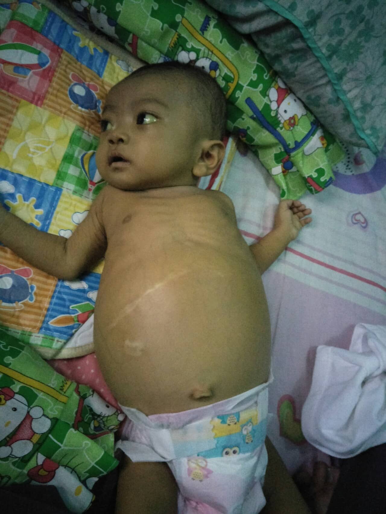
In children in the neonatal period, persisting jaundice, clay-colored stools and hepatomegaly. Jaundice in any child over 14 days of age should no longer be considered physiological and the child should be evaluated. More than half of children with biliary atresia initially have pigmented stools, which later become acholic, and as the disease progresses, signs appear cirrhosis of the liver and liver failuremanifested by palpable hepatomegaly, splenomegaly, ascites, signs of portal hypertension and developmental disabilities.
Causes and risk factors
The etiology of biliary atresia is unknown. Theories suggest many etiological and causal factors, both genetic and acquired. Since about 3% to 20% of children with biliary atresia have any associated syndrome or another congenital anomaly, and biliary atresia is more common in certain geographic regions. It is likely that some genetic component is present in the pathogenesis of the disease, although a single etiology has not yet been discovered. Only a few familial cases have been described, and no increase in morbidity was noted in twins.
The extrahepatic bile ducts first become visible as a bulge from the anterior intestine on day 20 pregnancy, and the intrahepatic bile ducts become visible on the 45th day, which are formed from primitive hepatocytes. The hepatic hilum is the junction between the accessory and intrahepatic bile ducts, and successful junction is critical to the development of an open biliary system. A non-syndromic isolated type of biliary atresia may result from improper remodeling in fetal life at the hepatic hilum. This is supported by the fact that there is a similarity in cytokeratin staining of the bile ducts in patients with atresia of the bile ducts and bile ducts of the fetus in the first trimester, which increases the possibility of atresia of the biliary tract due to impaired remodeling of the bile ducts in the hepatic hilum with preservation of fetal biliary ducts.
Other theories give preference to possible acquired, inflammatory and infectious causes of the pathogenesis of the disease. Rotavirus, type 3 reovirus are specifically referred to as their perinatal animal models causing biliary atresia; however, these results have not always been observed in humans.
There have also been studies showing immune damage to the ducts in patients with biliary atresia due to increased expression of the intercellular adhesion molecule ICAM-1 in the bile ducts.
Other studies support an acquired etiology with seasonal clustering of cases, especially during the winter months, and because 50% of children with this disease had pigmented stools at an earlier age, which then became clay colors.
Classifications.
Biliary atresia is not a single disease caused by a specific etiology, but rather a phenotype resulting from a different etiology. It is broadly classified into syndromic and non-syndromic isolated varieties. Davenport et al. grouped certain forms of biliary atresia based on similarities.
Read also:Ankyloglossia
- Syndrome of biliary atresia and spleen malformations.
Biliary atresia is associated with polysplenia, vascular abnormalities, including the preuodenal portal vein, interrupted vena cava, azygotic continuation, malformation of the heart, malrotation and transposition of internal organs. In this type, the malformation occurs at an early stage of embryogenesis and is the cause of other anomalies. Maternal diabetesappears to play a role, and women predominate here.
- Cystic biliary atresia.
In this type, obliteration of the biliary system with cystic dilatation is observed. The incidence is reported to be around 10% and he has a better prognosis.
- Cytomegalovirus IgM + ve associated biliary atresia.
This type accounts for about 10% of cases. These children have higher levels of bilirubin and AST, as well as more inflammatory infiltrates in the extrahepatic biliary apparatus on histology. This group of patients has a worse prognosis.
- Isolated biliary atresia.
This is the largest group, but the etiology is unknown.
Morphological classification.
Morphological classification is based on the level of obliteration of the bile lumen. The Japanese Association of Pediatric Surgeons has classified it as:
- Type I: obliteration of the common bile duct.
- Type II a: obliteration of the common hepatic duct.
- Type II b: obliteration of the common bile duct, hepatic duct, cystic duct without anomalies of the gallbladder and cystic dilatation of the hepatic hilum.
- Type III: obliteration of the common bile duct, hepatic duct and cystic duct without anastomosed ducts in the hepatic hilum; this is the most common variety.
Epidemiology
Noticeable regional variations of biliary atresia have been reported, with a higher incidence per Taiwan and Japan 1 to 5 per 1000 live births to lower rates in England and Wales from 1 to 5 in 20 000. Some small series also showed seasonal variability. Others showed a slight female predominance.
There are geographical differences between different types of biliary atresia, with the frequency of the syndrome biliary atresia and spleen malformations recorded in European studies is 10%, and in Asia - much less. Many cases of cytomegalovirus IgM + ve associated with biliary atresia have been reported in China.
Histopathology
Histological examination of samples of biliary atresia shows a variable liver fibrosis, proliferation of bile ducts, blockage of bile ducts, cholestasis, infiltration of inflammatory cells. Among all the features, bile duct proliferation is a highly sensitive and specific marker of biliary atresia.
Diagnostics
There is no single method that can accurately determine biliary atresia from other causes of conjugated hyperbilirubinemia with high specificity. Children undergo both serological and radiological tests, as well as histopathology, to make a diagnosis.
Laboratory research.
With atresia of the biliary tract, the levels of direct and indirect bilirubin are increased, and the conjugated part is increased more. Since alkaline phosphatase levels are elevated in children due to bone remodeling, the liver-specific alkaline phosphatase 5 'nucleotidase fraction should be measured. Gamma-glutamyl transpeptidase (GGTP) is present in the bile membrane of the tubules and increases in the case of obstruction of the biliary tract. GGTP provides diagnostic accuracy from 50% to 60% for biliary atresia. Serum transaminase levels are slightly elevated.
Visual research.
— Ultrasound.
Ultrasonography is a readily available non-invasive test that can provide valuable information about liver structure, vascular patency, ascites; and can also exclude other causes of obstructive jaundice, such as a common bile duct cyst. Ultrasonography shows gallbladder hypoplasia or lack thereof. This can be done both before and after meals; which will show a non-filling of the gallbladder. It can identify the symptom of a triangular scar; described as the presence of a solid proximal bile duct remnant before portal bifurcation. However, some authors disagree about the accuracy and specificity of this trait. Antenatally, infants with congenital biliary atresia can be detected by abnormal maternal ultrasound at about 20 weeks of gestation, which can facilitate early postpartum ultrasound and timely referral to specialists. Despite the simplicity and availability of ultrasound, the diagnostic accuracy of this method for diagnosing atresia is only 78%.
Read also:Perinatal encephalopathy (PEP) in children: what it is, causes, symptoms and treatment
- Hepatobiliary scintigraphy.
It uses a technetium-labeled diisopropylinodiacetic acid compound (DYSIDA). The presence of the isotope in the intestine excludes atresia of the biliary tract. The reliability of this test is impaired when conjugated bilirubin levels are high. It also has 10% false positive or false negative results.
Endoscopic retrograde cholangiopancreatography.
Not widely used due to limited availability of side-view neonatal scopes. As experience and availability increase, this method is likely to be used in situations where other tests have failed to confirm the diagnosis.
Duodenal intubation.
Aspiration of fluid stained with bile from duodenum excludes atresia of the biliary tract. This test is not widely used because it is invasive and unreliable.
Magnetic resonance imaging (MRI) and magnetic resonance cholangiopancreatography (MRCP)).
These tests provide greater accuracy, but are not easy to perform due to the cost and need for sedation, and the limited authorization at this young age.
Liver biopsy.
A liver biopsy can differentiate atresia with a high degree of accuracy from other causes of cholestatic jaundice. Signs suggestive of biliary atresia include proliferation of bile ducts, blockage of bile, multinucleated giant cells, focal necrosis of the liver parenchyma, extramedullary hematopoiesis and infiltrate inflammatory cells. Among these features, the proliferation of bile ducts is considered the most sensitive and specific sign.
Treatment
Surgical research is the only method for accurate and reliable diagnosis and treatment of biliary atresia.
- Postoperative cholangiography.
Postoperative cholangiography will make it possible to definitively diagnose atresia by impaired passage of the dye into the intrahepatic and extrahepatic biliary system.
- Portoenterostomy according to Kasai.
The standard surgical technique is the creation of a Roux-en-Y liver portoenterostomy (Kasai procedure), in which excision of the fibrous residue of the bile ducts is performed, the operation of the fibrous portal plate with dissection to the bifurcation portal vein. The loop connected in the Roux-en-Y style restores biliary-intestinal continuity and ensures the outflow of bile.
On rare occasions when gall bladder and the common bile duct is not patent, the possibility of portocholecystostomy may be considered. However, the anastomosis is not as flexible as the standard Roux loop, and revisions for re-obstruction of the biliary tract have been described with poorer long-term outcome. He has a lower frequency of cholangitis. In some cases, hepaticojejunostomy with type I atresia of the bile ducts has been described, but the results are inferior to the standard Kasai procedure.
Several drugs are used as adjuvants after surgery to improve biliary drainage, among them steroids and ursodeoxycholic acid are widely described. Steroids reduce the inflammatory response and promote bile excretion. One prospective, randomized, placebo-controlled study used a low dose of prednisolone (2 mg / kg / day), and it showed a significantly increased rate of recovery from jaundice in the steroid group, but did not provide any benefit in survival rate. Another START (Steroids in Biliary Atresia Randomized Trial) study did not show a positive effect of high doses of steroids on biliary lumen six months after surgery and had an earlier onset of side effects associated with steroids. Ursodeoxycholic acid is a hydrophilic bile acid and is typically present in an amount of about 1% to 4% of the total bile acid pool. It is known to promote the elimination of bile and is often prescribed in the postoperative period.
- Liver transplant.
Liver transplantation is an option suggested if liver cirrhosis is advanced or if Kasai's portoenterostomy has failed.
Read also:Dyslexia
Forecast
The main factors determining a satisfactory outcome after portoenterostomy.
- Age at initial surgery.
Liver fibrosis is a time-dependent factor. However, the age of portoenterostomy and the results of surgical intervention do not have a linear relationship. This was demonstrated in an age cohort analysis that showed that up to about 90 days of age the outcome of a portoenterostomy cannot be determined by age alone for cases of an isolated biliary variety atresia.
- Syndrome of biliary atresia and spleen malformations
Infants with biliary atresia and spleen malformations already respond to Kasai's surgery and have a poorer overall prognosis with a higher risk of death.
— CMVIgM + ve associated biliary atresia.
Carries the most unfavorable outcome and the highest risk of death in infants with atresia.
— Successful achievement of postoperative bile outflow.
Satisfactory bile outflow with good bile clearance after portoenterostomy gives better survival with the native liver. Nio et al. reported that most patients who survived more than 20 years after TELAhad normal or moderately elevated liver function test results after PE. Likewise, Uchida et al. reported that serum AST levels measured 1 year after PE is a strong predictor of liver dysfunction. Gupta et al. reported that there was a statistically significant decrease in GGT after portoenterostomy compared to preoperative levels in patients whose jaundice resolved.
— The size of the microscopic ducts at the hilum.
Several studies have shown that the size of the microscopic bile ducts in the cut residual bile duct has prognostic value. Others have denied this claim.
Complications
Postoperative complications include cholangitis, anastomotic leak, intestinal obstruction, portal hypertension and hepatopulmonary syndrome.
- Cholangitis.
The exact mechanism by which cholangitis develops is not known. However, the formation of a Roux loop with the restoration of bilio-intestinal blood flow leads to the colonization of the loop by bacteria and predisposes to ascending cholangitis. Cholangitis is reported in 50% of cases.
In the past, surgeons have tried several surgical techniques to reduce the risk of cholangitis, but none have proven beneficial.
In children with cholangitis, there is an increase in body temperature, exacerbation of jaundice and an increase in the activity of liver enzymes. Treatment should be prompt and aggressive with broad-spectrum intravenous antibiotics with good coverage against gram-negative bacteria.
- Portal hypertension.
Most children with biliary atresia due to liver fibrosis have high portal pressure. Because liver fibrosis is time-dependent, it correlates with age at portoenterostomy and bilirubin levels. This is a poor prognostic sign, as most children develop portal hypertension, variceal bleeding, and end-stage liver failure.
- Bleeding from varicose veins.
Varicose veins of the esophagus develops on average 2-3 years after Kasai portoenterostomy in 60% of children, of which about 30% will have bleeding. Endoscopic follow-up is recommended for every child with biliary atresia. Patients with active bleeding require sclerotherapy or bandaging.
- Ascites.
Ascites can be caused by portal hypertension, hypoalbuminemia, and hyponatremia. Spironolactone therapy is usually recommended.
- Intrahepatic cysts.
Biliary cysts or "lakes" can develop in the liver of patients with biliary atresia and cause recurrent attacks of cholangitis. Long-term use of broad-spectrum antibiotics is recommended. Cases that do not respond require liver transplantation.
- Hepatopulmonary syndrome.
This syndrome is characterized by cyanosis, dyspnea, hypoxia and the fingers of Hippocrates. Diffuse intrapulmonary shunting is thought to be due to vasoactive compounds of intestinal origin that bypass sinusoidal inactivation. Liver transplantation is usually required to reverse the process.
- Malignancy.
Reported rare cases of malignant changes in liver cirrhosis after portoenterostomy (hepatocellular carcinoma or cholangiocarcinoma).