Hyphema: what is it, causes, symptoms, treatment, prognosis
Content
- What is hyphema?
- Signs and symptoms
- Causes and risk factors
- Epidemiology
- Pathophysiology
- Diagnostics
- Treatment
- Forecast
- Complications
What is hyphema?
Hyphema is defined as an accumulation of red blood cells (erythrocytes) in the anterior chamber of the eye. By definition, blood should be clearly visible on direct eye examination or on slit lamp examination. Blood accumulates due to the destruction of vessels in the iris or ciliary body, usually due to trauma or concomitant diseases. The anterior chamber is the area bounded by the cornea in the front, the angle from the side, and the lens and iris in the back. This space usually contains transparent aqueous humor, which is produced by the ciliary body and excreted through the Schlemm's canal. The angle is an important anatomical site, since it is here that the trabecular meshwork and Schlemm's canal are located. Blockage of this area prevents the outflow of water, which leads to an increase in intraocular pressure.
Signs and symptoms
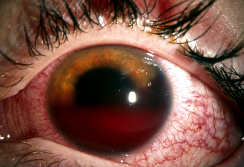
Loss of vision or loss of vision is often the first sign of hyphema. In patients with microhyphema (grade I), vision may be slightly blurred or normal. A person with a complete hyphema may not see at all (complete loss of vision). A person's vision can improve over time as blood moves by gravity lower in the anterior chamber of the eye, between the iris and the cornea. Many people experience better vision, but some may have other problems associated with eye trauma or complications due to the hyphema. Microhyphema, in which red blood cells hang in the anterior chamber of the eye, is less severe. The layered hyphema, when fresh blood is seen lower in the anterior chamber, is moderately severe. Complete hyphema (grade IV), when blood fills the chamber completely, is most severe.
Causes and risk factors
The most common cause is blunt trauma to the eye, although penetrating trauma and spontaneous hyphema also occur. Certain medical conditions and medical conditions can also put patients at risk of developing hyphema: leukemia, hemophilia, von Willebrand disease, sickle cell anemia and taking anticoagulants. Neovascularization of the eye, often associated with diabetes mellitusalso puts patients at risk.
Finally, postoperative patients may develop hyphema. The disorder can develop both during surgery and a week after surgery.
Epidemiology
The incidence of traumatic hyphema is 12 out of 100,000, of which 70% are in children. The disorder is most commonly seen in men between the ages of 10 and 20 and usually results from sports or recreational injuries. Children are usually injured during ball sports such as baseball, basketball, softball and football when the ball hits the front of the globe. Adolescents and adults are more likely to suffer from a high-energy blow to the eye, most often from an attack. Other reasons include paintball and airsoft guns and airbag deployment.
Read also:Marcus Gunn Syndrome
Pathophysiology
Blunt or penetrating trauma usually causes traumatic hyphema in the orbit. Bleeding occurs due to rupture of the vessels of the ciliary body and iris. When a blunt force is applied to the anterior part of the eyeball, an instantaneous increase in intraocular pressure occurs, creating a shear force through the ciliary body and the iris. However, penetrating trauma results in direct damage to the iris.
Spontaneous hyphema often occurs in patients whose health conditions predispose to ischemia, neovascularization, or vascular abnormalities. These patients usually have spontaneous vascular leakage. As expected, this is more common in patients with diabetes, eye tumors, blood clotting disorders, sickle cell disease, and those taking anticoagulants.
Diagnostics
It is important for the ophthalmologist to determine the cause of the hyphema. If there was an eye injury, you will need to find out the details of the incident in order to first determine whether whether the injury is an open trauma to the eyeball, i.e. something penetrated or severely damaged the outer layers eyes.
If it is a non-penetrating injury, the doctor looks at the medical history and then examines the eye to determine if it is a hyphema or other cause of redness, such as uveitis (which can lead to hyphema), conjunctivitis (eye infection) or subconjunctival hemorrhage (ruptured blood vessels in the eye).
If hyphema is suspected, the doctor checks visual acuity, measures intraocular pressure, and examines the eye using a slit lamp microscope and an ophthalmoscope. In some cases, a computed tomography (CT) scan may be required to fully examine the internal structures of the eye. You may also be screened for sickle cell disease or other conditions that increase the risk of complications from hyphema.
If an object is in your eye, do not try to remove it. If possible, secure it in place and see an ophthalmologist or hospital emergency department immediately.
Treatment
A person with a hyphema should see an ophthalmologist (a doctor who specializes in the diagnosis and treatment - surgical and non-surgical methods - of eye diseases) as soon as possible. In some cases, with severe bleeding or with a history of blood clotting in the victim (which increases the likelihood of bleeding and rebleeding), and if the victim is taking anticoagulants, it may be necessary hospitalization.
Read also:Waardenburg syndrome
Treatment usually involves bed rest with the victim on a bed with a raised headboard to allow blood to drain as quickly as possible. Often, the victim is given eye drops that dilate the pupil (such as atropine) to relieve inflammation in the eye and minimize the formation of scar tissue inside the eye (usually corticosteroids). To avoid aggravating the injury, the eyes are sealed with a protective bandage.
Doctors measure intraocular pressure at least once a day for the first few days. This examination is completely painless and is done using a device called a tonometer. An increase in intraocular pressure can be accompanied by nausea, eye pain, and blurred vision. To lower the pressure, an ophthalmologist may prescribe eye drops similar to those used to treat glaucoma. For several weeks after the injury, the victim should, if possible, refrain from taking aspirin and other non-steroidal anti-inflammatory drugs that can aggravate bleeding. Due to the fact that hyphema increases the likelihood of developing glaucoma throughout life, persons who have had hyphema should undergo an ophthalmologic examination annually.
If bleeding is severe or bleeding recurs, the ophthalmologist may prescribe aminocaproic acid or tranexamic acid, drugs that accelerate blood clotting. In rare cases, if recurrent bleeding leads to an increase in intraocular pressure, the blood is drained surgically.
Forecast
Most patients recover completely without sequelae, but complications are more likely in patients with other comorbidities, such as sickle cell anemia, and with an increase in size hyphema. For example, increased intraocular pressure is observed in 13.5% of grade I-II hyphema; while grade IV hyphae have a 52% risk. The prognosis of normal vision is also influenced by the degree of hyphema. Grade I hyphemas have about 90% of normal vision; whereas for grade IV, the prognosis for normal vision is 50% to 75%. The most common cause of visual impairment is corneal staining of the visual axis, highlighting a worsening prognosis for higher grade hyphema.
Complications
The two major acute complications of hyphema are acute intraocular hypertension and rebleeding. Acute intraocular hypertension is the most likely complication to be encountered in the emergency department. Since the blood is in the layers of the anterior chamber, it can block the trabecular network from adequate drainage of the combination of aqueous humor and blood
Read also:Keratoconus
Sickle cell anemia deserves special attention with regard to the hyphema. As noted above, elimination of the hyphema requires the passage of red blood cells through the trabecular meshwork and Schlemm's canal. However, the anterior chamber is relatively hypoxic, which causes crescent red blood cells in at-risk individuals. As sickleness worsens, red blood cells cannot pass through the Schlemm's canal, causing an increase in intraocular pressure.
Any patient with an intraocular pressure greater than 21 mm Hg. Art. should be treated in the same way as for acute non-traumatic glaucoma. It is necessary to consult an ophthalmologist to obtain treatment recommendations, as there is no well-defined treatment strategy. A number of medications can be used to suppress fluid drainage. These include topical beta-blockers, most commonly timolol, topical alpha-2 agonists such as apraclonidine and brimonidine. Carbonic anhydrase inhibitors are also commonly used, although caution should be exercised as the use of these drugs can worsen sickle behavior in patients with sickle cell disease. Examples include topical dorzolamide or systemic acetazolamide. Many different drug combinations are available under brand names. It is therefore advisable to discuss a targeted treatment approach with an ophthalmologist. About 5% of patients will have persistent or refractory intraocular hypertension requiring surgical removal of the clot.
Rebleeding can occur after 2-5 days and may increase the risk of permanent vision loss. Rebleeding occurs in about 30% of cases. The use of antifibrinolytics such as epsilon aminocaproic acid or tranexamic acid was once recommended in patients at increased risk. However, trials failed to demonstrate a reduction in rebleeding and an increase in hyphema resolution time.
Corneal staining with blood is a rare complication that usually occurs in patients with prolonged common hyphema. In one case series involving 289 patients, 2.1% of the hyphema were found to develop corneal staining with blood, and this occurred only in patients with complete hyphema. The recommended treatment to prevent blood staining of the cornea is flushing the anterior chamber.