Sturge-Weber syndrome: what is it, symptoms, treatment, prognosis
Content
- What is Sturge-Weber syndrome?
- Signs and symptoms
- Causes and risk factors
- Affected populations
- Symptomatic disorders
- Diagnostics
- Standard treatments
- Forecast
What is Sturge-Weber syndrome?
Sturge-Weber syndrome (SShV or Sturge-Weber syndrome, encephalotrigeminal angiomatosis, nevoid amentia) Is a rare disease characterized by an association of port-wine stains (flaming nevus) on the face, neurological abnormalities, and eye abnormalities such as glaucoma. SSW can be thought of as a spectrum of diseases in which victims develop abnormalities affecting all three of these systems (ie, the brain, skin, and eyes), or only two, or only one. Consequently, the specific symptoms and severity of the disorder can vary greatly from one person to another. Symptoms of Sturge-Weber syndrome are usually present at birth (congenital), but the condition is not inherited or occurs in families. Some symptoms may only develop in adulthood. Sturge-Weber syndrome is caused by a somatic mutation in GNAQ gene. This mutation occurs randomly (sporadically) for no known reason.
Sturge-Weber syndrome can be classified as neurocutaneous syndrome or one of the phakomatoses. Neurocutaneous syndromes or phakomatosis are general terms for a group of diseases in which growths develop in the skin, brain and spinal cord, bones, and sometimes in other organs of the body. In the case of Sturge-Weber syndrome, these growths consist of abnormal blood vessels.
Some publications break Sturge-Weber syndrome into three main subtypes. Type 1 consists of cutaneous and neurological symptoms. These patients may have glaucoma, Or maybe not. Type 2 consists of skin symptoms and possibly glaucoma, but there is no evidence of neurological damage. Type 3 includes neurologic involvement but no skin abnormalities. Glaucoma is usually not present. Type 3 Sturge-Weber syndrome is also called isolated neurological type.
Signs and symptoms
Sturge-Weber syndrome is a highly variable disease. Some patients may develop characteristic skin abnormalities, but not neurologic ones. Less commonly, neurological abnormalities develop without characteristic skin signs. Therefore, it is important to note that patients may not have all of the symptoms described below, and that each individual patient is unique. Parents should talk with their child's healthcare provider about their particular case, associated symptoms, and general prognosis.
- Facial anomaly.
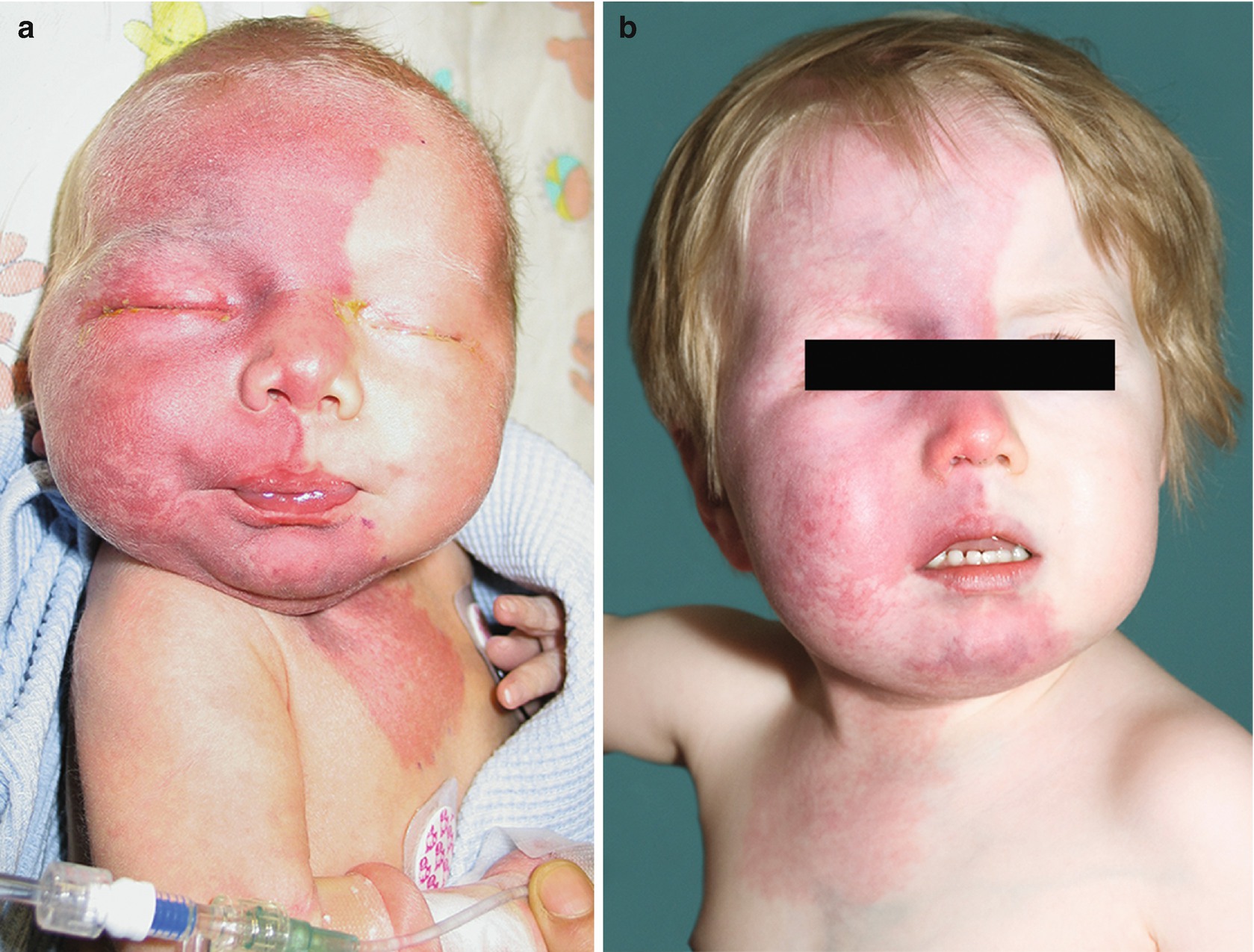
A congenital birthmark on the face (port port or flaming nevus) is often the most noticeable initial symptom. This mole can range in color from light pink to reddish to deep purple (see picture). photo above). The size of the "wine" stain can be different. Usually at least one eyelid and / or forehead on one side of the face is affected, but both sides of the face are rarely affected. In some children, the entire half of one side of the face is affected. Sometimes the discoloration may spread slightly to the other side of the face, or both sides of the face may be severely affected. In rare cases, a flaming nevus extends to the trunk and / or arms. The birthmark, which characterizes Sturge-Weber syndrome, is caused by an overabundance of capillaries just below the skin surface in the distribution of the trigeminal nerve. Capillaries are tiny blood vessels that form a thin network throughout the body, connecting arteries and veins, and are responsible for the exchange of various substances, such as oxygen, between cells and fabrics. If untreated, port-wine stains may darken with age, thicken and potentially form blood blisters (blisters) that can burst, causing spontaneous bleeding.
The abnormal blood vessels that make up the mole will vary in size, diameter, distribution and depth from one person to another and even from one person in different affected areas. This means that each person's flaming nevus is unique and can be very different.
- Neurological abnormalities.
Patients with Sturge-Weber syndrome can also experience a variety of neurological abnormalities. The degree of neurological damage can vary greatly from one person to another. Neurological symptoms are caused by abnormal growth of blood vessels on the surface of the brain (leptomeningeal angiomas). Epileptic seizures, which often begin in infancy or childhood, are common. Epileptic spasm is usually one-sided, opposite to the "port port", but sometimes affects both sides of the body. Epilepsy can vary in frequency and intensity, and sometimes worsens in severity and frequency with age. People may also experience muscle weakness or paralysis on one side of the body (hemiparesis), usually on the side opposite the nevus. Some children have developmental delays and mental retardation, ranging from mild learning disabilities to severe cognitive impairments; in other children, the disease does not affect intelligence or cognition. Cognitive impairment is common in patients with severe or uncontrolled seizures.
Read also:Amyotrophic lateral sclerosis (ALS)
Headaches may also occur, including migraine, and visual field defects such as loss of vision in half of the field of view of one or both eyes (hemianopsia). There is a risk stroke, stroke-like episodes, or mini-strokes (transient ischemic attacks). Stroke-like episodes can be associated with temporary (transient) weakness or paralysis of half of the body and visual field defects. Behavioral problems such as attention deficit disordermood disorders and poor social skills have also been observed in some children, especially those with lower cognitive function and higher seizure rates.
- Eye disorders.
Some babies are born with glaucoma, a condition characterized by increased intraocular pressure. Glaucoma usually affects the eye on the same side of the face as the port's mole. Glaucoma can potentially damage the optic nerve, the main nerve that carries signals from the eye to the brain, eventually leading to progressive vision loss. The same eye can also grow in size, so it seems that it protrudes from the orbit or enlarges it (buphthalmos).
Other ocular abnormalities may occur, including the development of angiomas in the membranes lining the inner surface of the eyelids (conjunctiva) layer blood vessels and connective tissue (choroid) between the white of the eye and the retina, as well as the transparent membrane that covers the membrane (cornea). The affected person's eyes may be of two different colors (for example, one brown and one blue). Additional eye symptoms may include an abnormal accumulation of fluid within the eyeball, causing the eyeball to enlarge (hydrophthalmos); degeneration of the cranial nerve that transmits light signals to the brain (optic atrophy); lens clouding or misalignment; retinal disinsertion; stripes that resemble retinal blood vessels (angioid stripes); and / or loss of vision due to organic damage to the visual cortex (cortical blindness). Patients with neurological abnormalities, but without a nevus, usually do not have vision problems.
- Endocrine disorders.
Some have also had endocrine disruptions, including central hypothyroidism and an increased risk of growth hormone deficiency. Central hypothyroidism characterized by insufficient activity of the thyroid gland due to insufficient stimulation of thyroid-stimulating hormone in a healthy thyroid gland. Central hypothyroidism in Sturge-Weber syndrome can be caused by taking anticonvulsants.
Additional symptoms may occur, including an abnormally large head (macrocephaly), overgrowth (hypertrophy) of certain soft tissues underlying the port port, and lymphatic malformations, which are non-cancerous growths consisting of fluid-filled ducts or spaces thought to be caused by abnormal development of lymphatic systems. These symptoms are consistent with a related rare condition known as Klippel-Trenone Syndrome (SKT), and most children with these symptoms are classified as having SKT. Researchers are unsure if SSB and SCT are related disorders that overlap, or if they are similar, but different, rare disorders.
Causes and risk factors
Sturge-Weber syndrome is caused by a somatic mutation in a gene GNAQ. This genetic mutation is somatic because it occurs after the fertilization of the embryo; in the case of SSV, most likely at an early stage of embryonic development. By definition, a somatic mutation can occur in any cell in the body other than the reproductive cells (sperm and eggs). Affected people will have some cells with a normal copy of the gene and some cells with an abnormal gene (mosaic pattern). This can be called having two different cell lines in the body. The variety of symptoms associated with Sturge-Weber syndrome is in part due to the ratio of healthy and abnormal cells in the body and the types of cells affected. Somatic mutations are not inherited or passed on to children. Researchers believe that somatic mutations GNAQ occur randomly for no apparent reason (sporadically).
Read also:Lockjaw
Genes provide instructions for making proteins that are critical to many bodily functions. When a gene mutation occurs, the protein product may be defective, ineffective, or missing. Depending on the functions of a particular protein, it can affect many organ systems in the body. Gene GNAQ creates a protein known as Gaq, which plays an important role in cell function, including the regulation of blood vessels. The specific pathway underlying impaired Gaq function in people with Sturge-Weber syndrome is not fully understood. More research is needed to determine the exact underlying mechanisms that cause the various symptoms of SSW.
As discussed above, symptoms are in part caused by abnormal development, growth, and proliferation of certain blood vessels. These blood vessel abnormalities often lead to secondary effects of the affected tissue, including a lack of oxygen in the affected body tissues (hypoxia), insufficient blood supply to the affected area (ischemia), blockage of the affected veins (venous occlusion), blood clots (thrombosis) and / or tissue death due to lack of oxygen (heart attack). Calcification of the affected areas of the brain can also occur.
Gene mutation GNAQ also cause a form of skin cancer (melanoma) that affects the eye (uveal melanoma). Mutations GNAQ, associated with uveal melanoma affect certain cells known as melanocytes. The mutation in people with uveal melanoma occurs in adulthood, rather than before birth, as in people with Sturge-Weber syndrome. Hence, the specific cells involved and the person's age when the mutation occurs in GNAQ, are extremely important and can cause various disorders or physical abnormalities.
Affected populations
Encephalotrigeminal angiomatosis affects men and women equally. The exact frequency and prevalence are unknown. One estimate is that the incidence is 1 in 20,000-50,000 live births. About 3 out of 1,000 babies are born with a flaming nevus, but only about 6% of people with a mole on their face develop neurological disorders associated with the disease. The risk rises to 26% if the nevus is on the forehead, temple, or upper face. The syndrome can affect people of any race or ethnicity.
Symptomatic disorders
Symptoms of the following disorders may be similar to those of Sturge-Weber syndrome. Comparisons can be useful for differential diagnosis.
Klippel-Trenone syndrome (KLT) is a rare disease that is present at birth (congenital) and is characterized by a triad of cutaneous capillary abnormalities (flaming nevus), lymphatic and venous abnormalities in combination with variable growth (hypertrophy) of soft tissues and bones. SKT most often occurs in the lower limbs and less often in the upper limbs and trunk. SKT affects men and women equally. The exact cause of the disorder is unknown.
Other disorders included in the classification of neurocutaneous disorders or phakomatosis, such as tuberous sclerosis, Hippel-Lindau disease, Wyburn-Mason syndrome, and neurofibromatosis, can have signs and symptoms similar to those seen in people with Sturge-Weber syndrome. Additional disorders that may have similar symptoms include type II Pascual-Castroviejo syndrome, Cobb syndrome, Maffucci syndrome, Gorham-Stout syndrome and Parkes-Weber syndrome.
Read also:Aphasia
In addition, in some children with megalencephalic-capillary malformation syndrome, symptoms overlap with SSM. However, most cases of megalencephaly capillary malformations are caused by mutations in the gene PIK3CAand if in doubt, genetic testing of the affected skin tissue can be performed.
Diagnostics
The diagnosis of Sturge-Weber syndrome (STS) is based on the identification of characteristic symptoms (for example, flaming nevus), detailed patient history, careful clinical evaluation, and various specialized tests. The diagnosis may be straightforward in an infant with a nevus, glaucoma, signs of brain damage, and neuroimaging findings consistent with the diagnosis of SSW. Diagnosis can be more difficult in infants who have a mole but no neurological symptoms.
- Clinical examinations.
A variety of imaging techniques can be used to identify and evaluate neurological complications, including skull x-rays or magnetic resonance imaging (MRI) with gadolinium. Gadolinium is a contrast agent used to improve scan results and produce more detailed images of tissues such as the brain or blood vessels.
Computed tomography (CT) can also be used to diagnose Sturge-Weber syndrome. Single-photon emission computed tomography (SPECT), which is a specialized computed tomography, helps to identify the affected areas of the brain, which may not be displayed on MRI or conventional CT. SPECT scans can be used in conjunction with other scans to evaluate the brains of suspected SSW patients.
Traditional angiography to assess the health and function of blood vessels is generally not recommended for individuals with suspected SSW, but may sometimes be needed to rule out a lesion with high blood flow velocity, such as arteriovenous malformation or arteriovenous fistulas. An electroencephalogram (EEG) is used to assess and localize seizure activity.
A complete ophthalmologic examination can reveal glaucoma and other ocular abnormalities potentially associated with Sturge-Weber syndrome. Due to the high risk of glaucoma, regular full eye examinations are necessary, especially in infants and young children. Follow-up examination should continue into adulthood, even if results remained normal during childhood.
Standard treatments
Treatment for Sturge-Weber syndrome is aimed at eliminating specific symptoms that appear in each person. Treatment may require a coordinated team of specialists. Pediatricians, neurologists, neurosurgeons, dermatologists, ophthalmologists, and other healthcare professionals may need to systematically and comprehensively plan treatment for an affective child. Psychosocial support is also important for the entire family.
Anticonvulsants are used to control seizures and drugs are used to treat glaucoma. Surgery for glaucoma or surgery may be needed to stop seizures that recur despite medication.
Low doses of aspirin are usually prescribed to people because it can reduce the risk of stroke. In addition, aspirin can improve blood flow in the regions of the brain located under angiomas. However, there is no evidence to support the effectiveness of aspirin.
Laser treatments can be used to lighten or remove birthmarks. Research on beta-blockers applied to the skin is underway.
Complementary therapy includes physical therapy for muscle weakness, special education for children with developmental delay or mental retardation, as well as other medical, social or occupational services.
Forecast
The symptoms of Sturge-Weber syndrome tend to get worse with age. However, most people with the syndrome have mild symptoms and are not life-threatening. The long-term perspective varies depending on the severity of the symptoms and how well seizures and glaucoma can be controlled and prevented. More severe seizures at an early age are associated with an increased risk of developing mental retardation. Adults with Sturge-Weber syndrome may have psychological problems that require additional intervention.