
Analysis of feces for dysbiosis: how to take it right and decoding
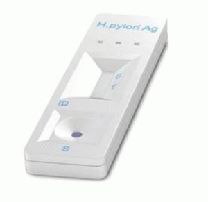
The analysis of feces for dysbiosis is taken to identify the composition of the microflora. With dyspepsia, the suspicion falls on an infection, or there is an imbalance in the intestinal balance. The color of feces and other clinical signs do not always give an unambiguous answer to the question of the causes of the imbalance in the body. Deciphering the analysis for dysbiosis allows us to unambiguously judge the nature of the onset of the disease: is it a manifestation of an acute intestinal infection, such as cholera, or is it a violation balance. In the express analysis, two criteria are used (explaining the name): the presence of its own proteins in the stool and the presence of bifidobacteria. If the named flora is absent, the cause is intestinal dysbiosis. The presence of protein indicates inflammation.
Stool examination is the only method of culture for dysbiosis, excluding smears obtained during endoscopy. The process is time consuming, for successful implementation you will need to follow several rules. Stool is collected in an amount of 1 gram. A clean glass vessel is used, tightly sealed. Air quickly kills bifidobacteria, providing incorrect test results for dysbiosis. It is not enough to collect feces, it is required to hand over organic matter on time - no later than 4 hours from the minute of collection. Storage in a refrigerator at a temperature of +4 degrees Celsius is permissible.
Content
-
1 Obligate and optional flora
-
1.1 Obligate microflora
- 1.1.1 Bifidobacteria and lactobacilli
- 1.1.2 Colibacillus
- 1.1.3 Propionobacteria
-
1.2 Optional microflora
- 1.2.1 Bacteroids
- 1.2.2 Staphylococci
- 1.2.3 Streptococci
- 1.2.4 Bacilli
-
1.1 Obligate microflora
-
2 The essence of the test
- 2.1 Biochemical test
- 3 Clinical signs
- 4 Where to get tested, how to choose
Obligate and optional flora
To decipher the analysis, you must have information about the composition of the microflora. The indicators change depending on the age. The result of a newborn will show numbers that differ from the analysis of an adult. Regardless of the quantitative indicators, it is customary to divide the microflora into three components:
- Obligate.
- Optional.
- Transient.
Obligate microflora
It is represented by the dominant species - lactobacilli and bifidobacteria, Escherichia coli and propionobacteria. Strains are introduced into this group according to two criteria:
- High numbers.
- Functions performed.
There are few propionobacteria involved in the production of vitamin B12. For this reason, they are assigned to the group of useful microflora. Representatives of the obligate part of the microflora:
- Bifidobacteria.
- Lactobacillus.
- Escherichia coli.
- Propionobacteria.
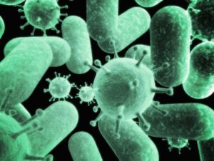
Microflora of obligate type
Bifidobacteria and lactobacilli
Normally, there are more bifidobacteria than lactobacilli, and together they make up the lion's share of the intestinal microflora - up to 98%. Bifidobacteria produce acetic, lactic acid as metabolites, substances from the following list:
- Lysozyme.
- Some alcohols.
- Bacteriocins.
These products greatly reduce the pH-factor of the environment, preventing the reproduction of pathogenic flora. Acetic and lactic acids contribute to the absorption of calcium, vitamin D and iron, ensuring the normal functioning of the musculoskeletal system. Bifidobacteria are involved in the synthesis of vitamins:
- AT 2.
- AT 12.
- TO.
- IN 1.
- PP.
- Folic and pantothenic acids.
- AT 6.
It is not surprising that with a shortage of bifidobacteria, complex avitaminosis rapidly develops, and a lack of calcium is acutely manifested. In children and adults, the population density ranges from 1 to 10 billion. units. The species composition is different:
- Natural feeding: bifidum predominates.
- Artificial feeding: longum and breve predominate.

The composition changes as they grow older. In adults, they find:
- infantis;
- longum;
- adolescentis.
Of lactobacilli, acidophilius, plantarum, rhamnosus are represented in the microflora. The density is usually an order of magnitude lower than that of bifidobacteria, the role in the body is similar:
- Formation of immunity.
- Increase the rate of regeneration of the mucous membrane.
- Activate phagocytosis.
- They start the synthesis of lysozyme, cytokines and interferons.
- Produce enzymes to break down lactose.
- Due to individual metabolites, the pH level is lowered to 5.5.
Colibacillus
Exceptionally normal strains of Escherichia coli are mentioned. Atypical ones do not belong to obligate flora. E. coli is often called Escherichia, after its discoverer. The density of the Escherichia population is equal to the number of lactobacilli, and bacilli appear in the first hours after birth. In a healthy body, they are found inside the large or distal parts of the small intestine.
Researchers tend to consider the mentioned microbe optional, for the simple reason that selected types of organism cause disease. Pathogenic and normal flora does not show morphological differences, with the exception of the presence of separate marker antigens. Escherichia coli functions:
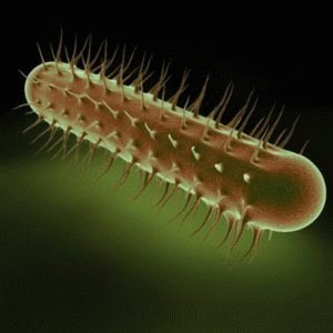
Escherichia coli bacteria
- Binding of oxygen harmful to bifidobacteria and lactobacilli in the large intestine.
- Participate in the synthesis of the vitamins listed above, with few exceptions.
- By producing a number of organic acids, they lower the pH-factor level to the threshold of unacceptability of the medium for the reproduction of pathogenic flora.
- Serve as a link in the exchange of cholesterol, bile acids.
- Regulate the absorption of calcium and iron.
The grounds for assigning Escherichia coli to the obligate group are the functions performed. The number of Escherichia is lower than that of bifidobacteria and lactobacilli, atypical forms are frankly harmful.
Propionobacteria
Propionobacteria belong to the obligate group not because of their high abundance. Microbes synthesize propanoic acid, which lowers the pH level, forming ulcers and burns of the mucous membrane at high concentrations. Sugar, lactic and pyruvic acids, glycerin, amino acids (with the formation of fatty acids) are used as a nutrient medium. Participate in the production of B12.
They are not found in free form, they are found in the digestive tract of humans and animals. The concentration correlates with the level of staphylococci (in the form of normal flora). The metabolite of propionobacteria is acetic acid.
Optional microflora
They include relatively small species that, when multiplying, cause a number of diseases:
- Bacteroids are the cause of purulent-inflammatory processes.
- Staphylococci - poisoning, septic processes.
- Streptococci - sepsis, endocarditis, tonsillitis.
- Bacilli (Clostridia) - diarrhea, colitis.
- Conditionally pathogenic flora manifests itself in different ways, does not show positive qualities.
Microflora optional type
Bacteroids
Rarely found in children under six months of age. A nursing baby is spared such a misfortune. Periodically participate in the final fermentation of nutrients - lipids, bile acids. They are antagonists of Escherichia coli, Shigella, Salmonella, are recognized as conditionally useful. Sowing of feces for bacteriodes is usually not performed, the process is very complex.
Already by the first year of life, during the analysis for dysbiosis in children up to 10 mln. units. As they grow older, the volume increases 10 times. There are many types of bacteroids in the body. Common ones are vulgatus, dorei, uniforms, ovatus. They cause abscesses and peritonitis, infect the heart, tonsils, cause periodontal disease. Most are resistant to tetracycline, gentamicin, streptomycin. Doctors usually use lincomycin or penicillin.
Metabolic products, including acetic and succinic acids, are regulators of the pH factor and lead to blocking the growth of pathogenic flora.
Staphylococci
Staphylococcus aureus is notorious for: meningitis, pneumonia, a number of diseases of the musculoskeletal system. In the first 2 years of life, 20% of children are carriers of Staphylococcus aureus. As they grow older, the number roughly doubles. The main shelter of the microbe is in the nasal cavity, is found in the armpits, in the digestive tract, on the head. The mortality rate for complications reaches 30%.
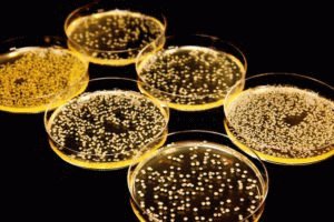
Staphylococcus aureus bacteria
Staphylococci do not perform tangible useful functions in the body.
Streptococci
With artificial feeding of children, the number of streptococci is 100 times higher than in infants. The bacterium is found in the gastrointestinal tract within a few hours from the moment of birth. Thermophilius is used as a dietary supplement, as a component for yoghurts and kefir. The principle of action is based on replacement: a harmless species of streptococcus survives from the occupied niche inhabited by representatives of clearly dangerous (for example, causing pneumonia and meningitis). Many representatives of streptococcus parasitize the teeth and gums, most of the species are hemolytic (dissolves red blood cells).
Bacilli
The most famous representative is the Clostridium. Some species cause botulism (the short-term presence of botulinum bacilli is often fatal). The immune system does not respond to the limited presence of bacilli, similar to the case of Staphylococcus aureus.
If botulism is a separate disease, acute clostridial poisoning lasts a couple of days, then spontaneously resolves. It is accompanied by nausea, colic.
The essence of the test
The rules for collecting feces are known. Bacterial sowing of the finished culture is carried out sequentially. For any strain of obligate and optional flora, there are special nutrient media. The scope of the study depends on the clinic where the decryption is performed. There is an accelerated method - express analysis.
- Intestinal bacteria grow on Levin's, Endo's, bismuth-sulfite agar, Ploskirev's media.
- Chistovich's medium is allocated for staphylococci.
- For the cultivation of bifidobacteria, Blaurocca's medium is used.
- Enterococci are grown in bile-esculin agar medium with sodium azide.
- Lactobacillus is a tomato medium.
- Yeast is Saburo's medium.
The test execution algorithm is known. The laboratory collects feces for dysbacteriosis, dilutions (solutions) are made, which the laboratory assistant drips onto the nutrient medium. Depending on the composition of the "feed", a certain strain germinates. The result shown by the microflora is analyzed quantitatively and qualitatively. The number of representatives of the obligate flora must fit into the allotted frame in terms of density, cause of dysbiosis is a pathogenic or conditionally pathogenic flora with an abundance exceeding the established marks.
Biochemical test
The above is a general principle used. But doctors realize that growing a strain for a week while the patient is suffering is not a way out of the situation. You will have to pass a feces analysis, but when assessing the presence of bacteria in the substrate, a special approach is used.
To develop the methodology, knowledge about the unique metabolites inherent in the strains was used. An example was the principle developed on the basis of the Research Institute of Biological Instrumentation in the 90s. Among the specific metabolites, Academician G.A. Zavarzin lists sterols, fatty acids, aldehydes. It remains to find the indicated ones in a convenient way. For example, using gas chromatography and mass spectrometry.
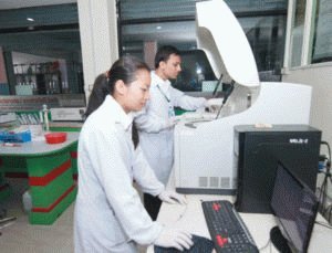
As a result, the biochemical analysis of feces for dysbiosis in infants takes about an hour. Patients will like the described method more than a weekly waiting for the result of the delivery of biological material. The advantage of this analysis is its high accuracy. The problem is the lack of equipment in the laboratories.
Clinical signs
Sometimes the answer to many questions can be given by... a chair. The appearance of the bowel movements will help speed up the diagnosis. Fecal color will tell you whether staphylococcus has settled in the stomach, or the cause of the malaise is Pseudomonas aeruginosa. Blood in the feces with dysbiosis means treatment for the first of these, although the manifestation resembles the symptoms of hemorrhoids.
It turns out that it is not necessary to donate feces, although with the analysis the picture will be complete. A detailed description of the observation will be required by the doctor to determine the treatment.
Where to get tested, how to choose
Colonies of bacteria on a nutrient medium look like even circles of different colors. For example, Staphylococcus aureus grows into a yellow colony, confirming the name. The analysis takes as long as it takes for the growth of the strains. On the official website of Gemotest, they promise to complete the work in 5-6 days. The issue price is 1,500 rubles. Feces can be donated free of charge.
The information received about the harmfulness of bacterial cultures makes patients wonder about the cost of the procedure, find out the details of the procedure. Moreover, in laboratories, not only a test is carried out to determine the sensitivity of flora to fungi (bacteriophages), individual clinics are testing resistance to antibiotics.
Antibiotics are able to be absorbed into the bloodstream, which is not beneficial, since the harmful microflora is localized in the intestines. Fungi pass into the colon, kill pathogens, leaving without consequences. From this point of view, mushrooms are preferable to antibiotics. An experienced physician looks at the analysis of additional places of concentration of bacteria. This is, for example, the gallbladder.
In some cases, the infection affects organs where, in a normal state, the appearance of living organisms - bacteriophages - is unacceptable. Then the only way out is antibiotics.
The express method is called so because of the collection of results in a matter of hours, preparation for the test is carried out quickly. Russian clinics often substitute concepts, calling it bacteriological biochemical analysis, which takes 5 days or longer.
The rules for collecting culture are important, but information about clinics where it is proposed to take an analysis for dysbacteriosis for carrying out the necessary method is more needed by patients. Preparation is more important than process. An exception is the case of living in a village, where there is no choice of laboratories, except for the regional or regional clinical one.
Dysbacteriosis can be treated with folk remedies. The review is offered to your attention.


