
Diagnosis of appendicitis: differential and instrumental
With pain in the right side of the abdomen, appendicitis is often diagnosed in a child or adult. But this is not the only case that causes acute pain and carries a threat to the patient's life. It is important to correctly define the disease, not to be confused with diseases of the gastrointestinal tract or the urogenital area. For the correct determination, a number of types of diagnostics are used. The main ones include the differential and instrumental method for diagnosing appendicitis. The features of these methods are described in the article.
Pathological conditions in the digestive tract appear for various reasons. Ailments are based on poor nutrition, bad habits, lack of proper attention to health, and the presence of chronic foci of inflammation.
State acute appendicitis causes the presence of pathogenic microflora. Staphylococcus, streptococcus, Escherichia coli are dangerous. With a decrease in human immunity, microflora develops, which leads to inflammation. Often, inflammation of the appendix of the cecum occurs in pregnant women for this reason, a decrease in the body's defenses. The adult sexually mature population often finds it difficult to empty the intestines.

This situation causes the accumulation of feces and the development of a microbial environment, leading to diseases.
Diagnostic methods:
- Laboratory research.
- Differential study or briefly DIF.
- Instrumental research.
When a patient is admitted to the surgical department, it is enough to feel the abdomen during examination. When pressing on the peritoneal area on the right side, in the navel, the patient feels a sharp pain. The sensation disappears after the specialist's hand is torn off. Some cases suggest a digital rectal examination. Using the described method, the overhang of the anterior rectal wall is detected. Examination of women involves a method of palpation by a gynecologist. In this case, the protrusion of the right vaginal fornix is determined. If necessary, the patient is referred for urine and blood tests to confirm the diagnosis. If there is an increased level of leukocytes in the analyzes, it is said about inflammation in one of the organs.
Early diagnosis in acute abdomen is important because diseases included in the acute abdomen pose a threat to the patient's life.
Content
- 1 Differential diagnosis
-
2 Instrumental diagnostics
- 2.1 CT scan
- 2.2 MRI
- 2.3 Thermography
- 2.4 Laparoscopy
- 2.5 X-ray irradiation
- 2.6 Ultrasound
Differential diagnosis
Differential diagnosis of appendicitis is used to exclude other possible diseases associated with the digestive system. The research algorithm is to distinguish the disease from others with similar symptoms. The study excludes the presence or determines such diseases as:
- Pancreatitis Often becomes the cause of erroneous diagnostic options. The difference from appendicitis is that the soreness is located in the upper abdomen. The patient has bouts of vomiting. The determining factor in the difference from pancreatitis is the study of urine for the presence of increased diastase.
- Acute cholecystitis. A similar symptom with appendicitis in soreness on the right. When feeling the abdomen, soreness may be felt under the ribs on the right side.
- Diseases of the liver, stomach, duodenum.
- Right-sided renal colic. A similarity to an attack of appendicitis is possible when a stone passes through the ureter. But then the pain radiates to the external genitals or the right thigh. There are bouts of urinary incontinence. Patients with an attack of renal colic are emotionally very excitable, rushing about, and with appendicitis, the patient remains calm.

When women with acute appendicitis are admitted, it is important to determine whether the attack is a manifestation of gynecological diseases, such as inflammation of the appendages, ectopic pregnancy. The patient must be examined by a gynecologist. If problematic situations are detected in the area of female genital organs, the patient is engaged in gynecology.
In children, it is important to differentiate an attack of appendicitis from ARVI, diseases of the genitourinary system and digestive tract organs, and various infections of childhood. The child is not always able to correctly describe the symptoms, so the essence of the method is extremely important. Differential diagnosis plays an important role in eliminating possible errors and incorrect treatment.
Instrumental diagnostics
Instrumental diagnostics of the definition or exclusion of appendicitis includes:
- Laparoscopy.
- Ultrasound.
- MRI.
- CT scan of the abdomen.
Some of these types of diagnostics are contraindicated in patients for individual reasons. The method is determined by the doctor, based on the indicators of the person's condition and individual characteristics.
CT scan
The aforementioned diagnostics allows you to view the object under study in the form of layer-by-layer images in full. This allows an adequate diagnosis to be established. This method assumes an accurate display of the appendix, detection of inflammation. In the case of acute appendicitis, the method determines the cause of the inflammation.
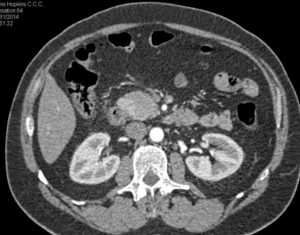
Organs on a tomographic image
Conducting CT with the use of contrast gives the most reliable results. The procedure involves preparation in the form of refusing food 12 hours before the examination, removing all metal objects and jewelry. Overweight is considered a contraindication for carrying out.
MRI
The MRI method does not give an accurate definition of the inflammatory process in the appendix, but it can be used if there are contraindications to other diagnostic methods. This is due to the fact that CT and X-ray studies give partial radiation, and an ultrasound study is not always informative in determining the inflamed process of the cecum. MRI is often used when a pregnant woman is admitted because it does not pose a threat to the fetus and mother.
Thermography
The specified diagnostic method is carried out using special equipment - a thermal imager. The device is sensitive to thermal signals of the body and determines which organ has the highest temperature, transmitting information to the monitor. The thermographic method accurately determines the location of the inflamed appendix, shows whether there are additional foci of inflammation in the body.
The advantages of the method:
- Safety;
- Low cost;
- No negative impact on health.
Laparoscopy
During the examination, the doctor makes holes in the peritoneal cavity through which a laparoscope with a small camera is inserted into the cavity.

The equipment introduced into the body allows a detailed examination of the peritoneal organs. If inflammation is detected in the appendix, a specialist can detect its condition, determine the likelihood of a breakthrough of the wall of the appendix, and study adjacent organs. Laparoscopic methods are informative, but not suitable for all patients.
X-ray irradiation
A study for appendicitis using X-ray irradiation is carried out together with differential diagnostics to exclude other diseases of the digestive system. This method allows the doctor to take pictures of organs showing inflammatory processes and neoplasms, if any. X-ray examination is carried out using contrast introduced into the body, which provides a clear image and makes the study more informative.
Ultrasound
One of the most commonly used research methods for acute appendicitis. The advantage of the method is the safety for the patient's health, the ability to see the state of internal organs in real time. But experts are increasingly abandoning the method when diagnosing appendicitis, since it does not determine the stage of development of inflammation. The malaise is determined only because of the visibility of the enlarged organ on the monitor screen. But the method allows you to exclude or detect other unpleasant situations in the abdominal organs that are similar to the symptoms of inflammation of the appendix.
If an inflamed appendix is detected, treatment is required as soon as possible, since the condition poses a threat to the patient's life. There is only one treatment option for acute appendicitis - removal. The operation is performed by a surgeon. However, forecasts with timely referral to a specialist and without the development of complicated options for an attack are optimistic. Risk factors are the development of peritonitis, late detection of the disease, preparation for surgery. The patient, in the absence of complications, is able to return to his usual life and work 3-4 weeks after removal.
With the appearance of symptoms of an acute abdomen, suspicion of inflammation of the appendix of the cecum, it is important to immediately seek help from a specialist without wasting time. If you cannot get to the surgical department on your own, you should call an ambulance. Diagnostics will allow you to determine the disease in time, prevent the development of aggravating conditions and in return to your usual way of life, work, sports or loved ones in the shortest possible time hobby.


