Features of the ultrasound of the testicles in men
Features of the ultrasound of the testicles in men

Ultrasound is a procedure scrotum scrotum studies using high-frequency ultrasonic waves, through which you can see its internal structure.
This method is the primary diagnostic measure which helps to identify the presence of abnormalities in male organism.
As we know, it is extremely important to identify the disease at an early stage of its development, because it affects our health and life expectancy.
The main indications for the study
Ultrasound of the scrotum do in certain diseases. Usually, the symptoms affect the genitals of boys, but did not rule out the disease organism as a whole. Indications for ultrasound of the pelvic organs are: orchitis, tumors, hydrocele, cryptorchidism, varicose veins of the spermatic cord pathology and organ injury scrotum.
If cryptorchidism testes are found in the abdominal cavity or non-existent in the scrotum. This disease is most often congenital, received in childhood or acquired due to a genetic disease.
The size of the testicles at the same time is much less than the age stipulated norms. In addition, there is poor blood flow in the pelvic area.
To identify the disease and children at an early age sent for a pelvic ultrasound, since it is dictated by the inability to identify the testicles in the scrotum.
Cysts in the testes generally are one-sided and have a small size. Distinguish congenital and acquired cysts.
First, in this case looks like a small outgrowth diameter 0.5 cm, while the latter are tumors which have arisen in the epididymis. In further such cysts can grow into hydrocele.
In this disease fluid formed between its shells. With the help of ultrasound can be seen as the accumulated substance is viewed testicle and appendage.
In case of contact infection in the male reproductive organs begin to develop orchitis. With the spread of the infection swelling increases, respectively, the body also increases in size. A hotbed of proliferation is a prostate or bladder, where infection and begins to migrate to the gonads.
At first, only affects the epididymis, but with the continuation of the inflammatory process, complications may occur. If the treatment of the disease in the acute stage of operation of the genitourinary system gradually adjusted, that in chronic inflammation the system may be paralyzed due to the potential atrophy of testis.
If the stronger sex is disturbed blood flow to the testicle tissue in the renal vein, in this case it begins to show varicose veins of the spermatic cord.
In most cases, varicose veins developed left-sided, and then without surgical intervention is not enough.
For right varicose veins additional examinations have to spend to exclude malignant tumors that can occur in the abdominal pelvic cavity. In addition to the US, to clarify the treatment of venous system of the scrotum, it is desirable to still make Doppler.
Statistics show that malignant tumors are extremely rare. This process is more prone to older people.
If the cancer originated in adolescence, it is the result of creation of metastasis, neoplasm that arise in the circulatory system. But most of the signs of testicular cancer are benign origin.
The tumor may develop as a result of diseases such as cryptorchidism and scrotum injury, and various pathologies.
Testicular tumors is quite difficult to identify because they are imperceptible. But as soon as the pain starts to sharply increase, the disease begins to progress. This can lead to testicular torsion or acute inflammation of the appendages.
With the help of a pelvic ultrasound can detect tumors even small size, examine their distribution, localized, depending on where the process went, that is, on one testicle or two.
According to its properties malignant tumors are very similar to benign, which contain the horny tissue.
After deciphering the final ultrasound diagnosis is made only after it is examined histological material obtained by the surgeon in the operating room.
Pelvic organs are extremely vulnerable to a variety of injuries. In this case the ultrasound helps to minimize the negative consequences of these injuries, it helps to determine the need for immediate surgery.
Especially it should be done at break eggs or a large hematoma. Remember that time the measures may prevent testicular atrophy and fester.
For sufficiently large hematoma will have to carry out a Doppler study to help determine the degree of timely surgical intervention for further treatment of the patient.
It is worth noting that an increase or change in color of the scrotum, fever does not always speak about the damage of the testes. The reason for such a state can become a sporting and sexual arousal. The main symptom of this is a sharp pain in the groin area.
As you can see, ultrasound of the testicles - the study of tissues, form, composition and other indicators of scrotal organs of men. Ultrasound examination can be carried out several times, the main thing - to carry out the correct preparation. At occurrence of any pain in the pelvis is necessary to consult a doctor immediately.
The procedure for conducting the procedure ultrasound of the testicles
To perform ultrasound of the testicles, men do not need such as specially prepared. It is only necessary to take a diaper, to lay it on the table, and the cloth to remove gel residue. It should be noted that the gel is bad enough removed from the hair, so it is desirable to shave the pubic area, if you do not want to feel uncomfortable. Ultrasound procedure male genitalia conducted lying.
Pre physician interrogates the patient and examines the area to be explored. In order to prevent retraction of the testes, the gel must be kept warm. If the subject area is painful enough, man, local anesthesia can be used. The survey makes it possible to determine the size of the testicles, and to study their structure. This procedure takes less than 10 minutes.
You should know that the average size of the testicles in the stronger sex in the length of about 4-5 cm and width - 2-3 cm.
In adult males the body has an average density of the echo, and the boys who have not yet reached sexual maturity, it will be low.
Not all patients can clearly define the boundaries of the epididymis, it has a kind of mace, located on the rear edge of the testicle. At an early age the appendage is impossible to determine.
Ultrasound of genitals is a simple, affordable and effective procedure.
The method is so popular among professionals and surgical therapeutic profile, it is not without it do any diagnostic process for the stronger sex with complaints of violation of sexual area. Such popularity of ultrasound acquired due to its high accuracy, the relative cheapness and availability.
A source: https://menportal.info/story/osobennosti-provedeniya-uzi-yaichek-u-muzhchin
How do ultrasound of the testicles in men
 Home »ultrasound of the genitourinary system"
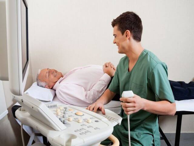
Testicular ultrasound helps diagnose scrotal organs without harm to the body. This method is recognized as informative and neinvaziynym.
Appointed during pathological and inflammatory processes occurring in the male genitalia. It requires no special training.
Decoding results of the study should deal only with the attending physician - the urologist, who can properly diagnose and prescribe the correct treatment if necessary.
Preparation for ultrasound of the testicles
Preparation for ultrasound of the testicles is conducting hygiene genitals immediately before the procedure. It is sure to survey recommended that you should have a disposable diaper and wipes. In order not to experience discomfort from the application of the gel, which is also bad wiped with the hair, it is better to shave previously investigated area.
How do ultrasound of the testicles in men?
Ultrasound examination of the scrotum organs held as follows:
- The patient must expose the lower torso.
- Take lying down.
- On skin specialist does gel to help slide the sensor and prevents air from entering. Air may distort the results.
- If a man feels pain during the examination, the local anesthesia.
- uzist doctor must first visualize a single egg, and then another. Required estimated size, structure.
Indications for study
A study of male organs do, if there are indications that:
- Inflammation of the testicles appendages.
- testicular inflammation.
- Inflammation of the testicles and appendages.
- A tumor of the scrotum.
- Veins of the inguinal canal.
- Dropsy.
- Injuries genitals, testicles.
- Infertility.
- The cyst is not inflammatory.
- Accumulation of pus in the scrotum.
Very often, in addition to the man may appoint Doppler study of the scrotum.
Norm
ultrasound study shows the functioning and development of the scrotum. The testicles are normal on ultrasound:
- They are round, smooth surface.
- The fine-grained structure.
- Ehoplotnost average.
- Determined high echogenicity line - mediastinum.
- Determination of the epididymis in the longitudinal plane.
- Albuginea surface testis and epididymis has the form vysokoehogennoy strip.
possible pathology
A study of the testes with ultrasound is considered a reliable method that helps identify the scrotal pathology:
- The absence of testicles in the scrotum, their location in the abdominal cavity. It refers to a genetic disease. In this situation, the testicles will be smaller granularity absent, weak blood flow or no existents.
- Tumors of the testes. Presented as neoplasms having heterogeneous structure. When it was revealed the formation of small size, it has all the chances of recovery.
- Calcifications.
- Cyst.
- Orchitis, which is accompanied by infections.
- Abscess.
- Hydrocele.
- Varicocele.
Uzi testicular inflammations
Ultrasound examination of the testicles is considered to be mandatory, not only preventive procedure, but also in inflammatory processes. It helps to diagnose the disease and diagnosis. Appointed after a preliminary medical examination. If necessary, it can be carried out several times during the course of treatment to correct the process if necessary.
A source: http://uzilife.ru/uzi-mochepolovoy-sistemy/uzi-yaichek-u-muzhchin.html
Ultrasound of the testicles in men and boys, as do

It is not infrequent appointment of the audit male testes through ultrasound. It should be noted that the study of the state takes place in conjunction with the epididymis, spermatic cord and scrotum as a whole.
Carefully and deliberately examine them is possible only through an ultrasound. Those who are assigned this type of inspection, do not worry about the harmfulness of ultrasound.
It has long been proven that these treatments are completely harmless for such a delicate part of the male body as the scrotum and testicles.
When administered ultrasound examination of the testicles?
In itself, the study of male testes with ultrasound diagnosis is the most high-quality and reliable.
See also:What causes polyps in the bladder in men
Waves of ultrasound instrument can accurately determine the state of inside of the scrotum, giving information on available pathologies. Another advantage of this analysis is the speed and ease of implementation of the procedure for the patient.
The main purpose of the diagnosis is the ability to use it to find out the cause of the disease of the testes and male intimate organs.
Ultrasound of the scrotum can not be appointed without the express valid reason. Thus, the diagnosis is assigned to the following reasons:
- if men are complaining of constant pain in the scrotum;
- if in the preliminary examination the doctor found the bulk of education, which should not be in the scrotum;
- with strong or injury during inflammation;
- erectile dysfunction;
- suspected infertility;
- noticeable change in the size of the testicles in the direction of increasing or decreasing;
- change in the state density of the testes;
- carrying out checks after operations on the organs;
- identified in the verification of suspected tumor;
- disorders of blood circulation in the scrotum;
- hernia in the groin area.
In some cases, your doctor may recommend additional procedures to conduct ultrasound - Doppler.
It is common these studies can help identify those who have serious diseases as Testicular torsion, hernia in the groin, poor circulation in the pelvic organs, the disease varicocele.
In carrying out any operation, whether the ultrasound diagnosis or ultrasound with Doppler, the patient does not require any special preparations.
What is an ultrasound of the scrotum?
Sonography is conducted prescribed by a doctor. Preparing for the US does not require any special knowledge and additional research. Before the procedure is recommended to shave the hair from the groin area.
This is due to the fact that during the investigation on the scrotum will be applied a special gel, which is very difficult washed out of the hair. In addition, doctors warn about the need to bring to the procedure of a cloth or towel for hygienic Spent doctor's chair.
It is mandatory to hold bath treatment before visiting the US.
The whole procedure for obtaining the status of the male testicles data occurs while the patient is in a state of lying on the couch. Investigation of the inner component is made scrotum through a specialized apparatus Handle device according to subject area and recording images inside of the scrotum on the ultrasound screen apparatus.
When this is applied to the genitalia special gel which prevents air between the skin and the instrument handle. It is important to bear in mind that the gel applied should be warm, not to allow the testicles to press inside.
The doctor examines the sensor alternately, first one egg, and then proceeds to the second. In some cases, the diagnosis of male reproductive organs is performed with local anesthesia.
This is due to the fact that the patient may present with painful touch sensor unit.
Among the disadvantages of this type of internal organs checks can be noted the unreliability of the data determined. Namely, if an ultrasound instrument screen, the doctor finds the tumor, it will be difficult to establish - is whether the tumor is malignant or benign is it.
How to carry out diagnostics at children?
At first glance, it is unclear for what reason can be assigned to a study of children with ultrasound.
However, teenagers can be sent to study the testicles to oversee the hormonal development and research for the functioning of the reproductive system.
In some cases it may be found in the earliest stages of the development of pathologies that impede the normal life of children and adolescents.
Very often in children occur injuries of the scrotum and the testicles or the sudden increase or decrease in shape. The basis for ultrasound examination purpose is the deviation from the norm in puberty. The reasons for the appointment ultrasound studies can act excessive hair growth, a tendency to obesity, impaired growth of the norm, the violation of the heart since birth.
Children and adolescents, as well as adult men, susceptible to diseases of the testes. Children can identify the following diagnoses:
- inflammatory process that develops into a dropsy;
- the inability of the testicles to descend into the scrotum;
- Testicular cyst;
- Testicular size does not correspond to normal;
- birth defects of the male organs.
All of the above diseases require diagnostic ultrasound apparatus through the application, and then treating the doctor can be assured with an accuracy in the diagnosis and may prescribe proper and effective treatment.
How is the examination of the boys?
The process of passing inspection procedures in children coincides with the passage of diagnosing the testes in men. First of all, once an ultrasound does not get one. The surgeon must make an inspection and find grounds for ultrasound procedures. In identifying the suspected pathology should, first of all, to make moral preparation for the upcoming boy studying his scrotum.
Psychological adjustment is needed before the procedure for older children and adolescents. For them to be uncomfortable providing for examination of their organs intimate strangers. In boys, toddlers, such problems should not occur. Ultrasound procedure takes place in the presence of parents and sufficient to bring a set of sanitary napkins and towels.
That procedure provides ultrasound of the testicles?
In itself, the study of the scrotum and the inside of the male genitalia takes at intervals of about 20-30 minutes. After that, the doctor is filled with indicators ultrasound machine. In the case where a doctor have been established deviations, is not correct, the patient will be assigned to follow-up study.
The physician must determine that the rate for men is an echo-positive component with a fine-grained structure. Sizes vary from egg 2.5-3 cm in width, 2.5 cm - volume, length - not more than 5 cm.
In the case of children and adolescents on the echo-positive data will be significantly different. According to statistics, indicators testicles norm in children and adults will be the same only for the achievement of a boy of 10 years.
In addition to identifying the norm in shape, size and density of the doctor examines the related details. Namely, recorded and venous vascular weave, blood circulation, the presence of cysts, neoplasms, tumors.
After carrying out research procedures with ultrasound the doctor puts a special performance in the table, and analyzes the deviations from the norm. If there are deviations or detect visible eye education, it may be appointed or re-examination already diagnosed and determined the course of treatment. After inspection the following diseases can be identified:
- the absence of testicles in the scrotum;
- benign or malignant tumors;
- concretions;
- orchitis;
- dropsy;
- spermatoceles;
- epididymitis;
- torsion and other diseases of male organs.
Examination of the testes and male internal reproductive organs is one of the most popular research surgeons. Doctors loved this procedure for its simplicity, speed, safety and accuracy of the data. Advantage for patients is the inexpensive cost of research, as well as an increase in the probability of a quick solution to the problems plaguing the genitals.
##
A source: https://AnnaHelp.ru/diagnostika/uzi-yaichek.html
How do ultrasound of the testicles in men and boys: norm and pathology

Ultrasound diagnosis of testicular - is one of the modern methods that allow you to get all the information about the scrotum and its organs, which include and testicles. Ultrasound of the testicles in men allows a diagnosis to be accurate to 99 percent.
anatomy of the testes
Egg - a man's body, which has an oval shape. This glandular body is fixed by a special spermatic cord. The structure of the testicle include: head, body and tail. Human anatomy is arranged so that the left testicle is slightly lower than the right. The scrotum has a very rich blood supply.
When administered?
Prescriptions for ultrasound can be a huge amount of:
- with suspected tumors;
- inflammatory processes;
- agenesis (a condition where there is no one or both testicles);
- when resizing regional lymph nodes;
- infertility;
- when a palpable tumors in the testis;
- if necessary, control dynamics;
- when biopsy;
- in the case of dimensional change and egg shape;
- atrophy and wasting;
- pain in the scrotal area;
- when egg Abuse result spermatic cord torsion;
- trauma of the scrotum.
boys
There are several options in the event that children prescribed ultrasound of the testicles:
- delayed psycho-emotional maturation;
- weight problems;
- at too low or high growth;
Ehoplotnost testicles in childhood acquired at the time of puberty. It is worth noting that it occurs in all the boys in different periods due to the different factors of genetic nature, ecology, as well as proper nutrition and possible pathologies.
Video 1. When are ultrasound of the scrotum?
What does the diagnosis?
- what sizes are testicles (normal, increased, or conversely reduced);
- what sizes are appendages of their heads;
- contours (smooth, blurred or not);
- check fabric on the echogenicity;
- evaluation of new formations (their size and density);
- the presence of free fluid, its volume;
- symmetry characteristic and vascular pattern.
Also for diagnosis in some cases apply dopplerografiju. The need for it arises:
- varicose veins (varicocele);
- at various traumas;
- with suspected torsion seed channel;
- in tumors.
Training
Before you begin to conduct a preliminary US conducted a series of hygienic procedures genitals. For ultrasound is necessary to prepare the disposable diaper and napkin.
Reference! Since ultrasound is used in the process of a special gel, in order to avoid discomfort to the best procedure to shave the area to be explored.
But if the procedure is planned studies of the prostate through the rectum immediately, the patient must adhere to certain rules.
How to do it?
Ultrasound of the scrotum and its organs is carried out (as a member of the US) in several stages:
- The patient lays bare torso below.
- Falls.
- In its gel applied to the skin, which improves sliding on the surface of the sensor and does not allow the air to hit, as it can affect the accuracy of the findings.
Feel pain in the process of US patient can only be, if he has testicular disease and appendages, bearing the inflammatory character.
Important! In most cases, orchiepididymitis ultrasound procedure is transferred for a certain period before the disappearance inflammation.
See also:Healthy grass men
If the patient experiences pain by ultrasound, it is assigned a local destination anesthesia.
The specialist carrying out the ultrasound sequentially examines first one testicle and then the other. Compulsory assesses the size and composition.
Norma results
In normal testicles are smooth and precise contours.
There is a homogeneous parenchyma.
Perhaps the presence of liquid sulfur, but only in small quantities.
It considered normal if seen only the head of the epididymis.
Sizes in men
Based on the ultrasound findings, set the average size of which have testicles in men:
average indications:
- a length of 4 or 5 cm;
- in width from 3 to 3.5 cm;
- thickness of 2 to 2.5 cm.
Dimensions boys depending on age
When ultrasonography children echogenicity is usually reduced, and men has an average value.
pathologies
Due to ultrasound became possible to diagnose the plurality of abnormalities of the scrotum and its organs. What does the ultrasound in the case of abnormal conditions of the testicles?
cryptorchism
Ultrasound in the presence of this pathology is assigned for more information about the "disappeared" testicle.
cryptorchism - a deviation from the norm of location, resulting in the gonads men present, but for some factors not had time to leave the abdominal cavity.
In most cases (90%) of the egg which has been "lost" in the channel groin.
Testis, which does not have the correct location area has smaller dimensions, the heterogeneous nature of the structure and its appendages are generally not visible.
varicocele
Varicose veins in the spermatic duct in men requires immediate treatment because it can cause infertility.
When an ultrasound varicocele clearly visible veins are extended and slightly changed their appearance and a diameter greater than 3 mm.
There are several stages of varicose veins:
- First. Veins are visualized only at elevated pressure or during upright survey.
- Second. Vienna, changing their appearance, do not disappear even in the horizontal position.
- Third. Vienna visualized in the zone located below the eggs, which decreased in size.
hydrocele
In case of this pathology in a zone situated between the two sheets of shell eggs, liquid accumulates.
As a result of ultrasound diagnosis of liquid occurs without effort that has accumulated between the shells.
cystic neoplasms
Spermatocele or, in other words, the seminal cysts may be of congenital or acquired in nature. In congenital cyst observed its small size, and the liquid inside it has a transparent appearance.
Reference! Acquired cysts formed as a result of inflammation or injury, which occurs as a result of occlusion of the duct.
Following the results of ultrasound it can be seen that the cyst has a rounded shape, clear and smooth contour, as well as anechoic structure.
Orchitis and orchiepididymitis
These abnormalities are the result of the defeat of microbes tissues due to reduced immunity. On US epididymis has increased size, reduced echogenicity and heterogeneous structure.
Male sex gland is greatly increased in their volumesAnd reduced its echogenicity. Very often, this anomaly is associated with reactive edema.
chronic orchiepididymitis
Testis has any size, non-uniform structure and uneven contours. Visible appendage, which is slightly increased. In the case of such an anomaly is assigned to the differential diagnosis.
Tuberculosis
An ultrasound can identify tuberculosis of inflamed epididymis, yet expert draws attention to other factors during the procedure. When tuberculosis testicle and its appendages there is a process to both sides and accumulate salt.
Oncological processes
How and what else tumors in other organs and parts of the body, cancer formation in the testicles, of course, require urgent examination and further treatment. Not passing discomfort and the feeling that there is a neoplasm, are reason enough to visit. According to the results of ultrasound will be clear whether a visit to the oncologist is needed.
gonioma
It is very rare to find testicular cancer, and mostly in the 95% of cases are tumors that arise even inside the womb. Given the structure of seminoma, allocate homogeneous and not homogeneous structure.
pathological nature neoplasm is generally in the right testicle. Tumors are very rare immediately (up to 3 out of 100 cases) from two sides.
On US tumor it is irregular and in some cases is from several formations having a heterogeneous structure. Diseased testicle size is a little deviation from the norm.
In that case If testicular cancer is suspected, a study conducted retroperitoneal area. As is mandatory biopsy.
Abscess
Testis formation has the form of a localized, sharp contours.
torsion
This abnormal condition in which the egg is turned around the vertical or horizontal axes. The result can be constricted blood vessels and / or nerves, which often leads to the development of ischemia and inhibition of functions of the prostate or even necrosis.
microlits
One of testicular pathologies during examination are small calcifications. They are in the nature of the primary process, the cause of the poorly understood. Calcifications can be diagnosed in conjunction with cancer, tuberculosis, or in case of problems with metabolic processes.
Contraindications
There is no such contraindications, which can interfere with the ultrasound. An exception may be only a scrotum injury.
Cost and where to do?
The cost of the study is 900 rubles, and the use of Doppler - 1500 rubles. Patients who have already spent US, divided only positive feedback.
conclusion
Ultrasound of the testicles - it's one of the most reliable methods of research used in urology, it has a high accuracy and low cost.
A source: https://MediGid.com/uzi/organy/malyj-taz/muzhchiny/yaichki.html
ultrasound of the scrotum
Ultrasound scrotum - a simple non-invasive research method to identify virtually all diseases both innate and adaptive nature. Examination of the primary diagnosis is considered - in most cases it is assigned after inspection and palpation, if there are suspicions in the presence of the pathological process.
When ultrasound is assigned male sex organs
scrotum study using ultrasonic waves will be recommended to the patient:
- under pain of different nature;
- presence of signs of inflammation;
- traumatic injuries of open or closed type;
- changing the appearance or shape - redness, swelling, induration;
- suspected tumors and hernias:
- increase in inguinal and retroperitoneal lymph nodes;
- changes in semen parameters;
- haematospermia - the presence of blood in the semen;
- slow or early puberty;
- erectile dysfunction, infertility.
The possibility of real-time procedure allows controlling a biopsy of the prostate. Ultrasound of the scrotum and small pelvis is indispensable for evaluating the prescribed therapy, as well as in the postoperative period.
What pathology reveals the US?
Ultrasound examination of scrotum can detect a large number of diseases in males in childhood and in adulthood.
Thus, the procedure makes it possible to determine the absence of the testes (testicles) in the scrotum that may be the result of cryptorchidism (undescended testicle into the scrotum) or another type of ectopia (abnormalities in the development).
During diagnosis can establish the location of the testicle, studied the inguinal canal, perineum and the surrounding areas hips as the body can be placed in these unusual for its location areas.
Modern equipment allows us to find the undescended testicle, even with its small size and the indistinct contours.
If the inspection of the newborn boy was found undescended testicle, it is mandatory appointed by the US, because after it is necessary to carry out the operation of the relegation of the testicle in the scrotum or remove it.
hernia, torsion of the testicle, infertility, and even malignant tumors - the negative effects can develop without proper medical care.
Possible localization undescended testis in a child
The procedure allows to determine the condition of the veins of the spermatic cord and to diagnose a varicocele - varicose them.
This is a fairly common pathology that can have a negative impact on spermatogenesis (development of male sexual cells). Also, ultrasound of the testicles in males revealed hydrocele - dropsy of testicle membranes.
The disease may be either congenital nature and acquired as a result of traumatic injuries or inflammation.
https://www.youtube.com/watch? v = _sT2aKUIBOg
The same reasons can cause cysts in the testes - spermatoceles. Their development can be triggered and embryonic remnants.
By diseases of inflammatory nature, detectable by ultrasound include orchitis - inflammation of the testicles and epididymitis - inflammation of the epididymis.
In some cases, the inflammatory process extends directly to two bodies that is known epididymo.
In the absence of proper treatment of these pathologies can lead to serious complications.
Ultrasound diagnosis of male genital organs shows tumors as benign as malignant nature, allowing the early stages to take appropriate measures.
Modern methods CFM (color Doppler) makes it possible to study the circulatory features in the scrotum organs and to identify abnormalities in the blood vessels art.
Preparation for the examination and holding
Adult male special training is not required, but will be here a child needs to take the time and explain the need for future action or even persuade the completion of a procedure.
The only thing to be done, if the survey is scheduled as planned - is to make the toilet of external genitals.
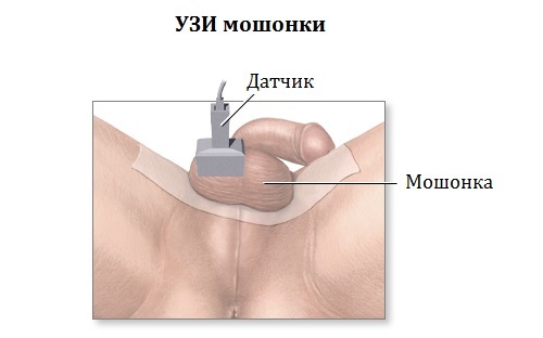
To conduct the study patient is asked to sit comfortably on the couch, after handing over his underwear, because how do Uzi to the skin of the scrotum.
The penis is fixed on the stomach or the patient holds his hand so as not to interfere with the movement of the sensor.
Holding ultrasound of the scrotum
In some cases, during the ultrasound Doppler, when it is necessary to evaluate the work organ blood vessels, the procedure may be carried out in a standing position. The doctor then applies a special water-based gel that promotes a more dense, qualitative and painless contact with the patient skin surface.
The study examined the scrotum in the longitudinal, transverse and oblique plane, which allows you to thoroughly examine all components of its structure and identify the pathological elements.
The procedure does not take much time - in most cases it does not last more than 15-20 minutes, and only in situations of dispute may be slightly delayed.
Testes of adult males normally have dimensions of 3-5 cm in length, thickness and width - 3.2 cm. Scrotum itself is a kind of a case for the testes, and it is formed by connective and muscular tissue about 8 mm wide. Passes in the middle of the seam, dividing it into two parts. Normally between scrotal wall and testicles may be a small amount of liquid (1-2 mL).
How to do pelvic ultrasound in men?
See also:Why disrupted sperm morphology and how is it treated?
The upper-side end testicular appendage located, consisting of a head, body and tail. The tail is gradually transformed into the vas deferens, and as part of the spermatic cord without going through the inguinal canal, enters the abdominal cavity.
Joining forces with the vas deferens, passes through the prostate and open in the urethra. Normally, in the investigation of ultrasound testicle structure is clearly visible, the head of the epididymis (body and tail with no abnormalities are not visualized) and the spermatic cord. When decoding is required compared to the right and left testicle and scrotal wall thickness.
Testicular in a normal state has a uniform consistency, with clearly marked dense tissue segment situated to the rear-outer portion of - the mediastinum. When Doppler ultrasound can examine the arteries that feed the bodies of the scrotum and spermatic cord venous network.
It looks like pathology scrotum on ultrasound?
The presence of abnormalities in the scrotum ehostruktury indicates heterogeneity, roughness and fuzziness bodies increase in size and the amount of liquid.
The presence of disease is determined by an increase in size of the appendages, and particularly the tail part. Epididymitis diagnosed by ultrasound increases the head of the epididymis and the change in its tissues.
Sometimes viewed the body and the body of the tail, and a decrease in echogenicity.
If the fluid accumulates in the scrotum, then we are talking about reactive hydrocele. Hydrops on ultrasound anechogenic determined fairly large area surrounding the testis, and sometimes extends to the inguinal canal.
Inflammatory processes in the testis and epididymis can often be similar to the manifestations of testicular torsion. Torsion characterized pinched artery testicle, and Doppler diagnosis will not be viewed pulsation in body vessels.
This pathology is considered to be very dangerous and requires immediate assistance - otherwise it can lead to loss of the testicle.
Epididymitis - inflammation of the epididymis testicle
If the ultrasound shows enlarged in size appendage, structural changes in it, but the body and tail are not visualized, the infection is likely to hematogenous or lymphatic nature.
Such localization is not inherent to infections, sexually obtained entering the appendage through the vas deferens, because of what is affected paramount tail appendage.
Advanced cases of these diseases can cause piotsele - the formation of a purulent cavity in the scrotum, a dangerous development of an abscess, myocardial testicle and even gangrene Fournier.
Varicocele is characterized by stagnation of the blood, and in most cases leads to atrophy of the testicles. As a rule, it is formed in the left testicle.
To identify pathology ultrasonography applies Valsalva sample - it makes it possible to determine the presence of reverse current in the veins of the spermatic cord.
Varicocele is diagnosed at the advanced of the veins of the spermatic cord - more than 3 mm and anechoic structures in its projections.
The procedure is the primary diagnosis for the detection of tumors, as organs of the scrotum are readily available for the research. The affected testicle is generally increased in size, and the tumor is irregular and heterogeneous structure. Normal testicular ultrasound helps identify the presence of disease, but it is not subject to the differentiation of tumors cells.
To determine more detailed characteristics of neoplasms applied Doppler scanning, allowing to estimate the structural features of cancer process.
Without the US does not do any search for the causes of male infertility. Diagnostics to determine factor in the change in the number of sperm.
The process concludes, based on violations testicular seminiferous structure and pathways that lead to infertility. One of the abnormalities, leading to a decrease in reproductive function in men is spermatoceles (seminal cyst).
On the ultrasound they appear as dark anechoic areas of small size (less than 2 cm), rounded shape with clear and smooth edges.
All of these diseases can be detected during short diagnostic, which takes less than 20 minutes. The relatively low price and ease of study will make it possible to protect themselves from further serious complications due to timely diagnosis and prescribed treatment.
A source: http://diametod.ru/uzi/moshonki
Ultrasound of the scrotum or testicles in men: how to make a Doppler, preparation and it shows
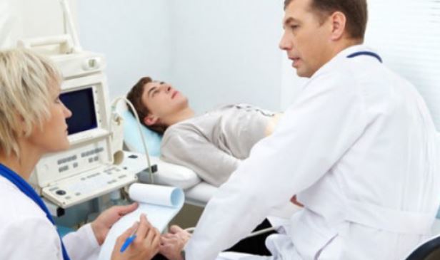
To examine the man for urological diseases and pathologies in organs of the reproductive system modern doctors often use ultrasound of the scrotum. The fact that the anatomical location of the body does not give other opportunities for its examination. How to perform the procedure ultrasonic diagnosis as to prepare it and what results can be obtained in a material of lower assort.
What are the advantages of ultrasonic scanning of the scrotum?
Ultrasonography of testes scrotum scanning means, and spermatic vessels using a special sensor. Such diagnostic method has several advantages:
- Absolutely painless and non-invasive techniques. The patient did not feel any physical discomfort, and the integrity of the dermis is not disturbed.
- High informative method. With the help of ultrasound can track the state of the testes and their anatomical structure. And if you use the Doppler, it is possible to see the functionality of the blood vessels of the scrotum.
- safety of the method. Ultrasound is an excellent and safe alternative to X-ray radiation.
How to prepare for an ultrasound of the scrotum?
For an adult male is no need to prepare for ultrasound of the testes in any special way. Suffice it to the doctor to conduct a standard hygienic shower and put on clean underwear. In order to comfort you can take with additional personal diaper, which will play the role of health-litter mat under your hips.
You need to take with you and the direction of the treating physician. If there are earlier results scrotal ultrasound diagnosis, you can take them with you to compare disease or healing process in the dynamics.
Regarding preparations for the ultrasound of the scrotum with Doppler or without children, here to spend enough soothing talk and explain to the boy that the procedure is harmless, painless and short. It takes only 5-10 minutes.
Contraindications to the procedure
Usually contraindications to this type of diagnosis have no men. The only exception is the sharp almost unbearable pain in the testicles and scrotum to the touch sensor. But it is likely not a contraindication, as a kind of warning, evidence of serious pathology of the testes or other organs of the scrotum.
The methodology of the
If you do not know how to do ultrasound of the scrotum, it should be understood that the entire procedure is as follows:
- The patient releases the scrotum from underwear and laid on the couch on a specially postelennuyu diaper on his back.
- Skilled diagnostician causes special gel per linear sensor apparatus and starts examination of the patient topically. First, the doctor determines the size of the testes, and then examines each of them in different projections - longitudinal, transverse and oblique cut.
- The last thing the doctor uses Doppler to assess the performance of the scrotum blood vessels. All results are recorded in the examination report and shall be delivered to the patient in the form of a definite conclusion.
interpretation of results
Learn how to do ultrasound of the testicles in men, it is necessary to understand what the expert gives his opinion only. Detailed identification is already doing the treating physician. However, it is useful to know what is the norm, and that - a deviation from it during the examination of the scrotum in men. Thus, the rate of such factors are considered:
- The edges of the testes are clear and smooth;
- The structure of the testes - homogeneous;
- The size of one testis - up to 25 cm2;
- Liquid egg around - a small amount;
- The upper part of the testis - visualized head;
- The lower part of the testis ─ visible tail and body;
- Above appendage can clearly see the spermatic cord.
Otherwise, any deviation from the norm to a greater or lesser extent, are already pathologies. All pathological conditions in this case can be divided into types:
- Abnormal structure of the scrotum. Here, the doctor can detect the absence of one or both testicles undescended testicle. In the case of undescended testis diagnostician can detect it in the groin. Most often, this pathology is diagnosed in children under the age of 2 years.
- The presence of a tumor. This may indicate to the detected ultrasound formation of irregular shape and inhomogeneous structures having blurred contours and inclusion of calcium. In applying the Doppler technician can detect blood flow to the existing formation that signals a possible malignancy. biopsy, which should show benign or malignant process is required to confirm the diagnosis.
- The presence of other entities. This group includes dropsy as fluid buildup in the male scrotum with one hand, or cysts. Typically, an ultrasound cyst visualized as black spot, having exactly clear contours.
- Inflammatory processes. These pathologies include orchitis, that is inflammation of the testicle. In the US in this case will be evident increase of seminal gland, testis blurred contours and heterogeneity of its structure. Also, a doctor can determine and epididymitis, that is inflammation of the epididymis testis. In this case the appendage is heterogeneous and very dark, and its dimensions are considerably increased.
- Pathological processes scrotum vessels. Typically, this is a varicocele. In this case we are talking about varicose veins of the spermatic cord. US in this case will show excessively dark and vascular cellular structure located on the background of the testes.
Despite these additional features decryption ultrasound examination, the patient should always be remembered that the final diagnosis is only treating physician, relying on other methods of examination of the patient -. Blood and urine tests, biopsy, etc., to be carried out as intended the doctors. It is only against the background of accurately diagnosis a person appointed by the competent treatment.
A source: https://ManExpert.ru/andrologiya/analizy/uzi-moshonki.html