MRI of soft tissue - the advantages of the technique and the features of the procedure
MRI of soft tissues is one of the most informative and reliable methods of diagnostic research.This technique makes it possible not only to assess the structural and functional state of soft tissue structures, but also to reveal even the most minimal changes in the pathological character!
Table of contents: What does MRI of soft tissue show? When is it prescribed?When is research not recommended?How correctly to prepare?How is the research going?Possible Risks Advantages ofTechnique What MRI of soft tissue shows is
MRI of soft tissues allows to assess the state of soft tissue structures, which is of extremely important diagnostic value!
Note: Soft fabrics make up exactly 50% of the total body weight of the patient!
In the course of this diagnostic procedure, the following organs are examined:
- nerves;
- blood vessels;
- lymph nodes;
- area of the fat layer;
- cross-striated muscle tissue;
- synovial tissue structures;
- loose intermuscular tissues;
- fascia;
- tendons;
- subcutaneous tissue.
The investigation of soft tissue structures by magnetic resonance imaging allows to evaluate the structural structure and possible anomalies in the field of diagnostics, to reveal the presence of the smallest tumoral neoplasms, processes of inflammatory character and other pathologies.
This procedure is indispensable for complexities with the diagnosis and questionable, contradictory results of other types of research of soft tissue structures.Assign this type of examination and during the preparation of the patient to conduct surgical procedures, as well as in the post-operation period.
Diagnostic results will assess the condition of soft tissues, according to the following criteria:
-
 the presence of tumor lesions;
 - inflammation and enlargement of the lymph nodes;
- condition of blood vessels;
- the degree of pressure of the revealed neoplasms on nerve endings, vessels and tissue structures;
- changes in the degenerative type;
- possible infringement of nerve endings and fibers;
- traumatic damage to muscle tissue;
- processes of inflammatory kind( degree of their manifestation and localization);
- presence of abscesses and phlegmon;
- possible purulent infiltration of tissue structures;
- bruising;
- stretching;
- damage to the integrity of individual tissue structures;
- presence of posttraumatic changes;
- presence of volumetric processes.
This procedure can also be recommended to monitor the effectiveness of the therapy and control the patient's condition.
When is it assigned?
Doctors recommend the study of soft tissue structures by magnetic resonance imaging in patients with the following clinical indications:
- pain in the area of soft tissue structures;
- inflammatory processes;
- vascular pathology;
- suspected of having tumor neoplasms;
- is neuritis;
- jamming of nerve endings;
- thromboses;
- thrombophlebitis;
- injuries;
- stretching.
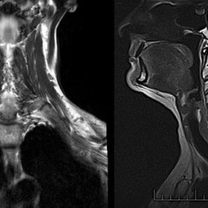
A study of soft tissue structures in the neck area is prescribed for the following diseases :
- vascular anomalies;
- traumatic neck injury;
- stenosis;
- osteochondrosis of the cervical vertebral column;
- pathology of the vocal cords;
- inflammation and enlargement of the cervical lymph nodes;
- pathology of the thyroid gland;
- disorders in the salivary glands;
- pathological changes in the larynx region;
- disorders of cerebral blood flow;
- narrowing of the cervical arteries;
- Migraine;
- arterial hypertension;
- cystic lesions in the cervical region.
Another popular and widely used method is magnetic resonance imaging of soft tissue structures of the upper and lower extremities. This diagnostic procedure is assigned in the following cases:
-
 swelling in the area of the upper or lower extremities;
 - disorders of motor activity;
- muscle tears;
- tendon injuries;
- extensive hematomas and hemorrhages;
- abscesses;
- phlegmon in the area of soft tissue structures;
- bone calluses;
- formation of scar tissue after previous traumatic injuries of soft tissue structures;
- myositis;
- diseases of inflammatory genesis;
- congenital anomalies in the development of upper and lower extremities.
Note: Magnetic Resonance Study of Soft Tissue Structures in the Facial and Gluteal Region is rarely prescribed to patients!
MRI of soft facial tissues can be recommended for the following health problems:
- neuritis of the facial nerve;
- severe headaches, permanent;
- guttural pathology.
The investigation of the soft tissue structures of the gluteal region by the magnetic resonance method has the following indications for carrying out:
- nerve pathologies in the region of the sacrum and coccygeal spine;
- disorders of circulatory processes in the gluteal region;
- pathology of plexuses of the neurovascular type;
- strong pain in the region of the buttocks and coccyx.
Important! the attending physician decides whether this type of study should be carried out in this or that way, on the basis of individual indications!
When is the study not recommended?
Diagnosis of soft tissue structures by magnetic resonance imaging is not recommended for use with:
- pacemakers, electronic devices, metal prostheses, implants, piercing, etc.;
- fear of being in a confined space( manifestations of claustrophobia);
- first 4 months of pregnancy;
- some mental disorders;
- epileptic disease;
- convulsive syndrome;The age category of the patient is under 5 years old
- ;
- obesity( weight from 120 kilograms and more).
Important !With caution this diagnostic procedure is assigned to small patients, women who are expecting a baby, and breastfeeding.
If during the lactation there is a need for carrying out this type of examination, during 2-3 days after the procedure a woman is forbidden to breastfeed a baby( for this period the child is transferred to artificial feeding).
Examination using contrast agents is strictly prohibited for patients suffering from diabetes, kidney and heart failure, increased tendency to reactions of an allergic nature!
Please note : to identify the possible individual intolerance and increased sensitivity of the patient to the substances that make up the contrast drug, preliminary preparation of a special sample will help!
How to properly prepare?
 Study of soft tissue structures by magnetic resonance method, as a rule, special, does not require advance preparation. The patient can eat whatever he wants, and take prescribed medications to him by the doctor in charge.
However, directly on the day of the study( especially if it is supposed to carry out diagnostics using a contrast medium), one should abstain from food!The last meal should be no later than 5 hours before the procedure!
Diagnostics by magnetic resonance method requires a fairly long stay in a stationary state( this is necessary for obtaining high-quality images).Therefore, in advance, take care that you do not interfere with anything and do not cause discomfort.
To do this, you need to visit the toilet in advance, if you have certain indications, take painkillers, put on comfortable, loose clothes from natural fabrics, without metal buttons and decorative elements.In the event that you experience excitement, fear of the forthcoming procedure, you can drink a mild sedative.
Note: when going to the diagnostics, take off all the jewelry.Use of decorative cosmetics is also not recommended, since it may contain impurities of a metallic nature!
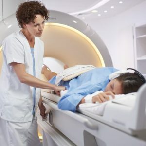
How does the study go?
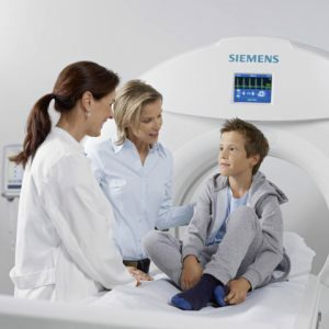
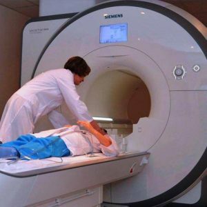 The device for carrying out diagnostics with a magnetic resonance method resembles a cylinder with a couch inside.The patient is placed on this couch, then his hands and feet are fixed with special fixators( such measures are necessary to ensure complete immobility of the patient during the procedure).
When using a contrast method of examination through a catheter, a special preparation is injected into the patient's vein.
Many patients are vainly worried before the examination.The tomograph is equipped in such a way that staying in it is as comfortable as possible.Inside the capsule there is a ventilation system to provide enough air, lighting and a special intercom system for communication with a specialist!
The procedure itself is completely painless, only mild psychological discomfort is possible.Typically, the duration of the diagnosis is about half an hour.If contrast research is used, then this time interval is increased by 2 times, which is due to the necessity to wait for a uniform distribution of the contrast medium.
In most cases, after the diagnosis is completed, the patient feels well enough and can return to the usual rhythm of life.There is no need for recovery after MRI.
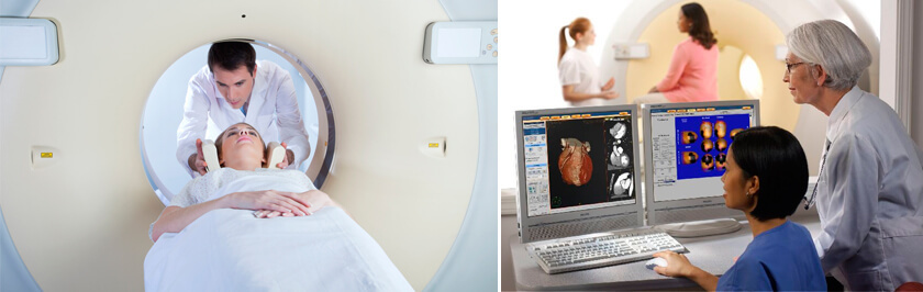
The interpretation of the results of the study, as a rule, takes from 1 to 3 hours.After completion of the diagnosis, the specialist who carried out the procedure gives the patient pictures stored on the electronic medium and his conclusion.
Important! The diagnosis of the diagnosis and the determination of the optimal therapeutic course based on the results of MRI is performed by the attending physician of the patient or by the specialist of the narrow profile !
Possible Risks
The procedure for studying soft tissue structures by magnetic resonance imaging is well tolerated.The appearance of undesirable reactions is possible only with the use of contrast agent .
If the patient has a high tendency to allergies or individual intolerance to this kind of drugs, the following symptoms may appear:
- sensation of itching and burning in the area of drug administration;
- strongly pronounced allergic reactions;
- general malaise, drowsiness, weakness;
- attacks of nausea;
- headaches;
- dizziness;
- rashes on the skin by type of urticaria.
Note that the above undesirable reactions are observed only in 1-2% of patients and, as a rule, they are due to the presence of certain contraindications, hidden from the specialist!
Advantages of
The study of soft tissue structures by magnetic resonance imaging is widely practiced due to the following advantages:
-
 Rapid diagnostics, without preliminary preparation and hospitalization of the patient.
 - Ability to consider soft tissue structures in different planes.
- High image quality, clear visualization of even the smallest elements.
- Ability to detect tumor neoplasms and other pathological processes at the initial stages of development.
- No need for radiation exposure to the patient or performing surgical interventions.
- The possibility of safe diagnosis in pregnant women, young children and the elderly.
MRI of soft tissues is a very informative procedure that gives the most accurate results.This method is used to diagnose, monitor the patient's condition after the traumas and surgical interventions, as well as assess the effectiveness of the treatment.MRI of soft tissue structures allows to reveal the slightest pathological changes and urgently take appropriate measures!
Elena Sovinskaya, doctor, medical reviewer