Scintigraphy: what is it, the preparation for the procedure, the testimony
 Scintigraphy is a highly informative, noninvasive diagnostic method related to nuclear medicine.With its help, visualization of organs and tissues is carried out.The features of anatomical location of the object are assessed, its functional state is determined, and various pathological changes are revealed.
To date, in the Russian Federation, the most important diagnostic facilities are large medical centers.
Table of contents: How is scintigraphy performed?General information about the diagnostic procedure Principle of operation of the device Contraindications Applied radiopharmaceuticals( RFP) What pathologies are detected in scintigraphy?How is scintigraphy performed?
Radio diagnostics is based on the introduction into the body of RFP( radiopharmaceutical) and the subsequent "reading" of ionizing radiation.The composition of the radiopharmaceutical is a molecule-vector absorbed by certain structures( including biological fluids), and a radioactive isotope-indicator.
General information about the diagnostic procedure
Subjects of investigation:
- bones;
-
 are light;
 - of the kidney;
- liver;
- brain;
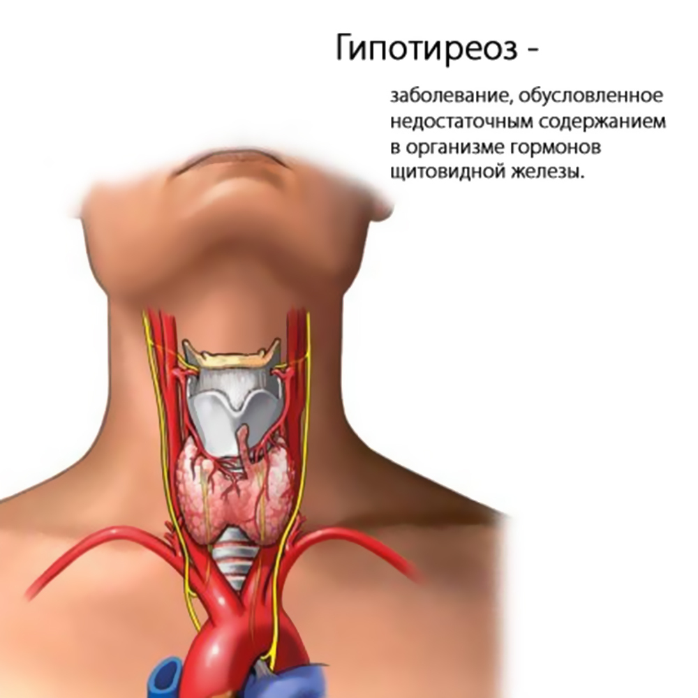
- thyroid;
- parathyroid glands;
- myocardium;
- main blood vessels( indirect radionuclide aortography);
- lymphatic system.
The procedure is absolutely safe for the patient.As a marker, isotopes of technetium are used, which are characterized by a very short half-life( 6 hours), and as a consequence, the minimum possible radiotoxicity of .The number of the radio indicator is sufficient to read the data, but too little to cause harm to the body.
Before the procedure of scintigraphy, the doctor finds out if the patient has chronic pathologies, and whether surgical operations were performed in the next six months.
Please note: does not require special preparation for the survey.After its termination, no complications develop.
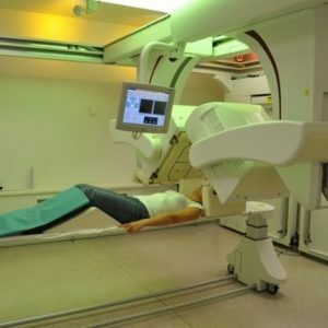
Scintigraphy includes three main steps:
- Introduction of radioisotope.
- Waiting.
- Read data.
RFP is administered as a solution intravenously.Then, the time interval during which the radio indicator is distributed in the test tissues is maintained.
Duration of waiting period for different organs:
- liver - up to half an hour;
- bone - 1,5-3 hours;
- thyroid - 30 min;
- lungs - 5 minutes;
- parathyroid glands - 3.5 hours.
The examination of the kidneys during scintigraphy is performed immediately.
Operating principle of
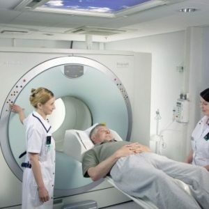
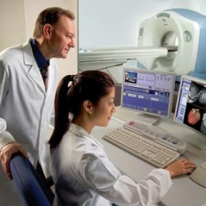
 The information is read by means of a special apparatus - gamma camera( emission computer tomograph). By itself, it is not a source of radiation.The scintillator converts gamma quanta into visible radiation particles.The number of photons is directly proportional to the amount of energy absorbed by the camera.Individual flares in photomultipliers are converted into electrical pulses, fixed by spectrometric devices.The difference in the pulse amplitudes makes it possible to distinguish the flashes of the marker from the background radiation.
During the radio diagnostics, static or dynamic images can be obtained.Two-dimensional images are sufficient for assessing the condition of bones, the thyroid gland and some other organs of the .Dynamic images are obtained by adding a number of static images;They are needed to study the functional state of the liver, gallbladder and kidneys.Visualization of contractile function of the myocardium is achieved with the help of ECG-synchronized scintigraphy.
Note: does not experience any discomfort or discomfort during the examination.Remote complications due to the introduction of a radioactive isotope were not recorded.
Contraindications
Radio diagnostics is not performed if on this day there was already conducted a study related to radiation exposure( including - conventional radiography or CT scan).Among the relative contraindications include the periods of pregnancy and breastfeeding.
Important: side effects develop in extremely rare cases.These include frequent urge to urinate, short-term increase or decrease in blood pressure, as well as individual hypersensitivity reactions to RFPs administered.
Applicable radiopharmaceuticals( RFP)
Various RFPs can be introduced before the start of the study.Each of them is characterized by tropism to various structures of the body.
A solution of pure technetium-99( Pertechnetate) is necessary for visualization of the thyroid gland.
In perfusion lung scintigraphy, macroembrages of albumin are administered, and in the study of the brain - hexamethylpropyleneamino oxime.
Diethylenetriaminepentaacetic acid is distinguished by its tropicity to the structures of the kidneys.
Tetrofosmin, labeled with technetium-99, is used to study the heart muscle, which is used to diagnose coronary artery disease and its possible complications.
Bi-and monophosphonates are used to visualize bone tissue;With their help during scintigraphy, metastasis and primary malignant tumors are determined, as well as changes that appear due to inflammatory processes and traumas.
What pathologies are detected in scintigraphy?
Some types of dementia, infectious diseases, acute acute disorders of cerebral blood flow( strokes) and Alzheimer's disease have been diagnosed in brain scintigraphy.Individual markers make it possible to visualize the distribution of dopamine, which helps to identify Parkinson's disease.The accumulation of indicators is directly proportional to the cerebral blood flow.Radiodiagnostics allows you to assess the dynamics of changes with medication and after medication on the vessels of the medulla.The average duration of the diagnostic procedure is 1 hour.
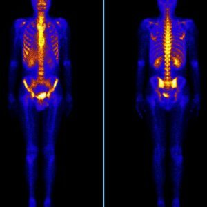 Osteoscintigraphy is the most reliable method of detecting bone metastases in the .With its help, secondary foci are determined( for cancer of the kidneys, lungs, breast, etc.) and primary tumors( chondro- and osteosarcoma).Through the analysis of images, the dynamics of the pathological process against the background of complex therapy is objectively evaluated.The duration of bone scintigraphy is 4 hours.
The study of the kidneys by means of an emission computer tomograph allows an objective assessment of their blood supply( speed and volume of regional blood flow) and functional( excretory) activity of .Nephroscintigraphy with indirect angiography reveals vesicoureteral reflux( reverse urine casting), nephrolithiasis( kidney stones), inflammatory changes and stenosis of the renal artery.The diagnostic procedure lasts less than half an hour.
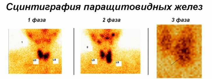
Scintigraphy helps to diagnose pathologies of the hepatobiliary system and liver failure. During the radiodiagnostics, the status of liver cells is evaluated, the functionality of the sphincter of Oddi, gallbladder motility, the patency of the bile ducts.During visualization, it is possible to determine the presence of bile from the duodenum in the stomach.The liver and gallbladder are examined for an average of 1 hour.The indication for conducting radiodiagnosis of parathyroid glands is osteoporosis( softening of bone tissue) and nephrolithiasis( kidney stones ).
 The aim of the study was to identify tumors that lead to an increase in the production of parathyroid hormone.Scintigraphy of glands lasts about 3 hours.
Please note: has already developed radio indicators for the diagnosis of individual forms of cancer.
With the help of lung diagnostics, primary pulmonary hypertension, microcirculation disorders against a background of some system pathologies, Takayasu's disease and such a dangerous condition as pulmonary embolism can be detected.Of particular importance is the ability to monitor the condition of the organ in dynamics.The lung examination takes about 20 minutes.
The study of the myocardium helps to identify transient ischemia in the background of atherosclerotic artery lesions, different forms of IHD, as well as the presence, location and volume of scars after previous heart attacks.Apply RFP, tropic to the unchanged( healthy) cells of the heart muscle.Scintigraphy is carried out in 2 stages - at first in a state of rest, and then - with a load.The method allows to determine the effectiveness of conservative therapy, endovascular and radical surgical interventions.The duration of scintigraphy of the heart muscle averages 2-3 hours.
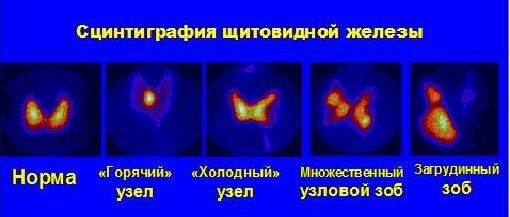
An investigation with radioisotopes helps visualize the anatomical structures of the thyroid gland. The presence and location of additional shares is determined.Scintigraphy reveals "hot" nodes, characteristic for toxic adenoma, as well as "cold" non-functioning formations, which give reason to suspect a malignant tumor.The procedure takes no more than 20-30 minutes.
Vladimir Plisov, medical reviewer