Progeria
 Progeria is a rare genetic disease, first described by Guildford, which is manifested by premature aging of the body, associated with its underdevelopment. Progeria is classified into a child, called the syndrome of Hutchinson( Hutchinson) -Gilford and adult-Werner syndrome.
With this disease, there is a marked retardation in growth from childhood, a change in the structure of the skin, cachexia, the absence of secondary sexual characteristics and hair, underdevelopment of internal organs, and the appearance of the old man. In this case, the mental state of the patient corresponds to the age, the epiphyseal cartilaginous plate closes early, and the body has child proportions.
Progeria refers to incurable diseases and is the cause of the appearance of serious atherosclerosis, which as a result develops strokes and various heart diseases. And as a result, this genetic pathology leads to a lethal outcome, i.e. It is fatal. As a rule, a child can live, on average, thirteen years, although there are cases with a life expectancy of more than twenty years.
Pediatric Hutchinson-Guildford
This disease is extremely rare in the ratio of 1: 4 million of newborns in the Netherlands and 1: 8 million in the United States. Moreover, the disease affects more boys than girls( 1,2: 1).
Consider two forms of Hutchinson-Guilford progeria: classical and non-classical.
Currently, more than a hundred cases of child progeria are described. And basically this disease affects the children of the white race. For Hutchinson-Guilford progeria, a polymorphous lesion is characteristic. Children who have this syndrome look quite normal at birth. But already a year or two there is a serious lag in growth. Usually such children differ too small growth and even lower body weight according to its length.
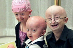
For children with progeria is characterized by complete baldness not only of the scalp, but also the lack of eyelashes, eyebrows from an early age. The skin looks weak and wrinkled as a result of the absolute loss of subcutaneous fat, there is cyanosis of the skin. The head is characterized by the disproportionate craniofacial bones, which resemble the face of a bird with a hooked nose, an abnormally small lower jaw, bulging eyeballs and protruding ears. These features, a large bald head and a small jaw, give the appearance of the child the appearance of an old man.
Other clinical manifestations of progeria include: abnormal and later teething, thin and high voice, pear-shaped thoracic and reduced in size clavicles. The limbs are usually thin, and the modified ulnar and knee joints give the sick child a "rider's pose".
Children even before the year have scleral seals, of a congenital or acquired nature, on the buttocks, hips and in the lower abdomen. Children with progeria are characterized by hyperpigmentation of the skin, which only increases with the years and hypoplasia of the nails, in which they become yellow, thin and convex, reminiscent of the watch glass. However, since the age of five, a widespread form of atherosclerosis with a large lesion of the aorta and arteries, especially mesenteric and coronary, develops. And much later, heart murmurs and hypertrophy of the heart appear in the left ventricle. Early occurrence of atherosclerosis in children, becomes the reason of short life of their life. But the main cause of death is myocardial infarction.
With progeria, cases of ischemic stroke are known. Such children in mental development are absolutely no different from healthy children, sometimes even ahead of them. Children with this diagnosis live on average about fourteen years.
With a child progeria of a nonclassical shape, the length of the body from the body lags slightly, over a long period of time the hair persists, and lipodystrophy progresses much more slowly;A recessive type of inheritance is possible.
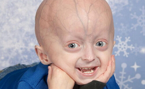
child progeria photo
Progeria causes
Until now, the exact reasons for the occurrence of progeria have not been clarified. Presumptive etiology of the development of this disease is a violation of metabolism in connective tissue, as a result of proliferation of fibroblasts by cell division and increased collagen production with reduced synthesis of glycosaminoglycans. Slow formation of fibroblasts is due to disturbances in the intercellular substance.
The causes of child progeria syndrome are mutations in the LMNA gene, which is responsible for the encoding of lamin A. It is a protein from which one of the layers of the core of the cell membrane consists.
In many cases, progeria occurs sporadically, and in some families found in siblings, especially when consanguineous marriages, and this suggests a possible autosomal recessive type of inheritance. When patients with skin studies were detected cells in which the impaired ability to fix fractures and damage to DNA and play a genetically homogeneous fibroblasts change atrophic epidermis and dermis, contributing to the disappearance of subcutaneous fat.
For progeria adults are characterized by autosomal recessive inheritance with a defective ATP-dependent helicase gene or WRN.There is an assumption in the binding chain of violations between DNA repair and connective tissue replacement.
It has also been established that the Hutchinson-Guildford progeria has impairments in carrier cells that can not completely eliminate DNA crosslinks caused by chemical agents. When diagnosing these cells with such a syndrome, they found out that they can not fully pass the fission process.
In 1971, Olovnikov suggested a shortened telomere size during the formation of cells. And in 1992 it was already proved on patients with the syndrome of progeria for adults. Analysis that binds Hayflick limit, telomere length and telomerase activity of the enzyme, allows to combine the natural aging process to form clinical symptoms child Hutchinson-Gilford progeria. Since this form of progeria is extremely rare, one can only conjecture about the type of inheritance, which has similarities with the Cockcain syndrome and manifests itself as separate traits of premature aging.
There are also statements about the belonging of the Hutchinson-Guildford proghery to the mutation, an autosomal dominant, which arose de novo, i.e. Without inheritance. It became an indirect confirmation of the syndrome, which was based on measurements of telomeres in carriers of the disease, their parents and donors.
Progeria
symptoms The clinical picture of children's Progeria different typical premature atherosclerosis, myocardial fibrosis, disorders of cerebral circulation, an increase in lipoprotein and cholesterol levels, prothrombin time assays, early heart attacks, skeletal abnormalities. In this case, there are pronounced disproportions in the face and skull, underdevelopment of the jaw and teeth, and hip displacement. Long bones with normal cortical structure and progression of peripheral demineralization undergo recurrent pathological fractures.
Joints are characterized by tight mobility, especially the knee with possible contractures of the hip, ankle, elbow and wrist joints. Radiographic studies show demineralization near joints with osteoporosis, varus and valgus deformities of the lower limbs. Also, tumors and thickening of collagen fibers are very common.
Werner's syndrome or adult progeria manifests from 14 to 18 years and is characterized by a lag in growth, universal graying with parallel progression of alopecia.
As a rule, the progeria syndrome develops after twenty years and is distinguished by early baldness, thinning of the skin on the face and extremities, characteristic pallor. Under too tight skin, surface blood vessels are looked through, and the subcutaneous fatty tissue and the muscles under it completely atrophy, so the limbs look disproportionately thin.
Then the skin over the protuberances of the bones gradually becomes thicker and ulcerated. After thirty years progeria patients develop cataract of both eyes, the voice becomes weak, high and hoarse, the skin is noticeably affected. This manifests itself in the form of sclerocerm-like changes in the limbs and face, dry skin, ulcers on the legs, calluses on the feet and telangiectasia. Such patients are generally of low height, with lunoobraznym face beak-shaped nose as a bird, a narrowed mouth opening and the protruding chin sharply full thin trunk and limbs.
In patients with progeria, the functions of sweat and sebaceous glands are disrupted. On the bony protrusions hyperkeratosis is formed, general hyperpigmentation manifests, the shape of the nail plates changes. And after various injuries on the legs and feet appear trophic ulcers. In addition to atrophy and thinning, patients notice significant changes in muscles and bones, calcification, generalized osteoporosis, osteoarthritis with erosions. Such patients are limited in the movements of the fingers and flexion contracture. For patients with progeria, deformity of bones is typical, as in rheumatoid arthritis, pain in the limbs, flat feet and osteomyelitis.
During the x-ray examinations, bone osteoporosis, heterotopic calcifications of the skin and subcutaneous tissue, ligaments and tendons are revealed. Also, cataracts are progressing slowly, atherosclerosis develops that disrupts the cardiovascular system. In most patients, the intellect decreases.
After forty years to progeria against a background of diabetes mellitus, dysfunctions of parathyroid glands and other diseases, almost 10% of patients develop tumor pathologies in the form of osteogenic sarcoma, astrocytoma, thyroid adenocarcinoma, breast and skin cancer.
Lethal outcome is usually a consequence of cardiovascular pathologies and malignant tumors.
In the histological analysis of the syndrome, progerias establish atrophy of the appendages of the skin where eczrin glands are retained;The dermis thus has a thickening, the fibers from collagen are hyaline, and the nerve fibers are destroyed.
The patients completely atrophy muscles, there is no subcutaneous fat.
The disease is diagnosed on the basis of clinical symptoms of progeria. If in doubt, the diagnosis determines the ability of fibroblasts to reproduce in culture( a reduced indicator for Werner's syndrome).For the differential diagnosis of progeria, Hutchinson-Guilford syndrome, Rothmund-Thomson syndrome and systemic scleroderma are considered.
Progeria treatment
To date, there is no specific treatment for progeria, it has not yet been developed. In general, the therapy is symptomatic with the prevention of complications after atherosclerosis and in the cure of trophic ulcers, diabetes mellitus.
An anabolic effect is prescribed for STH, which in some patients increases body weight and length. The whole therapeutic process is carried out by a number of specialists, such as an endocrinologist, therapist, cardiologist, oncologist and others, depending on the prevailing symptoms.
But in 2006, US researchers noted progression in the treatment of progeria as an incurable disease. They introduced a farnesyltransferase inhibitor into the culture of impaired fibroblasts, which had previously been tested for oncological patients. And this process returned the aging cells to normal form. This drug was well transferred, so now there is a hope that in the future there will be an opportunity in its application to prevent progeria even in childhood.
The effectiveness of Lonafarnib( farnesyltransferase inhibitor) is to increase the amount of fat under the skin, in body weight, mineralization of bones, which will eventually reduce fractures.
But, nevertheless, while this disease is characterized by unfavorable forecasts. On average, patients with progeria live to the age of thirteen, dying of hemorrhages and heart attacks.