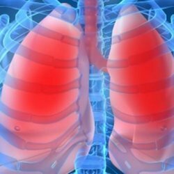
What is pulmonary alveolar microlith
 Alveolar microlithiasis of the lungs is a very rare and extremely difficult to diagnose disease. It was first described in 1933.There is this pathology approximately 20-40 times per 100,000 population. In the medical literature, only about 400 cases have been described, mainly among persons of Turkish origin.
In modern medicine, there is evidence that the risk of getting such a problem increases in a child whose parents are blood relatives. This pathology is recorded in different age groups, including newborns. The incidence of diseases among women and men is the same. Approximately a quarter of cases are diagnosed in childhood. In medical practice, it is not uncommon for several members of the family to be diagnosed at the same time. It was found that about 50% of the cases detected are due to relatives. This is one of the diseases called "family".Therefore, if someone in the family suffers such a disease, the rest of the relatives should be examined to exclude or confirm such a diagnosis. Timely examination allows you to select adequate treatment and slow down the development of the disease. The causes of the disease are not fully understood, modern medicine considers one of them a heredity, some experts consider the cause of occupational risks.
What is the problem?
Alveolar microlithiasis is characterized by the formation and accumulation of stones in the lung alveoli of very small stones. This is due to the excessive formation of protein and its accumulation in the alveoli. The protein concentrates the metal salts, these deposits reach sizes from 0.2 to 0.3 mm in diameter, sometimes they are larger - up to 3 mm. Microlits( bunches) contain calcium carbonate and phosphate, as well as other microelements: zinc, sodium, copper, potassium, magnesium. Microconcretions( microliths) in pulmonary alveolar microlithiasis differ in a peculiar concentric structure, they are located not on the walls of the alveoli, but in the alveoli themselves. Deposits can fill more than half of the alveoli and even the bronchi. At the same time, the lungs become dense and heavy, their weight sometimes reaches 4 kg, and the dimensions decrease. These changes lead to a decrease in blood flow to the lungs, the formation of respiratory failure and, as a consequence, the occurrence of various infectious diseases.
The main symptoms of the disease
The insidiousness of the disease lies in the fact that it does not appear at all for a very long time, develops slowly. Often, alveolar microlithiasis of the lung is detected by chance during an X-ray examination associated with another disease. Specific symptoms do not exist. The patient may be disturbed by shortness of breath, rapid fatigue, as the disease develops cough, shortness of breath, sputum, hemoptysis, characteristic for inflammation of the lungs or tuberculosis. In some cases, cyanosis of the skin and mucous membranes is detected, the nail phalanges of the fingers thicken and become like drum sticks. Then there is a feeling of heaviness and pain in the chest, pulmonary heart failure, palpitations with physical exertion, immunity decreases, and children are delayed development. Further development of the disease can occur in edema on the legs, pain in the right upper quadrant, which is caused by an increase in liver size.
Laboratory test results
Standard laboratory tests are usually inadequate. The general analysis of the blood most often shows the norm, but with the development of concomitant diseases there is an increased level of ESR, leukocytosis, the content of erythrocytes increases. In sputum and bronchial lavage fluid, microliths can be detected, but other diseases, such as tuberculosis, also give the same results. For correct diagnosis it is very important to identify the shape of microliths. The decisive sign of pulmonary alveolar microlithiasis is precisely their pronounced concentric structure. A biochemical blood test can reveal hypercalcemia, a slight increase in phosphate levels. But these changes do not give full confidence in the diagnosis, because they are not natural for this disease.
Instrumental survey
Modern medical technology allows more thorough and thorough examination of the patient. To make the correct diagnosis it is necessary to conduct a comprehensive instrumental examination, which includes:
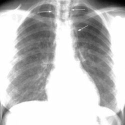
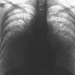
- X-ray. The images usually show numerous areas of darkening, usually located in the lower and middle parts of the lung. They look like scattered sand. Often, these blackouts are taken for tuberculosis foci. In some cases, sediments along the contours of the heart are noted.
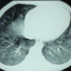
- Computed tomography.
- Perfusion lung scintigraphy with 99mTc( a method of functional imaging consisting in the introduction of radioactive isotopes into the body and imaging by determining the radiation emitted by them).
- Study of the ventilation function of the lungs. Currently, a computer spirography is being performed, which makes the procedure more informative for the doctor and more comfortable for the patient. The study of ventilatory function of the lungs reveals the development of a restrictive type of respiratory failure( this type of respiratory failure is characterized by a decrease in the lungs' ability to expand with inspiration and to subside during exhalation).
- Study of the gas composition of blood - a decrease in the partial pressure of oxygen in the arterial blood is observed.
- Electrocardiogram - signs of myocardial hypertrophy of the right atrium and right ventricle are possible.
- A biopsy of the lung is necessary to clarify and confirm the diagnosis.
So, in pulmonary alveolar microlithiasis, the examination program should be as follows:
- General tests of blood, urine.
- Biochemical blood analysis allows to determine the content of total protein, protein fractions, alkaline phosphatases, aminotransferases, phosphorus, calcium.
- Sputum analysis and bronchial washings are prescribed to detect the presence of microliths with a characteristic concentric structure.
- X-ray of the lungs.
- Computerized tomography of the lungs.
- Electrocardiogram( ECG).
- Spirography.
- Biopsy of the lung( transbronchial, if information is not enough - open).
Unfortunately, it should be noted that modern medicine does not have effective regimens for the treatment of pulmonary alveolar microlithiasis. Therapy is mainly aimed at eliminating the symptoms of pulmonary insufficiency and the treatment of concomitant infections. Drug treatment can slow the progression of the disease and alleviate the condition of the patient.
Medical assistance in the diagnosis and treatment of pulmonary alveolar microlithiasis is offered in pulmonology departments and centers in Germany.
Forecast is unfavorable. Patients die from pulmonary heart failure and secondary infectious diseases.