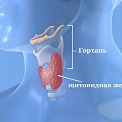
Methods of examination of the thyroid gland
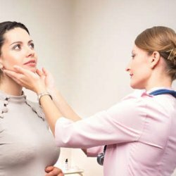
Thoracic examination and palpation of the thyroid
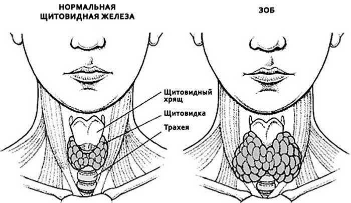
The uniform thickening of the anterior sections of the neck, and sometimes the characteristic "butterfly", corresponding to the shape and position of the thyroid gland, allows one to suspect its increase, called diffuse goiter. Such changes occur in the autoimmune nature of organ disease, or due to inadequate intake of iodine. Bumpy swelling of the front of the neck or the presence of a separate node are characteristic of the nodular form of goiter, which can be both benign and malignant in nature.
Finger examination( palpation) of the thyroid gland allows to determine its size and consistency, to identify individual nodes in thyroid tissue and the relationship with surrounding structures, and in the presence of a malignant tumor to reveal metastatic lesions of the lymph nodes.
However, one should not lay unreasonable hopes on this method. Even the most sensitive fingers of the doctor are not able to determine the node size of less than 1-1.5 cm, and often remain undetected and larger, if they are in the depth of the tissue of the lateral lobes of the gland. Therefore, in examinations of patients currently performed, palpation of the gland as an independent method is used mainly for preventive examinations of the population.
Study of the gland with ultrasound( US)
Ultrasound examination of the thyroid gland has now entered into a wide clinical practice. It allows to determine with great accuracy not only the shape and size of the organ, but also to reveal in the tissues individual formations with dimensions from 3 mm or more. The method is harmless and relatively cheap. It is currently used not only for the primary diagnosis of thyroid disorders, but also for dynamic monitoring of the processes occurring in it, for example, by changing the size of the node.
In addition, ultrasound is a subjective method that can be influenced by both the equipment used and the technique in which the researcher performs the research and his experience. Therefore, it is preferable, especially when conducting sonography in dynamics, to perform research on the same apparatus from the same specialist.
Computed tomography and nuclear magnetic resonance
This study allows you to obtain the largest amount of information on the shape, size and relationship of the gland with surrounding tissues and organs.
Serial thyograms of the thyroid gland are performed through 8 mm, and if suspected of concealed cancer, after 4 mm. In the study of patients with goitre of cervical-chested localization, one can not only estimate the depth of its spread in the mediastinum, but also the changes( constriction and deviation) of the esophagus and trachea.
However, because of the high cost and radiation burden received by the patient during the study, the method has limited application. As well as sonography, these examinations allow to receive only indirect signs of malignancy of changes of a thyroid gland.
Morphological Diagnosis of Thyroid Diseases Using TAB
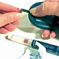
When a doctor has identified any formations in the thyroid gland, first of all he wants to know if this is cancer, and directs the patient to perform a fine needle aspiration biopsy - TAB.At present, there is a unanimous opinion that all nodular formations of the thyroid gland larger than 1-1.5 cm should be punctured and the final diagnosis is established when examining the cells of the obtained biopsy specimen. The method is simple and almost painless.
A thin needle is inserted into various parts of the node, which is controlled by ultrasound. Cells are obtained by creating a vacuum in the syringe while pulling the piston. Bioptate( taken tissue glands) is transferred to the glass, fixed, colored and examined by a cytologist who is looking for atypical( cancerous) cells in it.
Currently, this is the most reliable method of detecting malignant thyroid formations.
The method has a high diagnostic accuracy - more than 90%, provided that the puncture is performed by an experienced team, and the morphological study is carried out by a cytologist specializing in thyroid diseases.
Be Healthy!