Disease of lymphangioleiomyomatosis( leiomyomatosis)
 Leiomyomatosis is a pathological process characterized by the proliferation of smooth muscle fibers in tissues and organs that normally contain smooth muscle tissue.
The disease is very rare. The disease affects women of childbearing age from 18 to 50 years. The disease is characterized by progressive dyspnoea, pneumothorax, hemoptysis. Doctors are not very familiar with the clinical manifestations of leiomyomatosis.
Etiology and pathogenesis of the disease
Causes of the disease in our time have not yet been fully clarified. At the heart of the disease is a disseminated pathological process, characterized by tumor-like proliferation of smooth muscle fibers followed by transformation of lung tissue. There are assumptions about the hormonal dependence of the disease. This assumption is supported by the fact that women are usually exposed to the disease at a reproductive age, very rarely men. In the premenstrual period, during pregnancy, the disease worsens. The process stabilizes in postmenopause. It is also assumed that the disease can be caused by disorders of the immune system.
Versions of the appearance of the disease do not fully explain its cause.
Pathogenesis is still not fully understood. Some authors hold an opinion on the tumor nature of leiomyomatosis. There are also assumptions about the role of endocrine, genetic disorders. Many modern authors point to a big role in the appearance of the disease of dysfunction of the female sex glands. This is confirmed by the occurrence of the disease usually in women, increased respiratory failure in menstruation, and a decrease in progression after removal of the ovaries or the onset of menopause.
Symptoms of leiomyomatosis
The most famous manifestations of the disease are recurrent pneumohilotoraxes and slowly progressive dyspnea. In about 50% of patients, the disease begins with a sharp pneumothorax. Dyspnea may become worse, but after evacuation of air and the chyleous fluid decreases. Also in patients, there may be pain in the chest, and hemoptysis. In patients with leiomyomatosis, weakened vesicular breathing is usually observed.
At the initial stage of the disease, clinical manifestations of the disease may be absent, and for a long time the disease can be asymptomatic. The focal form of a leiomyomatosis passes asymptomatically and it can be revealed by means of a roentgen. Factors such as pregnancy, childbirth, the reception of contraceptives contribute to the activation of the disease. In such patients, the prognosis is usually unfavorable. The lethal outcome can come from two to ten years. Patients with leiomyomatosis live on average for about five years. There were cases in practice, with a fatal outcome after 17 years. The main and only cause of death is increased respiratory failure.
Diagnosis of leiomyomatosis
Laboratory diagnostic methods
- No significant changes are observed in the general blood test. In a few cases, there is an increase in ESR, eosinophilia.
- In general, urine analysis can sometimes show a slight proteinuria.
- In a biochemical blood test, hypercholesterolemia, increased levels of globulins, aminotransferases can be observed.
- When investigating pleural fluid, chylothorax is observed. Pleural fluid has a milky white color. After centrifugation, it retains turbidity, contains above 110 mg% of triglycerides, chylomicra.
Instrumental diagnostic methods
- When radiographing lungs, radiographs of chest cells in patients with leiomyomatosis show an increase in lung volume and an increase in the pattern of the lung having a mesh character. The focal form of the disease is characterized by darkening in diameters from 0.5 to 1.5 cm, having clear boundaries.
- On computer tomography of the lungs, cystic lung transformations characteristic for leiomyomatosis are evident. Cysts are multiple, diffuse. Cysts distinguish 2 types: small multiple types and large cysts. The thickness of the cyst walls is no more than 2 mm.
- When diagnosing the ventilation capacity of the lungs, the lung volume increases, which is associated with the appearance of multiple cysts.
- During the investigation of blood gases, arterial hypoxemia is detected. In this case, a decrease in the partial oxygen tension is observed. This fact is strongly pronounced after physical exercises.
- Electrocardiography reveals the movement of the electrical cardiac axis to the right, symptoms of myocardial hypertrophy in the right atrium, ventricle and other signs.
Pathomorphological diagnosis of
In this study, the following signs of the disease are identified:
- presence in some individual sections of the lungs of large air cavities;
- set of small nodules in diameter from 0,3 to 0,7 cm, they are whitish and filled with liquid, located subpleural;Severe compaction of lung tissue.
- hyperplasia of the lymph nodes;
- destructive changes in the walls of the bronchi, alveoli, blood and lymphatic vessels;
- in the interstitium of the lung diffuse proliferation of smooth muscle fibers;
- development of pneumohylothorax, hemohylothorax, due to destruction of the walls of the lymph and blood vessels of the lungs, and also due to the development of subpleural cysts;
- appearance of microcystic "cellular" lung.
In leiomyomatosis, extrapulmonary changes, such as angiomyolipomas, disturbances of retroperitoneal and mediastinal lymph nodes are very common.
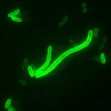
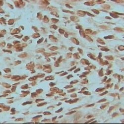
Histology
This diagnosis is based on transbronchial biopsy results. If it is not very informative, then the doctor prescribes thoracoscopic or open lung biopsy. The usual morphological picture of leiomyomatosis is characterized by proliferation of smooth muscle cells around the bronchovascular structures and in interstitium. Proliferating cells are similar to myocyte vessels. But they are more playmorphic, shorter, and in some cases there is the possibility of confusing them with fibroblasts.
Fiber analysis reveals a high content of glycogen and smooth muscle antigens in muscle cells. Currently, serological tests, in order to determine these antigens, do not exist.
In general, when young women have a clinical picture of recurrent chylothorax and pneumothorax, and there is also progressive shortness of breath, hemoptysis, chylopericard, ascites and lymphadenopathy, then it is necessary to suspect the disease with lymphangiomyomiomatosis.
Treatment of leiomyomatosis
Effective methods for the treatment of leiomyomatosis, other than lung transplantation, do not yet exist. Therapy is directed to treatment of respiratory failure. Immunosuppressants and corticosteroids are not effective. In some cases, removal of the ovaries is recommended. The prognosis of the disease is usually unfavorable.
It is possible that in the future, medicine will develop new effective methods for treating leiomyomatosis.