Idiopathic interstitial pneumonia
 The term "idiopathic interstitial pneumonia" is used to refer to a group of lung diseases with an unidentified etiology. The diseases of this group differ in the type of pathomorphological noninfectious inflammation and fibrosis in lung instillation. The difference is noticeable in clinical course and prognosis. Diseases can be acute and result in death, but they can be completely cured or form a chronic "cellular lung".
History of
For the first time idiopathic interstitial pneumonia was diagnosed and classified in 1935.In 1964, the diagnosis "fibrosing alveolitis" was first determined. Hence the synonymous name "idiopathic pulmonary fibrosis".There is also the name "cryptogenic fibrosing alveolitis".The latter name is most common in Europe.
In 1965, the disease was first differentiated into the following five species: giant cell interstitial pneumonia, lymphoid interstitial pneumonia, obliterating bronchiolitis with interscial pneumonia, desquamative interscisal pneumonia and conventional interscial pneumonia.
Over time, the first two options came out of the group, because their etiological factors were established.
In 1998, D. Myers and A. Katzenstein identified four variants of the disease: nonspecific, acute, desquamative and usual interscial pneumonia. This classification did not include bronchiolitis obliterans, since its causes are viruses or inhalation of toxins.
In the end, in 2001, according to the international agreement, 7 types of pneumonia of interest were accepted. The classification includes: lymphoid interstitial pneumonia, desquamative interstitial pneumonia, respiratory bronchiolitis, acute interstitial pneumonia, cryptogenic organizing pneumonia, nonspecific interstitial pneumonia, idiopathic fibrosing alveolitis.
Distribution of
In our time, only the distribution of idiopathic fibrosing alveolitis is reliably known. According to statistics, about 20 men from one hundred thousand and 13 women from the same number are ill. With age, the risk of disease increases. Most often they get sick after 60. Mortality is directly proportional to the age category. The older the sick, the higher it is.3 people per 100 thousand people die from this disease. In turn, the average life expectancy of survivors of this type of pneumonia is 2.5 to 5 years. It should be clarified that about 90% of cases of idiopathic interstitial pneumonia are a disease of idiopathic fibrosing alveolitis.
Diagnosis
Idiopathic interstitial pneumonia is classified according to the characteristics of the clinical picture, pathomorphological and radiographic signs. Patimonic violations for each individual form have not yet been identified. All patients have a decrease in lung volume due to physiological changes, as a consequence of the disease. In patients, the results of laboratory studies are characteristic of all types of idiopathic diseases.
Features of clinical and radiological manifestations of idiopathic fibrosing alveolitis
This type is characterized by increasing dyspnea and a non-productive cough of a paroxysmal nature with refractory to antitussive agents. In a quarter of patients, nail phalanges deform. At auscultation, "cellophane cracking" is heard from the lower sections to the upper ones. In later stages, there is a pulmonary heart.
In radiography, blackout is most often observed in the basal areas. This is the reason for the decrease in the volume of the lower lobes and the formation of cellular changes in the lung tissue. The accuracy of the diagnosis with an x-ray photograph is 50%.
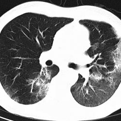
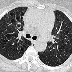
Computed tomography shows mainly bilateral changes associated with traction bronchiectasis. In most cases, there are phenomena of "honeycomb", less often - "frosted glass".Changes are seen mainly in the peripheral and basal parts of the lungs.
Features of clinical and radiological manifestations of nonspecific interstitial pneumonia
This disease develops very smoothly. Often people turn to a doctor 1.5-3 years after the first symptoms are felt. Dyspnea and coughing are not very pronounced and grow at a slow pace. The temperature and changes in nail plates do not appear more often than in 10% of cases. The disease is well treatable.
The x-ray image shows bilateral infiltrative changes in the lower parts of the lungs. Plots with the effect of "frosted glass" are located symmetrically. In most cases, this is the only sign of the disease. Repeated studies after treatment usually show a positive trend.
Features of clinical and roentgenological manifestations of cryptogenic organizing pneumonia
In this type of pneumonia, pathological changes occur in the alveolar courses and alveoli in combination with the polypoid bronchiolitis. Symptoms are similar to influenza. Cough can have a productive nature with the allocation of clear sputum. Rattles are heard. The shape of the nail phalanges never changes. To everything, in the diagnosis is involved and a blood test. GSK therapy leads to complete recovery of the patient.
When X-ray examinations are most often seen, one-sided darkening in the form of nodular shadows. The area of the lungs can be reduced to 25%.
Computed tomography shows subpleural and peribronchial seals in the lower lobes of the lungs.
Features of clinical and radiological manifestations of acute interstitial pneumonia
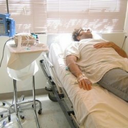
With this type of disease after the symptoms of viral infection, rapid onset of dyspnea occurs. Later, cyanosis develops. Over time, the patient requires artificial ventilation. Mortality exceeds 50%.
Radiographs strongly notice thickening of central and peripheral bronchi walls, "frosted glass".
Features of clinical and radiologic manifestations of desquamative interstitial pneumonia
The most common disease occurs in smokers and is characterized by dry cough and increasing dyspnea.
The x-ray photograph shows a knotty structure of "frosted glass" in the lower lobes of the lungs.
Features of clinical and radiologic manifestations of lymphoid interventional pneumonia
Symptoms of the disease are weight loss, fever, arthralgia, chest pain and sometimes anemia.
An x-ray image can show both diffuse lesions in the form of "frosted glass" and mixed alveolar-interstitial infiltrates.
Complete diagnosis of
In modern medicine, a complete diagnosis can only be done with surgical lung biopsy. For both types of idiopathic interventional pneumonia, both an open and a video-x-raycopy biopsy can be used. A complete diagnosis of this group of diseases allows to avoid not only the lethal outcome of the disease itself, but also the consequences of improper treatment with hard-to-transfer drugs that are used in connection with the complexity of the disease.