Tomography of the brain is the most reliable method of obtaining information for effective treatment
Contents:
- Indications MRI MRI
- modes brain brain
- Advantages of MRI brain
- Risks of the procedure MRI of the brain in children
- MRI contrast
- What is the diagnostic equipment?
- How is the MRI of the brain done?
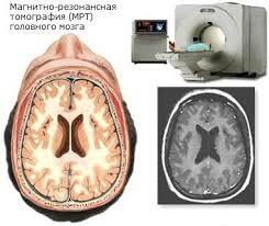
Effective treatment of brain pathologies is the accurate informative diagnosis. Such diseases manifest themselves, in most cases, very sluggishly. The patient may be troubled by headaches, nausea, dizziness, blurred vision. For the timely appointment of a doctor, a thorough diagnosis of the brain is necessary. In this case, the tomography of the brain is the most reliable method of obtaining the necessary information for a specialist.

Magnetic resonance imaging of the brain
Computed tomography of the brain is an X-ray examination method, which thanks to computer reconstruction provides images in the case of transillumination of the object around a narrow X-ray beam. For the study, special training is not needed. The whole procedure lasts a couple of minutes.
Magnetic resonance imaging of the brain provides an estimate of such diseases:..
- tumor, cyst,
- intracerebral aneurysms,
- acute ischemic stroke, etc.
When the ultrasound and CT -maloinformativnye research methods should do MRI brain and receiveNecessary quality of images. Indications
brain MRI
This inspection must be performed in case:
- Having skull injury which involves bone fracture or damage to the internal structures of the skull.
- If there is a suspicion of a tumor of the brain and adjacent tissue. For this pathology are characterized by such symptoms as persistent dizziness and headaches, weakness.
- If there is a suspicion of having a metastasis in the brain area.
- Diagnosis of demyelinating and degenerative pathology of neural tissue.
- If you want to confirm or rule out the development of multiple sclerosis( memory loss, attention span, poor orientation in space and time).
- Presence of a heart attack or stroke to assess the extent of brain damage.
- Control after surgery. Modes
brain MRI
To determine brain pathologies of different origin magnetic brain imaging is performed in the following modes:
- magnetic imaging head helps detected in brain tissue cysts, hematoma, ischemia zone tumor;
- Computer tomography of cerebral vessels;
- Examination of cranial nerves is performed with sharp pain, tingling, numbness - impaired sensation on the face;
- Tomography of orbits, eyeball structures;
- Magnetic tomography of the pituitary gland - with changes in the hormonal background.
- Tomography of the vessels of the brain and neck are performed in the presence of severe dizziness. Vascular pathology of the vessels of the head and cervical region is revealed.
- Magnetic - tractography is an additional study in the study of the brain, during which it is possible to diagnose the pathology of white matter caused by congenital metabolic defects, traumatic, autoimmune, toxic and radiation damage.
Advantages of MRI of the brain
What does the tomography of the brain show? You can get an exact answer to this question only when the merits of this procedure are determined:
- Objective diagnosis;
- There is an opportunity to study the structure of the brain and nerves;
- Ability to detect brain disease, cranial nerves, vessels, the presence of tumors in the early stages;
- Unlike other diagnostic methods that do not provide a complete assessment of the disease, a CT scan of the brain gives an excellent result and the ability to get a more complete picture of the patient's health status;
- The study can be carried out by introducing the patient into medical sleep( for adults and children suffering from claustrophobia);
- Tomography of the brain is performed using a high-field tomograph with modern software;
- Carrying out an MRI study excludes the presence of ionizing radiation;
- A snapshot of the MRI of the brain, as well as the entire diagnostic process, can be recorded on a disk.
Risks of the procedure
The conduct of the MRI of the head in some cases carries some risks:
- For an average patient, taking into account all safety rules, this study has virtually no risks.
- In case of sedation use, there is a risk of overdose. For this reason, the radiologist assistant constantly looks after the patient's vital signs;
- Although a powerful magnet, which is part of the scanner, does not carry any harm, problems during the procedure may still arise. This is due to the presence of devices implanted in the body of the patient, in which there are metals;
- The introduction of contrast material carries a high risk of an allergic reaction. The reactions presented are usually usually mild and quickly disappear when the necessary drugs are prescribed. In the event of an allergy, the nurse or radiologist should immediately help the patient;
- The most common form of MRI complication is recognized as nephrogenic systemic fibrosis. Its development can arise in the case of the introduction of large doses of contrasting material, which is based on gadolinium, to patients who have kidney function impaired.
MRI of the brain in children
A tomography of the brain is performed in the following cases:
- Frequent headaches and dizziness;
- Fainting states;
- Decreased vision, hearing of unexplained etiology;
- Convulsive conditions. Even if such conditions occurred once, a computed tomography of the child's brain is necessary;
- Delays in psychomotor and speech development;
- Changes in behavior.
A mandatory indication for the MRI of the brain for children is the fact that the child is engaged in sports that are characterized by a high degree of head injury( basketball, boxing, wrestling), or there have already been cases of brain concussion.
In most cases, small babies can not remain immobile for a long time in brain imaging. Therefore, in such a situation, specialists come to a decision about the MRI of the brain for children in conditions of drug sleep.
MRI with contrast
An MP-study is carried out using a special standard set of programs. In case of suspicion of a certain pathology( vascular pathology, trauma, epilepsy) add additional programs that are more sensitive in a particular pathology. Thanks to this, the informative quality and quality of the research, as well as its duration, are increased. There are situations when the study is accompanied by the addition of intravenous contrast. For example, an examination of the pituitary gland without the use of a contrast agent produces a false-negative result.
MRI of the brain with contrasting makes it possible to get an evaluation of all brain structures and the state of blood vessels supplying it.
Indications for MRI of the head with contrast:
- craniocerebral trauma;
- determination of thrombosis or stenosis of cerebral vessels( presence of hemorrhagic and ischemic stroke);
- changes in the development and structure of cerebral vessels( aneurysms and malformations);
- brain tumors;
- frequent headaches of an unexplained nature or migraine;
- scheduled operations or follow-up status monitoring.
Contraindications to MRI with contrast:
Contraindications to the procedure may be characterized as absolute and relative.
Absolute contraindications include:
- the presence of a pacemaker;
- implants made of materials that are exposed to magnetic field( implants from titanium to contraindications do not apply).
Relative contraindications include:
- claustrophobia( for those who suffer from this disease there are open-type devices);
- pregnancy( holding an MRI with contrasting to pregnant women is not prohibited, but before the procedure it is necessary to obtain consent from the gynecologist);
- epilepsy;
- overweight( different models of apparatus have different limitations on the maximum weight of the patient);
- implants, for the manufacture of which an unknown material was used;
- allergic to a contrast agent or kidney dysfunction
What does the diagnostic equipment look like?

Equipment for MRI
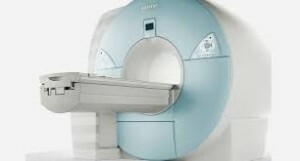
After it became a little understandable what brain imaging is, you can talk about the equipment that is used for this purpose. The standard equipment for MRI is a large cylindrical tube surrounded by a magnet. The patient is placed on a non-movable examination table. The table can slide inside the magnet. There are tomographs( systems with a short tunnel), the construction of which is arranged in such a way that the patient is not completely surrounded by a magnet.
There are devices open at the sides. Such tomographs are used to conduct a survey of people suffering from claustrophobia. Modern open MR tomographs make it possible to obtain very good photos of the MRI of the brain at various examinations.
However, if an open device is equipped with an old magnet, the picture quality may decrease. But not all studies can be performed on an open-type tomograph. Details can be obtained from your doctor.
The working computer system that processes the image is located in the adjacent cabinet with the scanner.
How is the MRI of the brain done?

MRI procedure
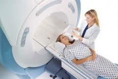
What is an MRI of the brain is understandable, but how it is carried out, no. This study can be performed on an outpatient basis and at the time of hospitalization. On the moving table, the radiologist assistant has a patient. The position of the body is secured by means of belts and special rollers. They help to lie still to the patient.
Around the head are placed devices that send and receive radio waves.
If there is a need for a contrast material at the time of the study, the nurse inserts a catheter into the vein. A vial of physiological saline may be connected to it. This solution helps ensure a constant washing of the system to prevent it from clogging up to the application of contrast material.
After completion of all preparations, the patient's table is placed inside the magnet, and the average medical staff and radiologist leave the treatment room at the time of the study.
When the test is completed, the doctor asks the patient to wait for some time when the screen will analyze what the MRI of the brain shows. There are situations when you need an additional series of pictures. For more information about this method of diagnosis, see the video MRI of the brain.
Today, computed tomography of the brain takes an important place in the identification of various diseases and pathologies. This procedure is absolutely painless, and also very effective. Remember! With the timely diagnosis of the disease, there is a great chance to get rid of the disease and not to think about the bad.
write the question in the form below:


