Tomography of the brain: appointment, methods and features of the conduction
Contents:
- Appointment, varieties imaging methods and indications for its conduct
- Preparation for imaging, highlights procedures and contraindications to its use
- Features of imaging children
brain diseases, as a rule, do not have too pronounced symptoms. Among the main signs - headaches, visual impairment, dizziness. To conduct adequate and effective treatment of such diseases, an informative and accurate diagnosis is necessary. Tomography of the brain is one of the most effective diagnostic methods to date.
Appointment, varieties imaging methods and indications for its conduct
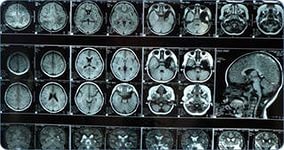
Various methods of computer tomography get the clear picture of all the structural changes in the brain. Thanks to modern equipment, images of slices are obtained after layer-by-layer scanning of the brain in a few millimeters.
Often computed tomography is performed using X-rays. The received black-and-white image allows to reveal brain tumors, traumas, strokes. Differently informative, the method still has a serious drawback - the patient receives a considerable dose of radiation during the procedure.
Magnetic resonance imaging is considered a more complex and expensive method. This study is based on the effect of nuclear magnetic resonance of hydrogen atoms. It allows to get good results in the diagnosis of inflammatory processes, multiple sclerosis and vascular disorders in the brain. The method is contraindicated only to those persons who use pacemakers, because the magnetic field causes a malfunction in the operation of these devices.
Positron emission tomography gives a color image of the layers of the brain, which allows us to examine the brain in the smallest details. This method is considered to be the most expensive and complex.
indications for imaging are the following disorders:
- disease or disorder of brain abscess
- , concussion
- intracranial hemorrhage and trauma in different parts of the skull
- vascular disease, in particular ischemic
- neoplasm or inflammation in organs and LOR sockets
- diseases of the salivary glands
- disease affecting the temporal part
- frequent headaches, tinnitus, dizziness
also using theFargo analyzes the results of surgical intervention - is determined by the condition of the brain and the further development of the tumor.
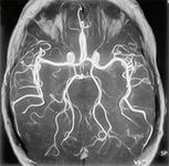
What does the CT scan show? In the first place it makes it possible to diagnose a number of diseases, such as benign and malignant tumors, meningiomas, hemorrhage, hematoma, infarction, stroke, encephalopathy, and various brain injury. In addition, this diagnostic method monitors the effectiveness of treatment. Tomography of cerebral vessels allows at the earliest stages to detect violations of cerebral circulation. The method gives a complete picture of structural changes, as a result of which the size and location of hematomas, blood clots, aneurysms, pathological vascular anastomies are determined with great accuracy.
Preparation for tomography, the main points of the procedure and contraindications to its use
No special preparation is required for the procedure. Basically it is based on the observance of the recommendations on nutrition. So, a few days before the diagnosis of the diet should be deleted, milk, fish sauce, sausage, black bread, meat, beans, peas, seeds, herbs, berries, mushrooms, as well as fresh fruit and soft drinks. Include in the diet can be poultry meat, broth, cheese, white stale bread, homemade cookies, coffee, tea, fruit juices without pulp.
There are no restrictions in taking laxatives, if you take them, you can not only continue using the drugs, but also increase the dosage slightly. The day before the diagnosis is allowed to use exclusively liquid food - broths, kissels, still drinks, clarified juices without pulp.
Highlights of the procedure:
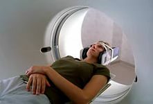
- during the procedure the patient is in a supine position on the back on a special tomograph table with fixed hands, head, chest. It is necessary to remove all metal jewelry, as well as dentures and hearing aids.
- if necessary, the patient is administered intravenous contrast substance
- of any sensations during the tomography. The patient does not test
- . At the request of the technologist, sometimes it is necessary to hold the breath
- . During operation of the scanner, noise can be heard, for this reason, a person is usually offered headphones.
You should also know that there are certain contraindications to CT scan, namely:
- intolerance of contrast, and allergic reactions to iodine
- pregnancy and lactation
- renal failure
- patient weight over one hundred and fifty kilograms
Patients suffering from claustrophobia, as a rule, Before the start of the diagnostic procedure prescribe sedatives.
Features of Tomography for Children
The most harmless for children's body is the method of magnetic resonance imaging. This study is assigned to a child in the event of frequent fainting, dizziness, headaches, noticeable lag in development, reduced vision and hearing.
The method is effective for recognizing diseases that affect the structure of the brain at the earliest stages.
Parents should prepare a baby for her before brain imaging of the brain. He must know how to behave and what awaits him, otherwise the child may be frightened, panicked, and then the study will turn out to be uninformative or may at all be interrupted. Therefore, it is necessary to assure the child that the MRI procedure does not hurt at all and is not terrible.
If your toddler is too active, and you are sure that he will not stand the time immobile, be sure to inform the doctor about it. Most likely, that in this case he will have to take sedatives, that is calming drugs.
For children less than five years old, an anesthetic procedure is usually performed, which is still not done without the written consent of the parents.
To a baby, magnetic resonance imaging is also done under anesthesia. In this case, the baby who is fed by mother's milk should be fed no later than two hours before the diagnosis.
Before starting the procedure, it is necessary to remove clothes and all metal ornaments from the child. Then the child is put on a special couch table, hands and head are fixed and sent to the tunnel of the scanning device. During the scanning process, the toddler should not move, but can communicate with parents outside the apparatus wall. That the child is not frightened by the noise of a working scanner, they wear special headphones. The procedure lasts usually about twenty minutes, sometimes a little more.
In conclusion, I would like to note that to date, there are many ways to examine the nervous system. It is not necessary to approach independently to a choice of methods of a tomography, only the expert can appoint the research necessary for you that guarantees that all possible nuances and contraindications to carrying out of a tomography of a brain will be considered.
write the question in the form below: