Nuchal hillocks: variants of norm and pathology
Contents:
- Anatomical features of occiput bone
- Occipital mound in adult
- Bruised
- Insect bite
- Atheroma
- Hemangioma
- Lipoma
- Osteoma
- Diagnosis and treatment
 The human skull is represented by a fixed joint of bones. Isolate the brain and facial parts of the skull. Each of them has its anatomical features, according to which it is possible to determine the sex, age of a person, and sometimes even racial identity. For each person there are their own variants of the formation of bones, which are determined by hereditary data and the influence of external factors. There may appear protrusions, indentations, deletion of the bone, on the occiput the occiput is formed. The shape of the skull is changed for the following reasons:
- rickets, borne in childhood;
- acromegaly - elevated level of somatotropin;
- trauma( CCT);
- infectious lesions;
- tumors are benign and malignant in nature.
Anatomical features of the occiput bone
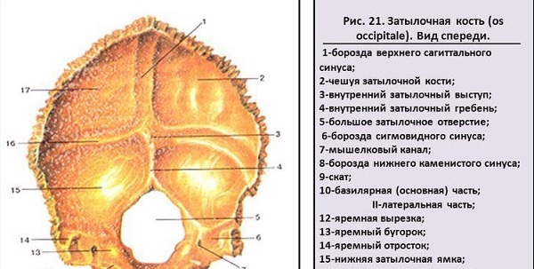
Large occipital forearm, receptacle of the medulla oblongata, is formed by four elements of the occipital bone. In front of the hole is the basilar part. In the childhood period, the sphenoid bone joins her through the cartilage. By the age of 20, their fixed fusion is formed.
Inside the cranial cavity, the surface is smooth, with a brainstem on it. Outside rough, with protruding tubercle. On the lateral parts there are two occipital condyles, each having its own articular surface. Together with the first vertebral bone, they form an articulation. At the base of the condyle, the bone perforates the sublingual canal.
Jugular notch, located on the lateral part, together with the same name of the temporal bone, make a jugular hole. Through it pass the cranial nerves and vein. The occipital part is represented by scales. It performs a cover function. In the center there is an occipital mound. It is unmistakably determined through the skin. A crest runs from the hillock to the large hole. On the sides of it there are pairs of nasal lines - these are points of muscle increment.
 Read whether a solid cone on the head can be dangerous: the causes and treatment of pathology.
Why there is a bump on the head of a child: diagnosis and treatment.
Occipital protuberance in adult
The Neanderthal man had a characteristic feature - the protruding occipital bone. In this manifestation is now very rare. It can be a characteristic feature of Australids, lappids, in the inhabitants inhabiting the Lancashire region in the UK.In another concept, this definition is used to characterize the protruding part of the skull having any cause. The most likely are:
- injury;
- insect bite;
- atheroma;
- hemangioma;
- lipoma;
- osteoma.
Contusion
Traumatic bone injury, accompanied by edema and build-up. If a cold compress is applied immediately after the injury, the consequences will be reduced. At the site of the injury, edema develops, a hillock appears, which hurts when you touch and turn the head. Treatment does not require a condition, it passes by itself.
Insect bite
The appearance of the cone is accompanied by unpleasant sensations in the form of itching, painful pressure. Often this is a type of local allergic reaction. Depending on the reactivity of the body, the hillock may have a different size. To get rid of use antihistamines, ointments to eliminate itching.
Atheroma
Sometimes a hard, painless formation appears under the skin, which tends to become inflamed when the infection gets. It is represented by clogged sebaceous glands. Treatment is performed surgically.
Hemangioma
If on the back of the head a cone of red color with translucent vessels, it is most likely formed by a benign vascular tumor. Usually this is a feature of intrauterine vascular insertion, with adulthood the tumor can begin to grow. There is a high risk of her trauma and bleeding. Using laser coagulation, surgical excision, cryodestruction, the tumor is removed. 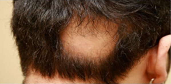
Lipoma
The appearance of a knoll on the head in an adult may be due to the development of a lipoma - a benign proliferation of connective tissue. The grease grows slowly, there is no danger to life.
Osteoma
A long-term benign tumor of bone tissue does not grow into neighboring tissues, it does not malignant. It is a mound in the form of an even hemisphere. It affects young people, but it grows for many years.
Osteoma can form the occipital mound in a person from very dense tissue. It has no bone marrow and gavers canals, which penetrate the usual bone tissue. Sometimes there is another type, in the form of a bone marrow formation, completely consisting of cavities. It is often formed on the bones of the skull and skeleton, it does not affect the ribs.
The bumps can grow from the outer plates of the skull, then they do not give any brain symptoms. If the process started from the inside of the skull, epileptic seizures, dizziness, memory impairment may occur.
The reasons for the development of the hillocks are not fully known. There is definitely a hereditary predisposition. To provoke growth can be traumas, presence of such diseases as rheumatism, gout, autoimmune processes, foci of chronic infection.
 All about brain tumors: types, symptoms, diagnosis.
Read why there is a burning sensation in my head: the main causes, diagnostics.
Learn about the common localizations of brain cancer, the methods of surgical treatment and the consequences after surgery.
Diagnostics and treatment
For the survey use radiographic methods. It is necessary to differentiate osteoma from osteomyelitis and sarcoma. Informative use of computer tomography, which will reflect the nature of education layer by layer. Histological analysis will show the absence of bone marrow, which is characteristic of osteoma.
Treatment is performed only surgically in the event that the hillock causes anxiety, it gives painful sensations. Sometimes this is only an aesthetic defect, when a person notices the occipital mounds in his mirror, in a photo, which reduces his self-confidence.
Preventive measures can not be carried out purposefully. A healthy lifestyle, the prevention of infections, the prevention of head injuries can eliminate the risks of osteoma.
write the question in the form below:

