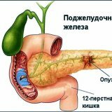
Cysts and fistula of the pancreas

Cysts and fistula of the pancreas are not a rare phenomenon. Cysts are capsules with a liquid inside. They are located on the gland itself, as well as on the tissues surrounding it. This disease occurs at any age, and regardless of sex. Cysts of the pancreas are a collective concept.
Cysts are divided into several types:
- Congenital. These include cysts that are formed as a result of the developmental defect of the pancreatic tissue, as well as the duct system.
- Purchased.
- Acquired cysts in turn are divided into retentional, degenerative, proliferative, parasitic.
- Retinal cysts occur as a result of stricture of the gland ducts, as well as when they are blocked by stones or tumors.
- Degenerative cysts develop as a result of damage to the pancreatic tissue during pancreatonecrosis, after hemorrhage, trauma or during the tumor process.
- Proliferative cysts are cavitary neoplasms. These are cystadenocarcinomas and cystadenomas.
- Parasitic cysts occur during the infection of oranginism with echinococcus and cysticerci.
Cysts, depending on the structure of its walls.
Distinguish false and true cysts of the pancreas depending on the structure of its walls. True cysts are congenital dysontogenetic cysts, cystadenomas and cystadenocarcinomas, acquired retention cysts. True cysts account for about 20% of all cysts of the gland. Its main feature is the presence of epithelial lining, which is on its inner surface. The sizes of true cysts are much more false. Some of the cysts for surgeons become a real find.
The walls of the false cyst are the compacted peritoneum and fibrous tissue. Unlike the real cyst, the false cyst does not have epithelial lining inside. From the inside, false cysts are treated with a granulation tissue. In the cavity there is a liquid with necrotic tissues. This liquid has a different character. As a rule, this purulent and serous exudate, containing a bloody admixture and clots, can also contain pancreatic juice. A false cyst is formed on the head, body, and tail of the pancreas. The amount of fluid that is contained in the cyst, sometimes reaches 1-2 liters or more. A large cyst often spreads in different directions. It can be placed forward and upward in the direction of the small omentum, while the liver pushes upward, the stomach downwards. The cyst can also go toward the gastro-colonic ligament, while pushing the stomach upward, and the transverse colum moves downward.
Large cysts.
Large pancreatic cysts usually flow without any symptoms. They arise in the event that the cyst greatly increased and began to squeeze the neighboring organs. Frequent symptoms in cysts are pain in the upper abdomen, dyspeptic phenomena appear, the general condition is broken, weakness appears, a person loses weight, body temperature rises. During palpation, a tumor-like formation in the abdomen is palpated.
The patient begins to appear dull, persistent pain, in some cases, pain paroxysmal. They are shrouded, bursting, and the patient must take a bent position or a knee-elbow position. The strongest pain sensations appear at pressure of a cyst on a solar plexus and celiac. But still, with huge cysts, the pain is slightly expressed, the patients complain of the sensation of squeezing in the epigastric region. Most often, dyspeptic phenomena are nausea, sometimes vomiting, and unstable stools.
During the study, the main feature is tumor formation. If a large cyst can be detected at the first examination. The boundaries are clear, the shape is oval or round, the surface of the cyst is smooth. Tumor formation depending on the localization is determined in the near-umbilical region, in the epigastric, as well as in the left and right hypochondrium.
Complication of the cyst.
The most vivid complications of the pancreatic gland cyst are hemorrhages into its cavity, purulent processes, various disorders that appear after the cysts are squeezed by neighboring organs, fistulas external and internal, ruptures followed by the development of peritonitis.
For the diagnosis, the clinical symptoms of the disease are taken into account, and special methods of investigation are conducted. In the blood and urine, there is an increase in the number of pancreatic enzymes. Computed tomography, including ultrasound scanning, helps detect a dense formation filled with fluid.
Treatment is performed surgically. Resection of the pancreas of the affected cyst is performed. Pseudocysts use draining operations.
Fistula of the pancreas.
Pancreatic fistula is pathological communication of the pancreatic ducts with internal organs or with the external environment. The fistula is external when its mouth is formed on the skin, and internal, when the fistula communicates with the hollow organs( small and large intestine, or stomach).The fistulas are complete and incomplete. With a complete fistula pancreatic juice is secreted through the fistula outside. An incomplete fistula is characterized by the fact that the pancreatic juice flows into the duodenum and part outward through the fistula.
Basically, fistula occurs with a stomach injury or after surgery on the pancreas, after opening its ducts. Internal fistulas appear due to changes in the pancreas that pass to the wall of an adjacent organ( with pancreatitis, perforation of the pancreatic cyst and penetration).
Complete fistulas are surgically treated. The main types of surgery are excision of the fistula, suturing the fistula formed either into the stomach or into the small intestine. The fistula is also removed together with the affected pancreas.


