Hemangioma: symptoms, diagnosis, treatment in children
Benign neoplasms are quite common in childhood.
Each tenth child aged less than one year is diagnosed with hemangioma.This is a benign tumor that originates from blood vessels.And although the process is benign, it is necessary to closely monitor the progression of hemangioma and start treatment in time.Otherwise, you should be afraid of complications.
Table of contents: Hemangioma: causes of development Hemangioma in children Types of hemangiomas Hemangioma on skinHemangioma: causes of
When asked why hemangioma develops, scientists still can not give an unambiguous answer.Probable cause is the effect of unfavorable factors during pregnancy during the development of mesenchymal fetal tissue.Blood vessels form from this tissue.The most aggressive adverse factor are infectious diseases of the pregnant woman, in particular, ARVI.
Facts about hemangiomas:
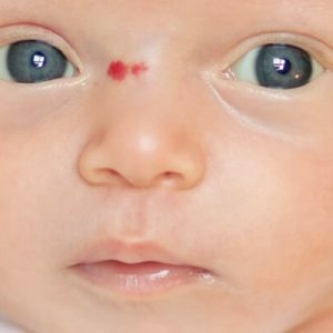
- Often found immediately after the birth of the baby or in the first weeks or months of life;
- Hemangiomas are more common in girls;
- Hemangiomas can be of completely different sizes: from a small point to a large spot.

Development of hemangioma in children
A characteristic feature of hemangioma is a change in its size. There are three stages in the development of hemangioma:
- Period of intensive growth;
- The period of stopping growth;
- Period of reverse development.
 It is difficult to predict how actively the hemangioma will increase in size.Sometimes the tumor increases even several centimeters per week.It is well known that in preterm infants, hemangiomas grow much faster than donors.Hemangiomas actively grow in the first months of life of the baby.When a child reaches the age of six months, the growth of the neoplasm slows down.This stage is called the period of growth arrest and lasts for several years.
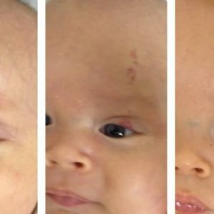
Further development of hemangioma is difficult to predict.Often there is a reverse development( regression) of the neoplasm.Gradually, the brightness of the spot decreases, white areas are seen on it.After six to eight months, the hemangioma is already becoming pale pink and smooth.By the third or fourth year of the child's life, only the area of depigmentation on the skin resembles the neoplasm.It is worth noting that regression is possible only in the case of simple hemangiomas.Cavernous as well as combined hemangiomas never regress.
Types of hemangiomas
Hemangiomas are most often localized to the skin, but may also occur in the internal organs. There are such types of hemangiomas :
- Simple( capillary);
- Cavernous( cavernous);
- Mixed;
- Combined.
Hemangioma on the skin
Hemangiomas have their favorite places.Most often they occur in the face, scalp, neck, mouth, hands.Much less often - on the external genitals, legs.
Simple hemangiomas
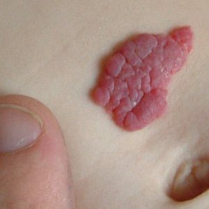
 In the structure of all hemangiomas, simple hemangiomas are approximately 95%.A simple hemangioma is a layer of small closely fitting capillary vessels.Sometimes the vessels are collected in lobules.The lumen of blood vessels is filled with blood.Simple hemangiomas are located on the skin and do not penetrate into the subcutaneous fat.The surface of capillary hemangiomas may be flat or knobby-tuberous.
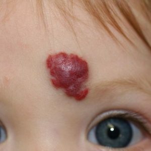
Looks like a simple hemangioma as a towering red spot on the skin, which can be of various sizes.If you press on the edge of the spot, you can see how it gradually turns pale.This is due to the clamping of the vessel and the expulsion of blood from it.But it is necessary to release the skin, as the spot is immediately filled with red.The spot has clear edges and is delimited from the surrounding healthy tissue.The skin can have one or several such neoplasms.
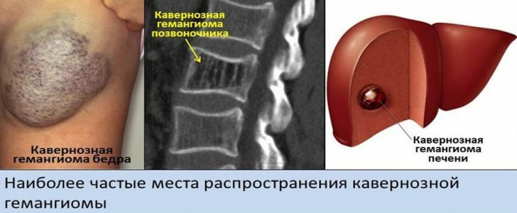
Cavernous( cavernous) hemangiomas
 Cavernous hemangioma consists of a multitude of cavities separated by partitions.This type of hemangiomas is located in the subcutaneous tissue.Cavernous hemangiomas account for about 3% of all hemangiomas.
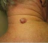
Externally, cavernous hemangioma looks like a voluminous formation that visibly rises above the skin.Surface of education is rough.Skin with cavernous hemangioma is not changed.But under the skin a tumor-like formation of bluish color is visualized.To the touch it has a soft-elastic consistency.If you press on it, the tumor decreases somewhat.But soon he regains his former form.Characteristically, when straining, crying and even coughing a child, the tumor briefly increases in size due to the influx of blood to it.
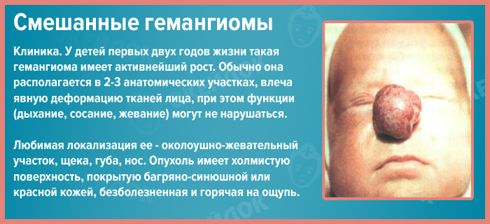
Mixed hemangiomas
Mixed are such hemangiomas that are combined with other tumors, for example, with lymphangioma, lipoma.There are such hemangiomas very rarely, approximately 0.6% of all cases of hemangiomas.
The color, consistency, appearance of the tumor will depend on the tissues that make up the neoplasm.
Combined hemangiomas
In the structure of all hemangiomas, combined hemangiomas are only 2%, but they represent the greatest difficulty in treatment.Combined hemangiomas have a subcutaneous and subcutaneous parts.External manifestations will depend on which component of the hemangioma prevails: capillary or cavernous.
Complications
Hemangioma grows fast enough and it is very difficult to predict its further impact on the body. Among the main complications of hemangiomas are:
- Bleeding.It develops when traumatizing tumor tissues.Especially dangerous is bleeding with hemangioma of the liver, as the amount of blood loss can be very massive.
- Ulceration.It develops mainly when the hemangioma is localized in the area of the lips, perineum, large folds of the skin.Characterized by the development of ulcers at the site of the tumor.
- Blood clotting disorder.It is connected with the fact that hemangioma, roughly speaking, is perceived by the body as a damaged vessel, because of which platelets are actively entering this area.Over time, the number of platelets in the blood decreases, which is fraught with poor blood coagulability.
- Inflammation and suppuration.Often associated with traumatizing the tumor.
- Disturbance of the function of organs affected by hemangioma( vision impairment in the hemangioma of the eyelid, hearing in the hemangioma of the ear).
Hemangioma of internal organs
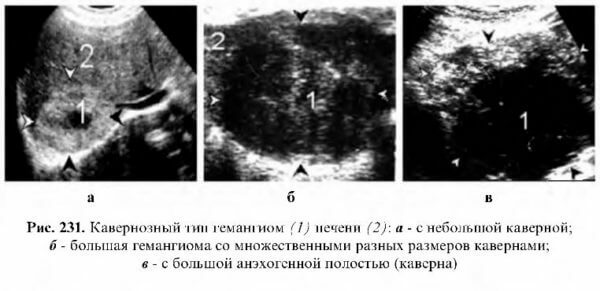
Hemangioma can form in the internal organs: the brain, uterus, lungs, kidneys.The most common is a liver hemangioma.The tumor is usually single, its dimensions are small.Hemangiomas of the liver are simple( capillary) and cavernous.Capillary hemangiomas are usually small and do not exceed a few centimeters.Cavernous reaches ten centimeters.
It is noteworthy that often the tumor does not bring any discomfort.So a person lives with ail for a long time.Approximately to the age of fifty, the size of the tumor increases and then the symptoms of the ailment already appear: dull pain in the right hypochondrium, nausea, flatulence, stool, jaundice.
Bone hemangioma
Bone hemangioma is a slowly growing benign tumor.More often the tumor is located in the spine, somewhat less often in the bones of the skull and pelvis, tubular bones.
Bone hemangiomas are usually asymptomatic, and therefore are detected by chance during a routine examination.Only in 1-1,5% of all cases of hemangioma of bones is accompanied by pain syndrome.Hemangiomas of bones do not always require active treatment, but constant monitoring by a doctor is necessary.The thing is that the increasing hemangioma of the spine, for example, spreads the bone elements, because of what vertebral fractures may occur.
Diagnostics
A doctor can suspect a hemangioma when examining the neoplasm.First, in favor of hemangioma, there is evidence of an elevated red spot.Secondly, with hemangioma, the spot fades with pressure on it and restores its shape and color after the pressure ceases.
Certain studies can be performed to confirm the diagnosis, as well as clarify the degree of skin lesions:
- Clinical blood test;
- ultrasound;
- Computer and magnetic resonance imaging.
Ultrasound is performed in the study of cavernous hemangiomas, as well as neoplasms of internal organs.This diagnostic method allows you to study the structure, depth, and size of the hemangioma.
If suspected hemangiomas of internal organs, computer or magnetic resonance imaging is performed.These methods allow us to identify new growths of the smallest size.In addition, only with the help of a tomography can you determine the presence of hemangiomas in the bones.
A clinical blood test is performed to determine complications and monitor the patient's condition during the course of treatment.Characteristic changes in blood in hemangiomas are a decrease in the number of platelets, and in addition, and erythrocytes with hemoglobin.
Treatment of hemangioma
 Treatment questions must be approached individually, taking into account the particular course of the disease in a particular child.It is often possible to hear such a view that hemangiomas should not be treated, because they can disappear by themselves when the child grows up.However, this opinion is too frivolous.Indeed, simple hemangiomas can regress, but this does not happen in every case.In addition, cavernous and mixed hemangiomas are not able to regress at all. Thus, the wait-and-see strategy can be applied only in the case of simple uncomplicated hemangiomas in the presence of signs of regression.
There are certain indications that the treatment of hemangioma should begin as soon as possible:
- Hemangiomas located in the head and neck region in the mouth, anogenital area;
- Rapidly growing tumors( an increase in its area twice a week);
- Complicated hemangiomas.
Surgical treatment: removal of hemangiomas
Surgical excision of the skin of the tumor is a common method for treating hemangiomas.However, at present, surgical intervention is rarely resorted to.First of all, because surgical intervention should be performed under general anesthesia.Surgical excision of the skin can be accompanied by blood loss, and after the operation there is a scar.However, surgical excision is preferred in deep hemangiomas, as well as in mature forms of the tumor.That is, when other methods of treatment are impossible.
Removal of hemangiomas by laser, cryodestruction
 Modern physical methods of hemangioma removal( cryodestruction, laser removal) have a lot of advantages in comparison with surgical treatment.Such manipulations are carried out on an outpatient basis, because the duration of the procedure is only 15-20 minutes, besides, it is not necessary to inject anesthesia into the child.
In cryodestruction, the skin is exposed to liquid nitrogen, which has a low temperature.The method itself is simple enough, it does not require any special preparation, it is carried out without anesthesia.Hemangiomas located on the skin are treated with liquid nitrogen for 20-30 seconds, hemangiomas on the mucous membranes - for 7-15 seconds.On the third or fourth day, a crust is formed on the treated area of the skin, after a month, the skin is completely healed.In large hemangiomas treatment is carried out in several stages.
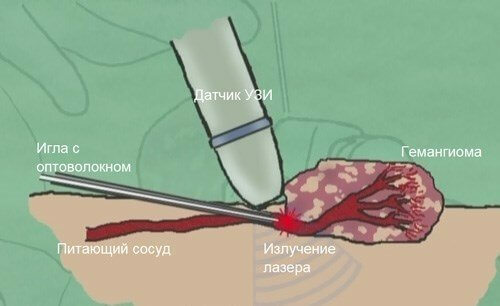
In the fight against hemangiomas, laser removal is successfully used.This method is used for tumors with a diameter of up to two centimeters.Under the action of the laser, the tumor is thermally destroyed.Advantages of the method are that the possibility of bleeding is excluded, since the laser beam cauterizes the vessels.In the site of exposure, a crust is formed, which disappears after two to three weeks.In its place a small scar is exposed.
Conservative treatment
Hemangiomas can be treated conservatively.One of the methods used in the fight against cavernous and combined hemangiomas is sclerosing therapy.A sclerosing substance - 70% alcohol is injected into the tumor.This leads to an inflammatory reaction and thrombosis of the vessel, which causes the blood flow to the hemangioma to cease.Soon, hemangioma is able to regress.Usually, several repetitions of procedures are required to achieve the desired result.
Hormone therapy is also used to combat extensive hemangiomas.To this end, the child is prescribed prednisolone.By the end of hormone therapy, angioma volumes decrease, and growth ceases, whitish areas of healthy skin appear on the surface of the hemangioma.If necessary, the course of hormone therapy can be continued after one to two months.However, with the help of such treatment, it will not be possible to achieve the desired cosmetic effect, that is, complete disappearance of the hemangioma.Therefore, we will have to resort to other methods of treatment.
In the treatment of hemangiomas, beta-blocker propranolol can also be used.The drug leads to narrowing of the vessels of the tumor, stimulating the replacement of the vascular wall with scar tissue.
In angiomas with complex localization, for example, in the area of the orbit or occupying a fairly large area, resort to radiation therapy.
In any case, the decision on the need for dynamic surveillance or active treatment is made by the child's surgeon.Therefore, if there is a hemangioma in the baby, you need to see a doctor and do not wait for self-healing.
Grigorova Valeria, medical reviewer