Tomography of the brain
Contents:
- Magnetic resonance imaging of the brain
- Magnetic resonance imaging of cerebral vessels
- Diagnosis of cerebral vessels
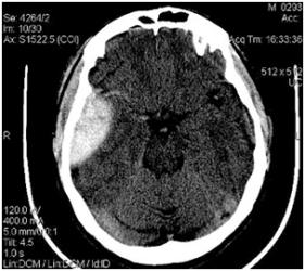
Computer tomography of the brain. The most mysterious and thoroughly unexplored human organ is the brain. With the advent of CT - computed tomography of the brain, it was possible to study this mysterious organ more thoroughly and in detail. CT of the brain is a new and modern method of examination on a computer multispiral tomograph, with which you can fix the slightest change in the brain and get their exact image. In focal and cerebral neurological symptoms, speech impairment, memory, hearing, vision and headaches, brain tomography is also used.
Brain tomography is performed to detect in patients:
- acute hematoma( subdural or epidural), hemorrhage;
- stroke and vascular disorders;
- inflammatory diseases( encephalitis, abscess);
- structural abnormalities( hydrocephalus, tumors);
- brain injury;
- fracture of the skull( base).
Most brain research is performed with contrast enhancement. The procedure is performed as follows: a special substance( 150 ml) is dosed into the vein and scan is immediately performed. Apply a contrast agent for the diagnosis of tumors, cerebral vascular lesions, cysts, aneurysm and atherosclerotic disorders.
Angiography( computer) - a kind of CT of the brain, with it diagnosed vascular lesions.
By conducting tomography of the brain, carefully monitor the treatment of various diseases, including tumors and abscesses. On the tomogram, the bones of the skull( base, arch), the sinuses of the nose, vascular sinuses and vessels are very clearly seen.
Indications and symptoms that require immediate brain imaging:
- headaches, often due to an unknown cause;
- is a syncope of a regular nature;
- head trauma( without loss of consciousness);
- vascular injury or suspected hemorrhage;
- head injury of a severe stage;
- passing the routine diagnosis to monitor the results of treatment.
Magnetic resonance imaging of the brain
MRI( magnetic resonance imaging) is the study and examination of internal tissues and human organs using nuclear magnetic resonance. This is a harmless method of treatment with the use of light rays that shine through the object in intersecting different directions and obtain a qualitative image of the object of investigation.
Patients who come to the brain scan, often complain of headaches and MRI - this is the main and basic standard in the study of this problem. This diagnosis is carried out for 10-30 minutes.
In the modern world, MRI is the leader among methods for diagnosing a variety of diseases in neurology, namely, tumors and changes in blood vessels and nerve nodes( pathologies).
When performing tomography of the brain, a clear picture is obtained, as the cranial bones do not screen on the image.
At the moment, computer-aided modern MRI diagnostic programs create a three-dimensional accurate image of all areas of the brain to detect a hotbed of the disease. Blood vessels of the brain are scanned with contrast and without it. They can also create a clear three-dimensional model, where the slightest vascular disturbances will be visible.
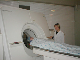
MRI is assigned to patients with such diseases:
- brain cancer;
- circulatory disturbance;
- tumors;
- concussion;
- abscess;
- disorders in the isthmus of the rhomboid brain.
MRI helps to obtain detailed information about tumors, in other words, benign and malignant tumors so that people do not fall into a state of depression, hearing and speaking about it, about its condition, condition and appearance.
The most important merit and merit of MRI is that this wonderful and clever device recognizes and scans the disease in the initial stage, when neither the doctor, nor the patient, nor any other devices and devices see it. And this is of great importance. Early diagnosis of the disease can save the most precious that a person has - life, and gives hope to cope with the disease and defeat it.
Magnetic resonance imaging of cerebral vessels
MRI of cerebral vessels with the development of science is the most reliable and reliable way of diagnosing pathologies of the vascular system, not involving the puncture of the artery. But in some cases paramagnetics( gadolinium) are used. Conduct studies with a tomograph, a magnetic field voltage of 0.3 Tesla( not less).
MRI of the brain vessels provides a real way for the doctor to get the thinnest cuts, and also to recreate a three-dimensional image of the blood supply in the area under investigation. It is possible to isolate individual vessels in the desired projection of the brain region under study.
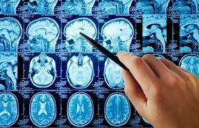
Tomography of cerebral vessels in patients under such conditions:
- dystonia( vegetative-vascular);
- tinnitus and dizziness;
- headaches( frequent character);
- stroke;
- ischemia of cerebral vessels;
- vertebrobasilar insufficiency.
MRI of brain vessels - diagnoses almost all cerebrovascular diseases of the brain. This method is ahead of radiography and computer X-ray tomography.
With the help of MRI it is possible to undergo a survey in different modes and under different programs. Your attending physician in the direction will indicate the desired plan for the examination.
Before starting the examination, you should consult with such physicians:
- neurosurgeon;
- by the radiologist;
- by the radiologist;
- neurologist.
Diagnosis of cerebral vessels
Diagnosis of vascular diseases is very often complicated by insufficiently expressed symptoms, flaccid and atypical course of the disease. Sometimes the symptoms of high blood pressure( intracranial) are combined with the symptom of the displacement of brain structures, with the characteristics of brain tumors.
There are a number of ways of early diagnosis of cerebral vessels:
- electroencephalography( reveals epilepsy, used for delayed speech development and damage);
- angiography is magnetic resonance( examines the brain envelope);
- computed tomography of vessels( reveals brain pathologies and congenital vascular diseases);
- non-invasive neuroimaging( indicates foci where hemorrhage occurred);
- echoencephalography( with the help of ultrasound the signal is fixed and displayed on the monitor, reveals the presence of tumors and traumatic injuries);
- ultrasound dopplerography( blood circulation analysis in the vessels of the brain);
- MRI( used in all cases of study).
The most effective methods of studying cerebral vascular diseases help to find the problem in time and prevent its development.
write the question in the form below: