Amebiasis: causes, symptoms, diagnosis, treatment and prevention
 Amoebiasis is an intestinal infectious disease that has a long course and is characterized by the appearance of ulcerative defects of the large intestine and lesions of other organs.Amoeba was discovered by the St. Petersburg scientist F.A.Leshem in 1875 when studying the patient's feces with bloody diarrhea.In Egypt R. Koch( 1883) isolated the pathogen from ulcers of the intestine and purulent cavities in the liver.Amybiasis, called "amoebic dysentery," was identified as an independent disease in 1891.
About the causative agent
Amoeba causing the pathology( Entamoeba histolitica) is of the Protozoa type.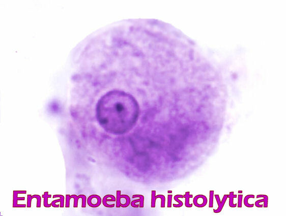 The life cycle goes through 2 stages:
- Vegetative;
- rest( cysts).
The vegetative stage can take the form:
- tissue - possessing high mobility with the ability of penetration( invasion).It is characteristic for an acute inflammatory process, with the presence of a parasite in the organs;
- great vegetative - in this form the amoeba is able to absorb red blood cells( erythrophage);
- luminal - an agent with characteristic low mobility, is defined in the large intestine and lives in convalescents.
Injected forms:
- is a pre-acute amoeba that is also inactive, found in the feces of a recovering patient.
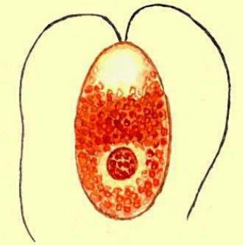
- is directly a cyst-a form living in auspicious conditions outside the host for several months.Life in the soil is about a week.Can withstand freezing to -20 ° C, but do not tolerate drying.
Note : vegetative forms are unstable in the external environment. In the acute phase of the disease in the feces of the patient, tissue and luminal forms of the pathogen are found.In healthy carriers and when the disease passes into the stage of decay, both the luminal and pre-cystic forms as well as the cysts themselves enter the feces.Cysts are the stable life forms of the pathogen outside its host.The formed cyst is surrounded by a colorless shell and outwardly resembles a sphere.Its nutria contains a glycogen-filled vacuole and four nuclei.If the cysts enter the small intestine, then a mature amoeba appears, which, when divided, yields 8 amoebas with one nucleus.They are capable of reproduction, transforming into vegetative forms that enter the large intestine.
How the infection occurs
The source of the infection is a person who differentiates into the environment various vegetative species of amoebas and cysts.Typically, the source of the disease become carriers of infection, patients with chronic forms of the disease or convalescent after acute form of amebiasis.Allocation can take years.During the day, sometimes up to 900 million cysts.Note : patients with acute amebiasis are not sources of infection, since they contain non-contagious vegetative forms. A person becomes infected when cysts get into the body with unwashed products and with dirty hands.Also, the danger poses contaminated dishes, things, laundry.Mechanical vectors are flies and cockroaches.The most common are men aged 20 to 50 years.Immunity to the disease is absent.Amoebiasis is registered worldwide, but is especially common in countries with a humid and hot climate.
In the intestine, the cyst form passes into the vegetative form, which penetrates into the intestinal wall, secrete enzymes that break down tissues and promote the formation of ulcers.Defects are formed from small erosions and abscesses( ulcers) having a structure of nodules with vegetative forms of amoebae contained in them.The ulcer is a collapsed bundle.The diameter of ulcerative formations can be up to 2.5 cm with dented edges and a purulent bottom.Ulcers can merge, affect the muscle shell and even cause perforation of the intestine.This is a very life-threatening complication that provokes peritonitis( inflammation of the peritoneum).Also, bleeding is possible as a result of vascular collapse.In the healing stage, narrowing of the intestinal wall and subsequent obstruction can develop.Note: ingestion of amoebas into the blood often results in liver damage, lung tissue, brain. An untreated, long-lasting amoebiasis is able to cause tumorous amoebiomes in the intestine - formations from the granulation tissue and body cells.
Types of clinical manifestations of the disease
WHO identifies three forms of amoebiasis:
- intestinal;
- is an extra-intestinal;
- cutaneous
Intestinal amebiasis: symptoms, course of the disease, complications
Intestinal amebiasis - is the most common form of disease. The period from the onset of infection to the development of complaints can last from 1-2 weeks to 3 months.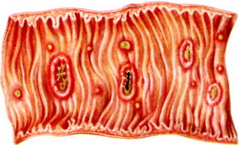
Symptoms of intestinal amebiasis
The disease occurs in mild, moderate to severe severity.It develops gradually. In patients with intestinal amebiasis, the following symptoms appear:
- low temperature,
- weakness,
- headache,
- increased fatigue,
- abdominal rasp, minor pain.
Important: is the main symptom of amebiasis - diarrhea . It has an abundant character, frequency - 4-6 times a day, like slime. With the progression of the disease, the stool frequency increases to 15-20 per day, its fecal appearance is lost, blood appears in the mucus( a kind of crimson jelly), cramping pains in the abdomen grow.The patient experiences constant painful desires on the bottom( tenesmus).The above symptoms of amebiasis persist for up to a week, then there comes an improvement.The disease passes into the stage of attenuation( remission).After a few weeks or months, there is a return of complaints, an exacerbation of pathology.So the recursive occurs.With continuous , the disease progresses as periods of increased and decreased complaints.Pay attention to : without treatment, amebiasis can last 10 years or more. Chronicity of the disease is accompanied by a pronounced weakening of the body( asthenia).Protein deficiency, lack of vitamins develops.Patients note a change in taste, tenderness of the mucous membranes of the mouth.On the tongue a raid is formed, the appetite is reduced.The skin becomes dry, facial features become aggravated.The abdomen is painful when touched, drawn.
Complications of intestinal amebiasis
With prolonged amebiasis complications of the cardiovascular system develop - heart rate increases, arrythmia periodically arises, manifestations of cardiac muscle malnutrition( deafness of tones).The defeat of the nervous system is accompanied by cases of depression, mood swings, apathy, insomnia, increased irritability. Severe complications of intestinal amebiasis:
- perforation of the intestinal wall;
- narrowing of the intestine( stricture);
- intestinal bleeding;
- Peritoneal inflammation( peritonitis) - amoebic pericolitis is especially dangerous( in 9-10% of patients), accompanied by a peritonitis clinic with fibrinous adhesion of the sites of the serosa of the intestine in places of ulcers, spikes.Purulent peritonitis - proceeds with sharp deterioration of state of health, abdominal pain, high fever, vomiting, flatulence;
- amoeba - tumor-like formation of the cecum and ascending part of the large intestine.It can cause intestinal obstruction;
- development of adenomatous polyp( of glandular tissue, in sizes with walnut);
- sometimes causes prolapse of the rectum;
- amoebic appendicitis( severe complication with high mortality - up to 80-90%).Usually manifested at the height of the acute course of the disease.
Extraintestinal amebiasis

Extraintestinal amebiasis can occur in various forms.The course of the disease and its symptoms depend on the lesions of the organ in which the amoeba has settled.
Hepatic amebiasis: symptoms, complications
May be expressed in the form of inflammation of the liver( hepatitis) or encysted abscess( abscess).Patients complain of pain in the right hypochondrium.On examination, attention is drawn to the increase in the boundaries of the liver, tightness of the edge, soreness.There may be jaundice, often with an increase in temperature.Pain can give in the right shoulder, strengthen with breathing, change of position.The temperature increase is not of a regular nature, it can vary throughout the day.The appearance of the patients is exhausted, the skin is dry and flabby, pointed features, flawed eyes, cheeks.Sometimes there is swelling of the legs.The abdomen is upset.Note the : when the process is chronic, the patient's exhaustion increases. Abscesses can be single or multiple.This form of the disease is a serious, fatal outcome much more often than with other forms.When the abscess breaks through the abdominal cavity, peritonitis occurs.It is possible to get the contents of the abscess into the cavity of the pleura.Then, as a complication of amebiasis, pleuropneumonia occurs or the abscess of the lung begins.Inflammatory processes of the lung tissue often go into a chronic form.
Symptoms of other forms of amebiasis
The amoeba can enter the brain through the vascular bed.In this case, the corresponding clinic of brain abscess develops.Patients are observed cerebral and focal complaints - convulsions, headaches, sensitivity disorders, paresis and paralysis.There are amoebae abscesses of the spleen, female genitals, kidneys, accompanied by complaints, characteristic of the major diseases of these organs.
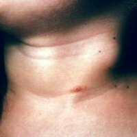
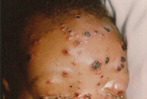
Cutaneous amebiasis
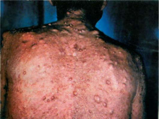
Amoeba in the skin leads to the development of erosions and ulcers in it.Locations - perineum, buttocks.Ulcers deep, painless with dark edges and strong, unpleasant odor.
Forecasts of the disease
Timely detection and treatment leads to a cure.Launched and untreated cases result in chronization, complications and death.
Diagnosis of amebiasis
 Preliminary diagnosis is established based on:
- epidemiological data;
- examining the patient and his complaints;
- data of urgent laboratory tests.
Laboratory diagnostics of amebiasis
In clinical blood analysis - an increase in the number of neutrophilic leukocytes( leukocytosis can reach very high values), an increase in the ESR. The main kind of diagnostics is parasitological research:
- kala - detection of tissue forms and large vegetative cellular forms;
- sputum, the contents of abscesses, bottom ulcer materials.
The presence of luminal forms and cysts in feces is not the basis for the diagnosis of "amebiasis".Note : a stool test should be performed no later than 10-15 minutes after the act of defecation.The analysis should be repeated several times. At the stage of attenuation of clinical manifestations, the study of feces is carried out after the patient receives a salt laxative.If there is no possibility of analyzing fresh material, then it is necessary to resort to conservation.The material is subjected to examination, both in untreated form, and with fixation by hematoxylin or Lugol solution.A longer-term method for the diagnosis of biomaterials in amoebiasis is the cultivation of amoebae on nutrient media.
Serological research
Immunological tests are considered an auxiliary method in determining the disease.The most revealing of them was the test of fluorescent bodies( RFA).The second most important is the complement fixation( RCC) reaction.It can also be involved in the study of the method of infection pathological secretions of laboratory animals.
Instrumental diagnostics of amebiasis
In the complex of diagnostic measures are applied:
- sigmoidoscopy - examination of the straight and part of the sigmoid colon.The condition of the mucosa, the presence of ulcers, erosive changes, amoebae, polyps, cystic formations, etc. are determined with the possibility to take the contents from the bottom of these formations for investigation;
- ultrasound - allows you to supplement the diagnostic data by examining the liver, assess the tissue structure of this organ, the size, the presence of abscesses and other formations;
- computed tomography( CT), especially its modern types.With its help, you can determine the size and number of pathological formations, as well as conduct a survey of the brain, lungs, other organs;
- radioisotope techniques are necessary in case of controversial cases for distinguishing amoebic abscess from bacterial;
- Irrigoscopy - X-ray examination of the large intestine against contrasting substances.
- microresonance tomography - is used in complicated cases and in weakened patients.
Treatment of amebiasis
Therapeutic measures for amebiasis consist of the use of three types of drugs that affect different groups of problems. These are:
-
 direct( luminal) amoebicides are medicines that have a harmful effect on the luminal forms of amoebas( Yatren, Khiniophon, Diiodhinon, antibiotics of the tetracycline group).They are used for the treatment of amoebiasis in carriers, in chronic forms, in recovered patients for the prevention of relapses;
 - tissue amoebicides - act on amoebas in tissues and mucous membranes( Emetin , Hingamin , Dihydroethetine, Ambilgar ). These drugs are chosen for the treatment of acute processes and extraintestinal forms of amebiasis ;
- universal amebicides - affect all forms of amoebas( Flagyl, Tinidazole, and Furamid).Affect the cellular level, destroying the parasite from the inside.They disrupt the information structures of proteins, stopping reproduction, and also promote the formation of radicals that have a harmful effect on amoebae.
Additionally, amebiasis is treated with drugs that restore normal intestinal flora.These include pro-biotics.Also, according to the indications, cardiovascular drugs, hepatoprotectors, immunostimulants and other medications are used, the need for which is determined by the doctor.
Preventative measures
The list of measures for the prevention of amebiasis includes:
- Early detection of sick people and carriers of amoebiasis, treatment of them.
- Dispensary monitoring of those who are sick.
- Sanitary and hygienic measures: food processing, water treatment, people hygiene.
For more information on amebiasis and amoebic dysentery, see this video review:
Lothin Alexander, radiologist