MRI of chest organs: what shows and the rules of preparation for the procedure

One of the modern and safe methods for diagnosing organs and tissues of the chest cavity is a chest MRT.It is carried out on a special apparatus that allows to identify various diseases that are not noticeable when using other methods, and also to track the dynamics of treatment.
Table of contents: General information Indications for use Preparation Features of the study How chest MRT is made Feelings during and after the procedure When results are available Are the consequences possibleGeneral information
 MRI, or magnetic resonance imaging is a non-invasiveEffective research, which is assigned to obtain an adequate assessment of the condition of internal organs and systems, including the structure of the tissues of the heart, blood vessels, lungs.
Important! The method is based on a magnetic field, radio pulses and special software for the computer, through which a detailed layered image of soft tissues, bones and organs is displayed on the screen.Subsequently, the doctor will be able to study it in detail, confirming or not confirming the presence of pathologies, even those that have not revealed a computed tomography, ultrasound or X-ray.
Thanks to the chest MRT, doctors have an opportunity to assess the condition of the internal surface of the organs, the walls of the chest cavity, the heart, the vessels, the pleura at any angles .Along with this, the device provides consecutive photographs of the organs of the cardiovascular system, as a result of which it is possible to identify problems in the work of the heart, its valves or large vessels.
In some cases, for the MRI, a contrast agent is injected into the patient's blood for better visualization of the vessels.
Indications for prescribing
Medically assigned patients can be assigned a chest MRT in several cases:
-
 in the presence of an oncology in this area in order to determine the amount of formation and the degree of influence on adjacent organs and tissues;
 - for suspected heart muscle or valve failures;
- to confirm the presence of chest bone disease;
- for assessing blood flow in the heart muscle after a heart attack;
- for suspected lymphogranulosis and other pathologies of lymph node development;
- for assessing the state of the membrane surrounding the myocardium - pericardium;
- for suspected pleural and mediastinal pathology.
Please note! To assess the condition of the arteries and veins of the chest, doctors use MRA, or magnetic resonance angiography.This is a special kind of MRI, which also helps to consider an aneurysm( artery wall swelling) or vascular bundle, that is, the rupture of their inner shell.
Preparing
For the maximum convenience of the patient, an assistant specialist can suggest that you wear a hospital shirt.The fact is that tightening clothes, as well as clothes with different metal inserts, can prevent a qualitative and reliable result.
There are no unified rules for eating and drinking on the day of the analysis, but every medical center that conducts an MRI adheres to its rules.Meanwhile, most experts agree that there is no need to make any changes to their diet in order for organs and systems to work in the regular mode.
True, staff ask patients to leave at home or at least outside the MRI cabinet, things that can affect the work of the magnet and, as a result, on the analysis itself.In particular, these are:
- jewelery, jewelry, bank cards, watches, pins, hairpins;
- metal lighters or other metal objects that can affect the magnet;
- body piercing;
- handles, folding knives;
- glasses;
- removable dentures;
- hearing aids, because under the influence of MRI, they may experience malfunctions.
Special attention is paid to the physicians of patients with metallic implants in their bodies. Some of them are absolutely safe for a person in the course of MRI, but they must be talked about immediately before the examination.In some cases, a radiologist or technologist may be required to have an MRI of the chest.
At the same time, the following devices are contraindicated for MRI:
- cochlear implant;
- separate types of clips that are used for brain aneurysm;
- built-in pacemaker;
- stents, which are installed inside the vessels.
Do not make MRI and individuals with a weight of more than 150 kg, as well as those with a diameter of the circumference of the trunk is more than 150 cm.
In addition, there are a number of devices or elements that can be dangerous to humans at the time of the survey.It all depends on their type and the strength of the machine's magnet.These are:
-
 installed ports for drug administration;
 - artificial heart valves;
- prosthetic limbs;
- installed neuromuscularis;
- metal joint endoprosthesis;
- pins, stents;
- metal plates, screws, surgical brackets;
- other electronic devices, including the driver of the heart rate.
Please note! Most often, metal elements that are used in orthopedic surgery, fears of doctors in the conduct of the survey does not cause.Nevertheless, they must always be mentioned.In extreme cases, physicians can prescribe diagnostic radiography to obtain an accurate answer to the question of whether this or that element affects the results of the MRI.
Claustrophobia, or the fear of confined spaces, is not a contraindication to the survey.Nevertheless, people suffering from it can be offered sedatives, however, as well as those who have various neurological pathologies.Children and infants receive sedatives on a mandatory basis.
Important! MRI of chest organs are not attempted by pregnant women unless the benefit exceeds the possible risk.
Features of the study
A special apparatus is used for the examination, which is a tube surrounded by a magnet.Inside this tube there is a movable table, on which the patient lies before the examination.The standard apparatus has an elongated tube inside which a person suffering from claustrophobia may experience discomfort. Along with it there are also devices of the so-called open type, in which the side walls are open .They are irreplaceable for people with excess weight.True, they are not suitable for all studies.In addition, in some cases their results may be distorted.
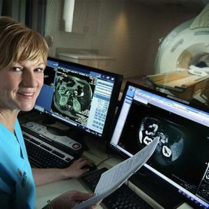
At the time of the MRI, radio waves and signals picked up by sensors are generated.Subsequently, these signals are processed by a computer program, resulting in a series of images depicting a thin section of tissues.
How is chest MRT performed
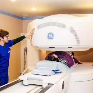 Survey can be performed on an outpatient basis or in a hospital .To do this, the patient lays down on the mobile table, and the radiologist assistant fixes his body with straps and rollers, since the reliability of the results depends directly on the ability of a person to remain stationary throughout the study.
Around the chest, devices with wires are placed, which generate radio waves.If a decision was previously made to introduce a contrast agent, at this stage a catheter is inserted into the vein, through which it enters the blood.
Then the table slides inside the device, and all the medical personnel moves to the next room where an image is directed to the computer.
Important! The start of the patient procedure is notified by the clicks and tapping that the magnet emits at the time of operation.If they bring discomfort, they will most likely be offered headphones or earplugs.To avoid unpleasant situations, the physician can maintain contact with the patient by using the intercom device.
The procedure lasts from 15 to 60 minutes, depending on the number of shots required, the age of the device, the need for additional procedures, such as angiography.
Sensations during and after the procedure
Most often, at the time of the examination, the person does not feel anything, but there are exceptions:
- A slight increase in temperature in the chest area.If it causes inconvenience, medical personnel should be informed about this.
- Pulsation or feeling cool at the time of contrast agent injection.
- Metallic taste in the mouth after administration of a contrast agent.
If no sedatives were taken by the patient before the procedure, no experts give advice on the day regimen after chest MRT.
Please note! Some people may experience nausea, local pain, itching in the mucous membranes, urticaria, and other manifestations of allergic reactions in response to the contrast agent.They should be immediately notified to the doctor, after which he will be able to provide the necessary assistance.It is because of this that lactating women are not recommended to undergo MRI, and if so it is advised to refrain from feeding the baby within 24 to 48 hours.
When the results are available
In some centers, patients can be informed of the preliminary results of MRI immediately after the procedure, but most often they take from one to several days of .This is explained by the fact that the technologist is directly conducting the procedure, and the radiologist is studying the pictures.Subsequently, he issues an opinion on the basis of which additional examinations or treatment are prescribed.
Are the consequences possible
Usually, after the examination, patients do not experience any side effects.Meanwhile, exceptions are sometimes possible, such as:
-
 exacerbation of neurological diseases, manifestation of aggression, irritability, which is due to the fact that a person for a long time lies motionless;
 - slight increase in body temperature;
- cough in people with chronic respiratory diseases;Heart rate enhancement
- .
Important! It is not recommended to carry out an MRI of the chest with the introduction of a contrast agent to people with renal insufficiency, since this can adversely affect their condition.
In view of the fact that it is difficult for physicians to predict the consequences of examination in patients with metal elements in the body, including bullets, debris, infusion pumps, defibrillators, chest MRTs under their personal responsibility. The fact is that these elements can move or cause burns under the influence of a magnet.In addition, the magnet can cause malfunctions in the above-mentioned devices.
MRI of chest organs is a modern highly effective and painless study that gives a complete picture of the state of organs, tissues and bones of this area.The main thing is to listen to the recommendations of specialists before and during its implementation, and then they will be able to get a reliable result.
Elena Sovinskaya, doctor, medical reviewer