Ultrasound of the vessels of the brain and neck: indications for conduction and diagnostic value
Modern procedure of ultrasound of the vessels of the head and neck allows you to learn complex information about the status, structure and functioning of vascular branches in the body( veins, arteries, capillaries, etc.).These branches are located behind the cavity of the skull, regular and complete nutrition of the brain depends on them, as well as the organization of outflow and blood flow to it.
A study is necessary when the patient has a number of neurological pathologies and symptoms.Also, the diagnostic procedure is prescribed for people who fall into the risk group of neurological disorders, diseases of the cardiovascular and nervous system.
The procedure is painless and does not require special preparation.We will become acquainted with it in more detail.
Table of contents:Methods of performing the procedure on the vessels of the head and neck
So, there are 3 main types of examination of vascular branches in these areas: Doppler, duplex and triplex scanning. All these methods are built on the same principle, but still have some functional differences.
Dopplerography of the vessels of the head and neck
 This method has other names - UZDG, blind doppler, etc.Inspection of the vessel is carried out in a two-dimensional plane.From it you can get a complete picture of the natural structure of the vessels and their structure, however, information about the speed and quality of the blood flow will be limited.
For the procedure, ultrasound sensors are placed on the classical zones where people have large cervical arteries and veins.In some people, the main artery is displaced from birth or in the process of trauma, so you should determine its location before fixing the device.If large veins are also located in atypical sites, their research is usually skipped.
Duplex scanning
Another name is a duplex study.This area of ultrasound diagnosis provides a complete picture of blood circulation in the veins and arteries.The monitor of the device displays the pictures of the tissues of the area under investigation( head, neck), and the vessels are clearly visible on their background.
Triplex scanning
The method is based on a duplex, but provides information in a more convenient form.The speed of blood circulation is noted in various shades.For example, the red color indicates the blood flow velocity towards the ultrasound device, and the blue gamma is responsible for the outflow of blood from it.
In what cases is the ultrasound of the vessels of the head and cervical department
given Let's get acquainted, what people are planned to be surveyed at least once a year.
Risk group for ultrasound of vascular diagnostics is patients:
- with risk of brain stroke;
- at the age of 40-45 years;
- men with a history of diabetes mellitus;
- with high cholesterol, triglycerides;
- with a low level of lipoprotein density, as evidenced by the results of the lipidogram;
- smokers who are diagnosed with heart disease and vascular malformations;
- with cervical osteochondrosis;
- with hypertension, at which there is a regular arrhythmia.
It's important to know! Planned vascular diagnostics are prescribed before operative intervention on vessels and heart.This will allow the surgeon to make sure that the artificial supply of blood does not damage the brain.
Indications for the event may be subjective complaints of the patient, which may indicate a vascular pathology:
-
 dizziness, vertigo, headaches;
 - uncoordinated movements during walking;
- noises and ringing in the ears, other hearing pathologies;
- suddenly appeared problems with eyesight, points before the eyes, the appearance of "blind zones", etc.;
- disorders of healthy sleep: anxious sleep, sharp cries, severe awakening, insomnia, etc.;
- memory impairment, a drop in concentration and attention;
- diffuse;
- loss of strength and general weakness;
- violation of muscular activity of the jaw and face and body, sensitivity;
- MRI or CT confirmed the pathology of the brain substance;
- after a previous myocardial infarction, stroke.
How to prepare for the procedure for the examination of the cervical and head vascular bed
No special preparations are required for the event.
Prior to the procedure, consult a therapist and a neurologist about the general condition of the body, establish an indication for ultrasound.Also with them it should be determined whether it is necessary to cancel preparations for blood vessels and heart, which are taken by the course.
Please note! On the day of the session, you should avoid drinking beverages that expand the blood vessels: coffee and coffee drinks, black and strong tea, alcohol, energy, drinks with ginger, guarana or ginseng, etc.
4-5 hours before the event, it is recommended to stop eating, since after meals the blood flow in the head decreases, which can distort the picture.Infants and preschoolers on the contrary better feed an hour before the procedure.It is desirable to organize a child's sleep during the study.
You should stop smoking 2 hours before the scheduled time.
Immediately before the session, the patient takes off his outer clothing and all ornaments( earrings, chains, hoops, etc.).The whole area of the head and neck should be freed to attach the sensor.Long hair should also be picked up in a bun or tail.
Stages of ultrasound examination of vessels
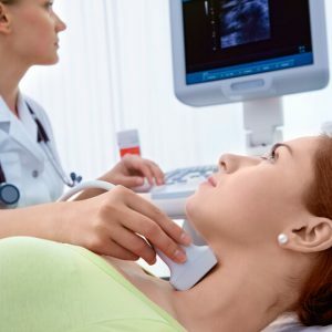
After releasing the study areas from clothing, jewelry and hair, the patient lies on a medical table or a couch( lying on his back).The head is located in the area of the ultrasound unit.
 A specialist sonologist begins the study from the field of carotid arteries.He directs the patient's head away from himself to ensure full access to the cervical region.With the help of sensors and the device, the doctor examines the lower part of the carotid artery( the position of the sensor is cut downwards).Further, the sonologist advances up the cervical region, setting the device over the area of the lower jaw.
This will determine the direction of the vessel, its depth and the place where it diverges on the branches of the carotid arteries.Then, using a special color regime, the doctor fully examines the main artery and separately each branch.Color helps identify areas where blood circulation is difficult, and also see vessels with a pathological structure of the walls.
In the case of detection of defects and pathologies, the doctor conducts an additional examination to determine the type of disease, the degree of damage to the vessels, possible progression of the disease, etc.
After examining the main artery, the left and right are also examined.
If necessary, the sonologist also examines the vertebral arteries by placing an ultrasound probe along the neck.The vessels are located between the cervical vertebrae and near them.
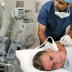
During transcranial examination( head area), the ultrasound probe is placed on the scalp, first applying a special gel to the zone of the temple, the occiput, and above the orbits.
Through the ophthalmic region, the sonologist determines the nature of blood circulation in the arteries of the ophthalmic and suprapubic zones.Here you can determine the pathology of its influx directly into the brain, which can be caused by blockage of arteries inside it.
Ultrasound on the temporal bone determines the structure of the vessels at the base of the brain.These include the artery( anterior, middle and posterior), the veins of Galen and Rosenthal, a straight sinus.
Through the occipital region, the sonologist determines the state of the vessels inside the cranial cavity( the main and vertebral arteries, the Galen vein, the direct sinus, etc.).
What is the purpose of the study, deciphering the results of
 Doppler ultrasound and other ultrasound methods allow the doctor to understand how a vessel is formed, to know the features of the structure of the arteries and veins, to see the depth of their extent, the degree of branching, etc.Information about the circulation of blood in the body, such as speed, strength, volume, etc., also becomes available.The procedure also allows to establish whether there are obstructions to the blood flow, such as emboli, thrombi, plaques( cholesterol, atherosclerotic).It is also possible to identify inflammation or lesion of the walls of blood vessels, determine the primary symptoms of their pathology, aneurysm of the arteries.Investigated spasms of the walls of the vessels allow us to determine their tone and reserve possibilities for restoring blood flow.
Having received data from the monitor, the neurologist determines the type and degree of pathology, the features of progression, the correspondence of the results with the subjective symptoms of the patient.On the basis of information, it can be concluded that the disease progresses further, the possibility of its complete cure, possible complications and consequences.
In order to decipher the results, investigate such indicators: the features and speed of blood circulation at different periods of heart contractions, the thickness of the walls of arteries and veins, indices( pulsatory and resistance).The results also encoded data on the structure of each vessel and the presence of formations in them, their structure.
If the echogenicity of the artery is unstable, the vascular walls are densified, and the stenosis is less than 20%, one can speak of non-stenosing atherosclerosis.
When stenosing the form of ultrasound should show the presence of special atherosclerotic plaques.Also, data show whether these plaques cause embolism, which can soon lead to a stroke.
Deformation and compaction of diffuse vascular walls can speak of vasculitis.This is also evidenced by the pathology of separation of the layers of the vessel.
If ultrasound showed the presence of an unknown vasculature or fistula between the venous and arterial bed, then we can talk about arteriovenous malformation.
If a diabetic has signs of macro- and miangiopathy, a process of decompensating the disease can be established.
As we see, ultrasound of the vessels of the head and neck is necessary for the timely diagnosis of the most complicated and life-threatening diseases and pathologies.The procedure is absolutely harmless and is available in every modern clinic, so do not delay planned inspection for years.
Chumachenko Olga, medical reviewer