Heart diseases: classification, diagnosis
 Heart defects are pathological changes in which there are congenital or acquired defects in heart valves, aorta, pulmonary trunk, interatrial and interventricular septum.These changes cause disruption of the normal functioning of the heart, which leads to an increase in chronic heart failure and oxygen starvation of body tissues.
The incidence of heart disease is about 25% of the number of the rest of the heart pathology.Some authors( d. Romberg) give personal data with higher values - 30%.
Table of contents: What are heart defects, classification Why do heart defects arise?Video "Heart defects":
What are heart defects, the classification of
Among the many classifications of malformations, the following distinguish:
- - the main cause - rheumatism, syphilis, atherosclerosis;
- congenital - an unambiguous answer to the question of the causes of their occurrence is not, the problem is still under study today.Most scientists agree that the pathological process triggers changes in the human genome.
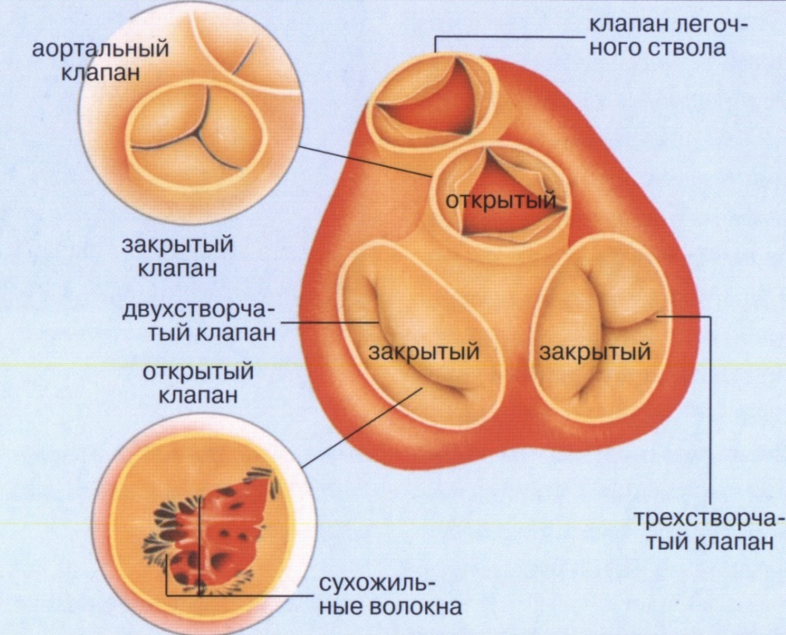
Defects affecting the valves:
- bivalve( mitral);
- tricuspid( tricuspid);
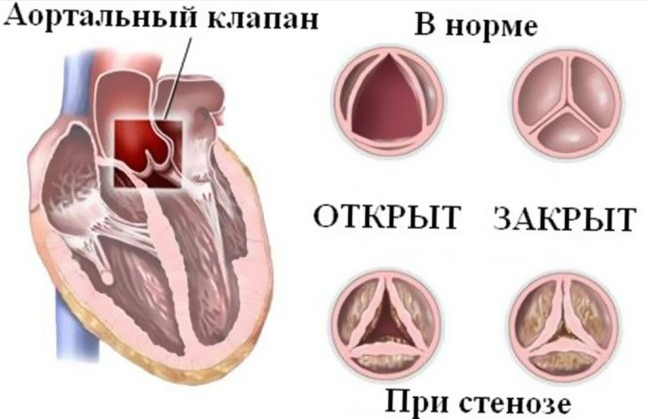
- aortic;
- of the pulmonary trunk.
Defects of partitions:
- interventricular;
- interatrial.
By the type of lesion of the valvular apparatus, heart defects can occur in the form:
- deficiency( incomplete closure of valves);
- stenosis( narrowing of the holes through which the blood passes).
Depending on the degree of existing chronic circulatory failure, the following may appear:
- compensated defects( the patient is able to live, study and work, but with limitations);
- decompensated pathology( the patient is severely restricted in the ability to move).
The form of gravity provides for defects:
- light;
- average;
- heavy.
By the number of defects formed, the defects are:
- simple( with an existing single process);
- complex( a combination of two or more defects, for example simultaneous presence of insufficiency and narrowing of the hole)
- combined( a problem in several anatomical formations).
Important: , some physicians in their practice have drawn attention to the fact that men and women have their own peculiarities of the course of painful processes.
For women( girls) are more common:
- non-penetration of the botulinum duct.As a result of the pathological process, a relatively free communication between the aorta and the pulmonary trunk is formed.As a rule, this non-spreading exists normally before the birth of the child, then it closes;
- defect of the septum between the atria( there remains a hole allowing blood to flow from one chamber to another);
- defect of the septum designed to divide the ventricles, and non-proliferation of the aortic( botallic) duct;
- triad Fallot - a pathological change in the septum between the atria, combined with a narrowing of the hole of the pulmonary trunk and augmented( hypertrophic) enlargement of the right ventricle.

In men( boys), the following is usually found:
- aortic aperture narrowing( aortic stenosis) in the area of the aortic valve flaps;
- defects in pulmonary vein connection;
- narrowing of the aortic isthmus( coarctation), with an available open botulinum duct;
- atypical location of the main( main) vessels, the so-called transposition.
Some types of vices occur with the same frequency, both in men and in women.
Congenital malformations can develop early in utero( simple) and late( complex).
In the formation of fetal pathologies at the beginning of a woman's pregnancy, there is a defect between the aorta and the artery of the lung, the non-opening of the existing aperture between the two atria, and the formation of a narrowing( stenosis) of the pulmonary trunk.
In the second, the atrioventricular septum may remain open, and a tricuspid valve defect with its deformation, complete absence, atypical attachment of the valves, and the "Ebstein anomaly" also occur.
Note: very important classification criterion is the division of defects into "white" and "blue".
The white defects of are pathologies with a more tranquil course of the disease and a rather favorable prognosis. With them, venous and arterial blood flow through their bed, not mixing and not causing hypoxia of tissues under sufficiently measured loads.The name "white" is given by the appearance of the skin of patients - characteristic pallor.
Among them are:
- defects with stagnation of oxygenated blood in a small circle of blood circulation.Pathology occurs in the presence of an open arterial duct, a defect of the interventricular or interatrial septum( enrichment of the small circle of blood circulation);
- defects with insufficient inflow of blood into the lung tissue( depletion of the small circle of circulation), caused by constriction( stenosis) of the pulmonary artery( trunk);
- defects with a decrease in arterial blood supply, causing oxygen starvation of the human body( depletion of the great circle of blood circulation).This defect is typical for the constriction( stenosis) of the aorta at the site of the valve, also for narrowing the aorta( coarctation) at the site of the isthmus;
- defects without dynamic circulatory disorders.This group includes pathologies with an atypical location of the heart: on the right( dextrocardia), on the left( sinistrocardia), in the middle, in the cervical region, in the pleural cavity, in the abdominal cavity.
The blue defects of occur with the mixing of venous and arterial blood, which leads to hypoxia even at rest, they are characteristic of more complex pathologies.Patients with cyanotic skin color.With these painful conditions, the arterial blood is mixed with venous, which leads to a lack of oxygen supply to tissues( hypoxia).
To this type of painful processes are:
- defects with delayed blood in the lung tissue( enrichment of the small circle of blood circulation).Transposition of the aorta, pulmonary trunk;
- defects with insufficient blood flow to the lung tissue( impoverishment of the small circle of circulation).One of the most severe cardiac malformations of this group is the tetralogy of Fallot, characterized by the presence of a narrowing of the pulmonary artery( stenosis) to which the defect of the septum between the ventricles and the right( dextransposition) aortic position, combined with an increase in the size of the right ventricle( hypertrophy), are attached.
Why heart defects develop
The causes of pathology have been studied for a long time and are well monitored in each case.
Reasons for the appearance of acquired defects
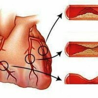
Occur in 90% of cases due to rheumatic fever, which complicates the structure of the valves, causing their damage and the development of the disease.For a long time already doctors who treated this disease had a saying: "rheumatism licks joints and gnaws at the heart".
Also acquired defects can cause:
- atherosclerotic processes( after 60 years);
- untreated syphilis( to 50-60 years);
- septic processes;
- trauma of the chest;Benign and malignant neoplasms
- .
Note: most often valve acquired defects occur before the age of 30 years.
Causes of congenital malformations
Factors that cause the development of congenital malformations are:
- genetic causes.Hereditary predisposition to the disease was noted.Breaking in the genome or chromosomal mutations cause a violation of the proper development of heart structures in the intrauterine period;
- is harmful to the environment.The effect of ionizing rays on a pregnant woman, poisons of cigarette smoke( benzpyrene), nitrates contained in fruits and vegetables, alcoholic beverages, medicines( antibiotics, anti-tumor drugs);
- Disease: measles rubella, diabetes mellitus, impaired amino acid metabolism - phenylketonuria, lupus.
These factors can cause problems in the heart of a developing child.
What happens to the heart and blood circulation with acquired defects
Acquired malformations develop slowly.The heart includes compensatory mechanisms and tries to adapt to pathological changes.At the beginning of the process, cardiac muscle hypertrophy begins, the chamber cavity increases in size, but then decompensation is slowly formed and the muscle becomes flabby, losing the ability of the "pump" function.
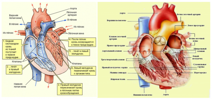
Normally, blood during the contraction of the heart is "pushed" from one chamber to another through the hole with the valve.Immediately after passage of a bloody portion, the valve flaps normally close.When the valve is insufficient, a certain gap is formed through which the blood is partially thrown back, where it merges with the new "portion" that has come up.There is stagnation and compensatory expansion of the chamber.
When the hole is narrowed, the blood can not pass in full, and the rest of it complements the incoming "portion."Just like in case of insufficiency, with stenosis stagnation and stretching of the chamber occur.Over time, the compensatory mechanisms are weakened, and chronic heart failure is formed.
Acquired heart diseases include:
- mitral valve insufficiency - due to development of scar processes after rheumatic endocarditis;
- mitral stenosis( constriction of the left atrioventricular orifice) - fusion of valve flaps and a narrowing of the opening between the atrium and the ventricle;
- aortic valve failure - incomplete closure during relaxation( diastole);
- narrowing of the aortic orifice - blood at the time of contraction of the left ventricle can not all go to the aorta and accumulates in it;
- Insufficiency of the tricuspid( tricuspid) valve - the blood during the contraction of the right ventricle is thrown back into the right atrium;
- stenosis of the right atrioventricular orifice - blood from the right atrium can not exit all into the right ventricle and accumulates in the atrial cavity;
- Pulmonary valve insufficiency - blood during the contraction of the right ventricle is thrown back into the pulmonary artery, causing an increase in pressure in it.
Video "Mitral stenosis" :
What happens to the heart with congenital malformations
 The exact cause of congenital malformation is unclear.In some cases, the development of these pathologies contributes to some of the infectious diseases that the future mother is having.Most often - measles rubella, which has a teratogenic( damaging fruit) effect.Less commonly - influenza, syphilis and hepatitis.Influence of radiation and malnutrition was also noted.
Sick children without surgery for a number of vices are killed.The earlier the treatment, the better the prognosis.There are many kinds of congenital heart diseases.Often observed and associated defects.Consider the main, common diseases.
Congenital heart defects can be:
- defect( non-healing) of the interventricular septum is the most common type of pathology.Through the available hole, the blood from the left ventricle gets to the right and causes an increase in pressure in the small circle of the circulation;
- defect( non-healing) of the interatrial septum is also a frequently observed type of disease, it is more often observed in women.Causes an increase in the amount of blood and increases blood pressure in a small circle of blood circulation;
- open arterial( botalla) duct - non-proliferation of the duct connecting the aorta and the pulmonary artery, which leads to the discharge of arterial blood into the small circle of the circulation;
- coarctation of the aorta - narrowing of the isthmus with an open arterial( botallic) duct.
General principles for the diagnosis of heart defects
Determining the presence of a defect is an understandable procedure, but requires special attention from the doctor.
 To make the diagnosis it is necessary to conduct:
- a thorough interview of the patient;
- examination for "cardiac" symptoms "
- listening( auscultation of the heart) to detect specific noises;
- percussion to determine the boundaries of the heart and its shape.
This is usually enough to find defects caused by the disease.
But the examination is necessarily supplemented:
- with laboratory diagnostic data;
- radiography and ultrasound of the heart;
- electrocardiography;
- by other methods if necessary( angiography, dopplerometry).
A timely examination of a pregnant woman in many cases helps to determine the presence of congenital heart disease even in the early stages of fetal development.
Stepanenko Vladimir, Surgeon