Hypochromic anemia
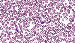 Hypochromic anemia is one of the common names for all forms of anemia that are characterized by a deficiency of hemoglobin, where the quantitative color index in the blood is less than 0.8.In general, hypochromic anemia disrupts the formation of hemoglobin due to the reduction of red blood cells, even in small amounts. This is due to iron deficiency, hereditary pathologies in the formation of hemoglobin and chronic lead poisoning.
Among hypochromic anemia, iron deficiency anemia, thalassemia, a pathological change in the synthesis of organic compounds( porphyrins) and anemia that occurs in certain chronic diseases are recognized.
Hypochromic Anemia Causes
Lack of iron is the main and frequent cause of hypochromic anemia. About 10% of women who are of childbearing age suffer from iron deficiency hypochromic anemia, and 20% have a latent form of iron deficiency.
Blood loss also relates to the causes of anemia. In one milliliter of blood contains about 0.45 mg of iron, so during the period of menstruation a woman loses almost 30 mg on average. In the body, with improper iron production simultaneously with food, even during an insignificant menstrual cycle, its balance can be disturbed and give rise to the appearance of iron deficiency anemia hypochromic. Also, various kinds of gynecological diseases can cause such anemia. But in the postmenopausal period for many women, as well as men, the first place among the causes of iron deficiency are gastrointestinal blood loss as a result of various types of tumors, ulcers, hemorrhoids of the esophagus, diverticulosis, intestinal polyposis.
In addition, the first signs of cancer of the colon and large intestine is precisely the lack of iron in the body. Therefore, in the presence of iron deficiency anemia, all patients after forty years are subject to mandatory examination for the definition of tumors with this localization. Sometimes there is some connection between iron deficiency anemia hypochromic and Crohn's disease, as well as ulcerative colitis of nonspecific etiology and reception, for example, aspirin and prednisolone. In addition, the causes of these anemia are donation, nosebleeds, hemorrhagic vasculitis, frequent blood tests for various types of research.
The state of iron deficiency anemia very often appears in pregnant women as a result of increased iron intake, which is necessary for the development of the placenta and fetus.
Hypochromic anemia often develops during puberty, when there is insufficient iron in the body, compensated only by its inferior intake. Almentary factor is considered a rare cause of iron deficiency anemia. And in the emergence of thalassemia, as one of the forms of hypochromic anemia, is a reduced synthesis in the beta-polypeptide chain, which are formed by two pairs of genes.
A heterozygous form with one genetic defect usually occurs without any clinical manifestations. But the same form in which pathological changes occur in the second and fourth gene, is subject to the development of microcytic hypochromic anemia with mild or moderate severity. Basically, such patients are carriers of this disease. The development of severe anemia is promoted by the homozygous form of the disease, in which an anomaly occurs in three of the four genes that damage the beta-thalassemia chain. Almost 25% of this form of anemia occurs in blacks.
Hypochromic anemia symptoms
The clinical picture of hypochromic anemia is characterized by increased fatigue, decreased appetite, inability to perform some physical activity, dizziness.
For iron deficiency anemia, tissue sideropenia is also characteristic and muscle weakness is well expressed, which is caused by a reduced level of enzymes present in the muscles. As a result, the nails become brittle, and the skin is dry, hair often falls out and angular cheilium appears. In many patients, the taste changes, which is marked by the desire to eat toothpaste, clay, chalk. Tissue deficiency of iron is expressed by spoon-like nails, dysphagia appears, incontinence during coughing or laughing, secretion in the stomach is disturbed.
In hypochromic anemia, the concentration of hemoglobin decreases more than in erythrocytes, and therefore the color index decreases. Microcytosis is often noted, but anisocytosis and poikilocytosis sometimes occur. In the bone marrow, an increase in the percentage of cells found in the erythroblastic series with a certain delay in maturation is detected. This form of hypochromic anemia is diagnosed without special difficulties on the basis of hematological and clinical indicators.
Symptomatic of hypochromic anemia in tumors is manifested as anemia of unclear etiology. Patients very often complain only of weakness, and some patients in the anamnesis have even expressed effect from therapy with application of iron-containing preparations. And only X-ray examination, as well as blood tests, reveal the complete picture of the disease. But, unfortunately, sometimes a doctor calms down with negative radiographic findings, and therefore misses tumors in the early stages.
Sometimes, with erosive gastritis, there is severe blood loss, which is not compensated by iron-containing drugs. In addition, the hernia in the food hole of the diaphragm, flowing without pain symptoms and disorders on the part of the gastrointestinal tract, but characterized by all signs of hypochromic iron deficiency anemia, can intensify and become intolerable. In other cases, patients complain of an overcrowded feeling behind the chest after eating, pain in the heart, like with angina pectoris, vomiting.
But Crohn's disease should become a signal of moderate hypochromic anemia, which is characterized by vague inflammation, diarrhea and pain in the abdomen, fever, and blood loss from the digestive tract.
Chronic hypochromic anemia is detected with ankylostolidosis, which can last quite a long time. At this point, pain in the epigastric region, nausea, vomiting and diarrhea are characteristic. Also, the blood from the intestine continues to leave, so there is a loss of both iron and protein, leading to hypoproteinemia.
Hypochromic anemia in children
In children, as in adults, there are two main forms of hypochromic anemia - iron deficiency and latency. The latter form is characterized by an isolated iron deficiency in tissues without anemia. This iron deficiency anemia is very common among young children. This disease mainly develops due to a lack of iron during multiple pregnancy or prematurity, and also if the child refuses to eat.
Iron deficiency itself causes a number of disorders in the digestive tract, which exacerbate this deficiency. A huge role in this imbalance is the nutrition of the child. Infants and young children lag behind many peers in both speech development and psychomotor development. But, starting from two to three years, children are observed relative compensation, in which the amount of hemoglobin increases to normal, but the latent lack of iron can be present.
Conditions for the appearance of hypochromic anemia in children is the period of puberty, especially for girls. This period is characterized by the need for iron in increased quantities as a result of the appearance of menstruation and increased growth of the body. Very often at this moment, reduced immunity and poor nutrition, which can be associated with losing weight.
hormones play a role. So, for example, androgens help the process of erythropoiesis and actively utilize iron, but estrogens practically do not manifest themselves. With increasing iron deficiency anemia, lethargy, irritability and apathy increase. Children develop complaints of dizziness and frequent headaches, which lead to memory impairment. Also hears strong dyspnoea and muffled heart sounds. On the electrocardiogram, all signs of changes in the walls of the myocardium are hypoxic and dystrophic. The limbs of the child are always cold to the touch. Many children with moderate and severe hypochromic anemia experience an increase in the liver and spleen, especially if there is a deficiency of vitamins and protein, as well as active rickets in infants. There is a decrease in the secretion of gastric juice, impaired absorption of trace elements, vitamins and amino acids, as well as reduced immunity and nonspecific protective factors.
Hypochromic Anemia Treatment
Substitution treatment with iron preparations is considered a fundamental method in the treatment of patients with iron deficiency anemia. To do this, use Lactate, Sulfate or Iron Carbonate. More commonly, iron sulphate is prescribed 300 mg three times a day. Very often for the treatment of hypochromic anemia, drugs such as Ferroplex, Feromide, Ferrogradum, called combined are also used.
All saline iron preparations strongly irritate the mucous surface of the stomach and intestines, so almost five percent of patients have adverse reactions of the body in the form of nausea, vomiting and diarrhea. This discomfort must be removed by reducing the dose of the drug, reducing the duration of treatment, and sometimes even canceling the drug, which results in poor indicators in the treatment. Some patients try to eat foods that contain iron, but it still can not replace iron-containing drugs, especially cure hypochromic anemia. Therefore, on average, iron therapy to achieve the desired result should be about six months. This is very important for replenishing the missing amount of iron in the body, with its subsequent stock in it.
For some clinical indications, intravenously prescribed iron-containing drugs, and with severe form of hypochromic anemia, infusion of erythrocyte mass is used. But the most important in the treatment of this disease is, first of all, the elimination of the factors of its development. This applies to the therapy of the underlying disease that caused the onset of this pathology, and then a specific treatment is prescribed. It always depends on the form of hypochromic anemia.
For the treatment of homozygous thalassemia hypochromic, transfusions of erythrocytes are used from an early age. Basically, thawed types of erythrocytes are used. Initially, a course of shock therapy is performed, which includes up to ten transfusions for two or three weeks, and an increase in hemoglobin reaches 120-140 g / l. After that, the appointment of infusions is reduced. This therapy of hypochromic anemia with the use of transfusions improves not only the general condition of the patient, but also reduces significant changes in the skeleton, the size of the spleen, reduces the incidence of infections in these children in severe forms, and improves their physical development. Such treatment prolongs patients life for a long time. But sometimes, after applying this therapy, there are some complications in the form of pyrogenic reactions, increased hemolysis and seizures. In addition, transfusion therapy can cause hemosiderosis of many organs. Therefore, when treating this form of anemia, Desferal is prescribed. It timely removes from the body an excess amount of iron. The dose of this drug depends on the age of the patient and the transfused amount of red blood cells. Young children are injected intramuscularly at 10 mg / kg, adolescents - 500 mg per day. Desferal is also recommended for use with ascorbic acid of 200-500 mg, which enhances its effect.
In some cases, when the spleen is palpated, significantly enlarged in size, and thrombocytopenia and leukopenia are associated with all the symptoms of the main anemia, a splenectomy is indicated.
Treatment of thalassemia homozygous etiology, as a form of hypochromic anemia, does not give serious remissions, but significantly improves the life of patients. When treating heterozygous thalassemia, it is important to remember that all iron preparations are highly contraindicated. Since this form of hypochromic anemia always has an excessive content in the body of iron. Therefore, in patients who will take iron-containing drugs, the condition will be extremely worsened, unlike those patients who will not receive them. And this as a result can cause severe decompensation and death of patients from various manifestations of hemosiderosis.