Echocardiography of the heart with Doppler analysis and other methods and norms
Content:
- Advantages and disadvantages
- Doppler analysis
- Preparation and implementation
- Decoding the results
Echocardiography (EchoCG) is a method of examining the heart and is based on an ultrasound scan of the breast.
Thanks to this method, a study of various diseases of the body is carried out.
It helps to assess the diameter of the heart and its substructures, the width of the myocardium of the ventricles, atria.
The procedure reveals the mass of the heart, as well as many other parameters.
Echocardiography doctors and ordinary people sometimes can give another name - this is ultrasound.
Therefore, when asked how the ECHO of the heart differs from Ultrasound of the heart, doctors say - nothing.
This is the same procedure.
The doctor may send for echocardiography if:
- the client has a murmur in the region of the heart;
- people with ECG abnormalities;
- a person notices failures in the functioning of the heart;
- increased body temperature, signs of SARS;
- during the diagnosis, deformation of the heart muscle was found, as well as an increase in the mass of the organ.
Echocardiography can be used in the following cases:
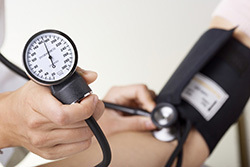
- Patients with very high blood pressure.
- Patients with heart defects in the family tree.
- If there are signs of pain in the left chest area.
- If shortness of breath or swelling of the limbs is present.
- The patient has fainting.
- The patient has dizziness.
- Found signs of tumor formation in the heart.
- After suffering a heart attack and other diseases.
Echocardiography is the safest and most unique way to detect heart problems and defects. It can be carried out to people of different age categories (children and adults), it can even be prescribed to pregnant mothers. It is prescribed to determine fetal anomalies and correct the situation as soon as possible so that the baby remains safe and sound. Echocardiography is safe for the baby and his mother.
Such a procedure is necessary for pregnant women in such cases:
- If a pregnant woman has had heart defects in blood relatives.
- There have been cases of miscarriages.
- If a pregnant woman has diabetes mellitus.
- If rubella has been transferred during pregnancy.
- If in the first or second trimester, the expectant mother used antibiotics or other drugs.
Cardiac echocardiography - advantages and disadvantages
ECG Is an electrocardiography, and echocardiography is an echocardiography of the heart.
What is the difference between them, and is it there at all:
- An echogram of the heart works thanks to special devices - a transducer (the doctor puts it on the client's chest in the region of the heart). The machine reads the ultrasonic waves that pass through the walls of the body and displays them back. The converter receives the necessary pulses, after which they are processed by the computer.
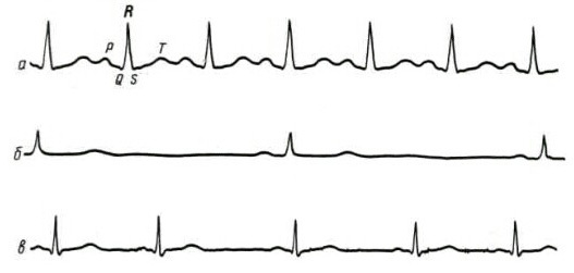
- Electrocardiography works in a different way: sensors are attached to the client's chest cavity. They determine the electrical activity of the heart. The sensors are connected to a special machine, which after measurement shows a special graph of the strength and nature of the electrical signals.
 Thanks to echocardiography of the heart, doctors determine the degree of its work and the work of the whole organism as a whole. It can also reveal how weak the heart is. Electrocardiography can only measure the level of signaling, and whether there are active electrical impulses that are sent by the "motors" of the body. Results are always shown as a graph.
The results of the echogram of the heart are recorded in photographs. Thanks to the electrocardiogram, it is possible to determine heart rhythm defects, to identify arrhythmias, tachycardia and bradycardia. An ultrasound examination will help determine how well the heart is functioning, the state of its valves, whether there are blood clots in the body, or deformation of the heart.
There are also certain similarities between echocardiography and electrocardiography:
- Both diagnostic methods will help to assess the diameter of the heart chambers. An example is the detection of an increase in the diameter of the atrium.
- Both methods will help to identify pathology in the location of the "engine" of the body.
- Swelling of the heart, thanks to echocardiography of the heart, electrocardiography can also detect inflammation of the tissues that surround the heart muscle.
ECG of the two options is the most affordable method. But, unfortunately, he is not always able to identify and show the exact picture of the problem. And the echogram of the heart, on the contrary, will help to reveal a clearer picture. Echocardiography of the heart can reveal precise structural abnormalities and provide accurate images.
The method is more reliable in identifying the state of heart health. One of the big advantages of an ultrasound of the heart muscle is the doctor's ability to monitor the chambers of the heart. One of the significant disadvantages is that this method can only be used in private medical institutions, and it is much more expensive than ECG.
Doppler echocardiography and other ultrasound techniques
Doppler echocardiography and other diagnostic methods reveal a large number of problems.
For example:
- Heart failure.
- Skolsky-Buyo disease.
- Heterogeneous cardiac tumors.
- Vegeto-vascular dystonia.
- Inflammation of the heart muscle.
- Forms of ischemic disease.
- Arterial hypertension.
- High blood pressure.
- Congenital or acquired heart defects.
- Blockage of blood vessels.
There are several ways to perform ECHO.
Examination of the heart is done by the following methods:
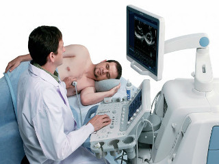
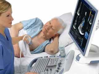
- Transthoracic echocardiography Is the most well-known method, as it has been used for many years. This method of finding heart problems is performed over the chest using a special sensor. It is pressed against the body of the examined person in the region of the heart. During the session, the patient's body should be on the side or on the back.
- Transeal echocardiography. This method uses an ultrasound scan of the heart. This method is carried out using a special sensor through the esophagus. The sensor is installed there for the best approximation to the patient's heart, and this helps to see its parts. With a conventional ultrasound, it is impossible to observe the parts of the heart.
 There are also different modes of checking the heart.
Movable M-mode. With echocardiography in M mode, the transducer delivers waves in one selectable axis. During the study, the heart is displayed and the data is shown on the monitor screen. The heart can be viewed from above in real time.
If you change the directions of ultrasound, it will be possible to check the stomachs, the vessel that comes out of the left stomach and supplies oxygenated blood to all organs of the patient, as well as the two upper sections hearts. This study of heart functions can be used for both a child and a person of age, because the procedure is absolutely safe for health.
Doppler echocardiography. Doppler echocardiography is performed to determine the speed at which the blood is moving and the turbulence in the blood flow. The informational data obtained during the conduction carries a message about heart defects and filling of the left stomach.
Calculation of the object's speed and its change, as well as the difference in signal frequencies are the basis of this test method. The frequency changes when red blood cells collide with sound. Doppler shift denotes the amount of this change. This signal can be perceived by the human ear and it can be reproduced with the help of an echo apparatus in the form of sound that we can hear.
B-method. Using a 2D echocardiogram, treating physicians obtain images in two fields of view. When conducting a wave in the form of ultrasound, the frequency of which is 30 times per 1 second, it is conducted along an arc of 90 degrees, that is, the scanner field is normal in relation to the four-chambered heart and its position. You can change the direction of the sensor using a picture that is displayed on the screen and with its help you need to analyze the structure of the heart.
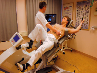
Stress echocardiography. It is used to study the work of the patient's heart with the use of physical activity during this method, according to these indications, carry out and produce the necessary load with doses that are determined in advance.
Using pharmacological drugs, they cause agitated heart work. They immediately investigate the changes that occur with the muscles in the heart during a stress test. In the absence of a decrease in blood supply, the minimum percentage of risk for cardiovascular complications is often indicated.
The procedure can be biased in the nature of the assessment of use, echocardiography programs display images on the screen that are recorded at different stages of examinations. Such an analysis of the work of the heart at rest and under stress provides a comparison of the readings.
This method of observation allows you to find hidden malfunctions of the heart, which are invisible at rest. Most often, the examination process takes about 45 minutes; for each client, separate loads are selected, which depend on age and health.
To prepare the patient for such a study, it is necessary to perform such actions as:
- Wear loose-fitting clothing that will not hinder movement.
- Stop exercising and eating large amounts of food 3 hours before the stress echo.
- Then, 2 hours before the study, drink a glass of water and have a small snack.
Cardiac echocardiography is absolutely safe. Problems can appear only in connection with the peculiarities of the patient's anatomy, they may be combined with the inability to leak ultrasound using the transesophageal technique. Destruction of breast cells, overweight, bulky breasts in women or large hair cover in men can become a hindrance.
In what situations is it not allowed to carry out an ultrasound of the heart:
- If a person has stomach diseases such as stomach ulcers and gastritis.
- If the patient has a tumor of any degree. Transesophageal ultrasound of the heart in these cases is not allowed. In this case, transthoracic echocardiography can be done.
The cost of this treatment in private hospitals varies from 2,300 to 3,000 rubles. The price will also depend on the qualifications of the doctor, the current equipment, the location of the clinic building that provides these services, as well as the honor of the clinic.
Not every state polyclinic can offer this method of heart examination. In Moscow, you can undergo echocardiography, and it will be more expensive in comparison with Voronezh. When comparing prices for ultrasound and ECG examinations, in the case of an ECG, the patient will have to pay about 700 rubles. Often, an electrocardiogram in state-type clinics is carried out completely free of charge.
Echocardiogram of the heart - preparation and conduct
 An echocardiogram of the heart is performed on an outpatient basis. Before the examination, observing an interval of several hours, the patient should refrain from consuming any food or even water.
In addition, the day before the study, it is prohibited to abuse caffeine and products containing it, as well as to stop taking medications containing nitroglycerin. If a denture is available, all dentures must be removed prior to examination.
Transesophageal echocardiography of the heart is performed as follows:
- The patient is placed on a medical couch, he takes a comfortable position. A catheter is then inserted to give general or local anesthesia. The patient's throat is washed with a special anesthetic composition so that irritation does not occur.
- Further, special equipment is connected to monitor the work of the cardiac system and the respiratory system. When using general anesthesia, the human body is also provided with oxygen, artificially supplied by a special device.
- The examined person is turned to the left side, a special mouthpiece is placed into the oral cavity, and then an endoscope is inserted. When using general anesthesia, the endoscope is inserted into the person's throat through the esophagus and advanced further. A special sensor at the end of the device projects everything seen on the computer screen. The heart muscle is examined by a specialist from various angles.
A transthoracic echocardiogram of the heart does not provide for any planned preparation steps.
In the office, the patient needs to take off his clothes from the upper body, fit on the couch. Then a gel is applied to the left side of the patient's chest so that the ultrasound waves are more correctly transmitted to the monitor. Then the doctor installs the sensors and notes the displayed data.
After the study, all data is analyzed, and a written opinion is drawn up, which is given to the patient. With the obtained results of echocardiography of the heart, the patient needs to contact a cardiologist to clarify the diagnosis and prescribe the correct treatment.
Norms of echocardiography of the heart for decoding the results
The norms of echocardiography of the heart are compared with the results of the study, and the cardiologist explains the formulation of an accurate diagnosis.
In order not to wind yourself up once again, below you can familiarize yourself with the parameters that are considered the norm when conducting an EchoCG:
- The cavity of the pancreas at the end of the relaxation phase is from 0.9 to 2.6 (average value is 1.7).
- The LV cavity in the final relaxation - from 3.5 to 5.7 (mean value - 4.7).
- The diameter of the aortic orifice is from 2.0 to 3.7 (mean value 2.7).
- The plane of the left atrium is from 1.9 to 4 (mean value is 2.9).
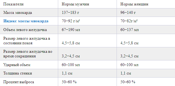 Above are the main parameters that doctors pay close attention to when making a diagnosis.
In a normal state, the wall thickness of the pancreas should be 5 mm, and its dimensions at rest should be
Only a professional cardiologist is able to correctly decipher and explain the results of the study to the patient, based on the norms of cardiac echocardiography. Independent reading and decoding of the results, although there is a place to be, will definitely not give a complete picture of the diagnosis to a person, and even more so it is impossible to independently prescribe the necessary treatment.
You can only familiarize yourself with the average values to calm your own nerves before consulting a specialist. Only a qualified doctor with a certain work experience can correctly read the results research and answer all the questions that have arisen, as well as prescribe the correct treatment of the arisen diseases.
But don't worry in advance if the average values don't match. There is a possibility that the equipment on which the study is carried out records indicators opposite to other points. This only indicates that the equipment is rather outdated.
Modern devices provide the most accurate indicators that guarantee the patient receiving the necessary treatment in accordance with the diagnosis and various recommendations.
Once again, I would like to note that independently decoded results can lead to an incorrect diagnosis. The correct result can only be obtained by consulting an experienced cardiologist, who will also prescribe the appropriate treatment.
Partial use of site materials (no more than 30% of the content of the article) is allowed only with a hyperlink on www.med88.ru, full use of the article (more than 30% of the article content) is possible only with written permission edition.