Diagnostics of diseases of the heart and blood vessels, as well as features of the detection of coronary heart disease

Content:
- Modern methods
- Diagnosis of ischemic disease
Diagnosis of heart disease is very important, because more than 40% of deaths worldwide are provoked by pathologies of the cardiovascular system.
The risk group includes not only elderly people, but also children, adolescents, youth under 30 years of age.
To prevent the appearance of heart disease, an annual cardiological examination is carried out, modern methods of which make it possible to identify diseases at the initial stage and choose the necessary treatment.
It is quite difficult to independently determine diseases of the cardiovascular system.
They show their characteristic features from time to time, alternating with an improvement in the general condition.
Symptoms can indicate heart problems:
- dyspnea;
- dizziness;
- chest pain;
- swelling;
- high temperature;
- blood pressure surges;
- nausea, vomiting;
- accelerated rhythm;
- excessive sweating.
The manifestations of heart disease are preceded by heavy physical exertion, stress, emotional stress, fatigue, climatic changes, or taking medications.
Diagnosis of heart disease is necessary to obtain data and confirm the diagnosis, which provides for the presence of pathologies of the cardiovascular system in the patient.
For this purpose, the doctor chooses and prescribes suitable methods for examining the state of the heart:
- ECG. The use of special electrodes for the transmission of heart rate and regularity. Helps to identify pathologies and damage to the myocardium, metabolic disorders, potassium, magnesium, electrolytes.
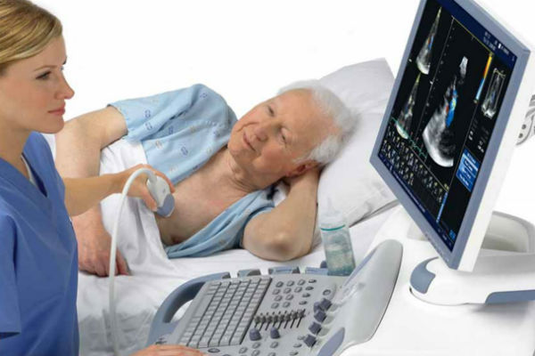
- Echocardiography. Obtaining an image of the work of the cardiovascular system due to the registered acoustic signal, which is transmitted to the screen during the ultrasound examination.
- Tomography. The method is based on the lumen of blood vessels for the subsequent determination of their biochemical composition and structure.
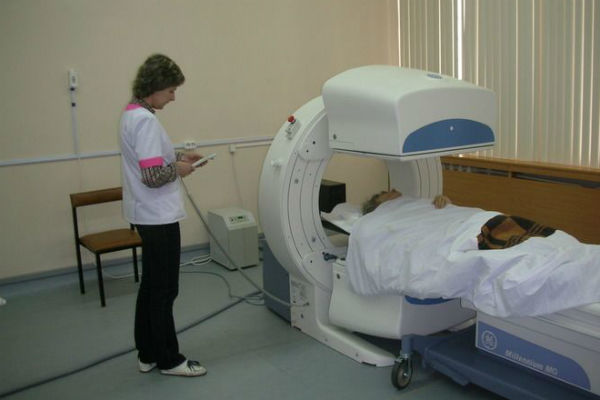
- Myocardial scintigraphy. Intravenous administration of a radioactive staining enzyme for organ imaging.
- Angiography. Radiography with the introduction of an intravenous injection or a pre-installed catheter with a contrast agent for vascular examination.
- 24-hour blood pressure monitoring. Obtaining the results of changes in blood pressure in 24 hours.
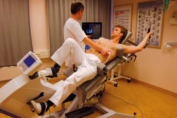
- Stress echocardiography. Assessment of the state of cardiac work in critical conditions created artificially using pharmacological and physical influences.
The results obtained are processed in the laboratory or announced by the cardiologist at a subsequent appointment. Further treatment is prescribed depending on the age, sex of the patient, stage of the disease and its form.
People are forced to see a specialist by pain and tingling sensations in the chest area, an increase or decrease in blood pressure.
Types of diagnostics of cardiovascular diseases
- Blood test from a vein
- ECG
- Echocardiography
- Halter
- Ultrasound
- SMAD
- Coronary angiography
- Duplex vessels
- Checking blood vessels for blood clots
What you need to know before your first visit to your cardiologist:
- medical history;
- the presence of pathologies from the cardiovascular system in close relatives;
- first manifestations;
- conditions for the onset of symptoms.
 At the initial examination, the cardiologist will definitely listen to all complaints and measure blood pressure and pulse, listen to the heartbeat with a stethoscope. Based on the results obtained, the doctor will prescribe a complete cardiac examination.
Tapping or percussion is widely used at the initial stage of examination of diseases and pathological processes in the work of the heart. The essence of percussion is quite simple.
By means of physical impact on the outer surface of the chest, the doctor provokes the appearance of various sounds, by which he determines the natural location of the muscular organ. The beating of the heart is accompanied by a dull sound.
The percussion procedure of the heart determines the boundaries of its dullness:
- Relative. The sound is muffled, dull. The edges of the organ, covered by the lungs.
- Absolute. Central open area of the anterior surface of the heart. Dull clear tone.
Allows you to determine the procedure for tapping the chest and quantitative values corresponding to the norm of the border of the heart:
- Right. The dimensions are the result of moving to the left along the 4th intercostal area on the right. Corresponds to the right ventricle.
-
Left. The left ventricle is outlined when exposed to the left 5th intercostal space towards the chest. Additionally, pressure is applied
1.5-2 cm. - Upper. Movements are carried out from top to bottom strictly along the intercostal spaces on the left. The end point is the third section between the ribs and defines the left atrium.
The displacement of the heart to the right side indicates an increase in the myocardium, hypertrophy, and pathology of the right ventricle. Removal to the left indicates arterial hypertension and a change in the work of the left ventricle. If there is a uniform increase in the boundaries, the patient is sent for additional examination. Possible causes: pericarditis, liver cirrhosis, pneumonia.
Auscultation of the heart is a traditional diagnosis of heart disease by indirect listening to the heart with a special device - a stethoscope. It is used during the initial examination prior to hardware diagnostics and laboratory tests.
The heart auscultation procedure involves listening to the tone during myocardial contraction using the appropriate points on the body:
- top of the heart;
- between the second and third ribs on the right and left;
- fifth rib on the right;
- between the third and fourth ribs on the left.
In a healthy person, the doctor determines only two tones, quickly replacing each other, the alternation of a pause and the appearance of a sound takes the same time intervals. A weak first tone indicates myocardial damage, heart failure, an increase in organ weight, or dysfunction of its valves.
Strengthening of the initial sound can be caused by stenosis of the right ventricle, tachycardia. Too quiet a second tone is provoked by aortic insufficiency and low blood pressure. Loud and clear sound - acceleration of valves, hypertension.
Disorders of the cardiovascular system are observed not only in adults. Pathological changes are increasingly affecting young patients.
What heart diseases are observed in children:
- congenital defect;
- rheumatism;
- arrhythmia;
- violations of the heart muscle;
- heart failure;
- myocarditis.
They require examination and consultation with a cardiologist, followed by the appointment of diagnostics and treatment, if necessary, the child is placed in a hospital.
Diagnosis of diseases of the heart and blood vessels: modern methods and self-diagnosis
Diagnosis of diseases of the heart and blood vessels is very important, since diseases of the heart and blood vessels occupy a leading position in the list of the most common diseases in the population.
In addition, according to statistical indicators, cardiovascular diseases rank first in terms of mortality. Fortunately, the level of modern medicine makes it possible to quickly and accurately undergo diagnostics and, thereby, prevent the development of the disease at an early stage.
 Instrumental methods for examining a patient are:
- electrophysiological, electrocardiographic, and X-ray studies;
- Magnetic resonance imaging;
- echocardiography;
- positron emission tomography;
- cardiac catheterization.
Electrocardiography is a method of detecting electrical impulses of the heart using an electrocardiograph. The impulses are marked on a special moving belt.
At the end of the electrocardiography, the doctor receives an electrocardiogram (ECG), having studied which, he can determine the pacemaker of the heart (sinus node or bundle of His), assess the general condition of the conducting system, heart rate, and also identify some heart diseases (for example, heart attack myocardium).
Diagnosis of diseases of the heart and blood vessels by electrocardiography is completely safe for the patient and proceeds as follows. Small electrodes are placed on the chest and limbs of the examinee, which determine the intensity and direction of the heart currents. As a result, curves with teeth, segments and intervals are displayed on the ECG, which can be used to judge the work of the heart.
What diseases look like on an ECG
- Heart attack
- Sinus rhythm
- ECG decoding
Exercise can be an indirect indicator of coronary artery disease or coronary artery disease (for example, people with angina pectoris do not tolerate physical activity). According to the results of stress tests, one can speak of diseases that do not manifest themselves at rest.
With physical exertion, the heart may not have enough oxygen due to narrowed coronary arteries, which normally deliver oxygen-rich blood to the organ. If the lungs are examined in a timely manner, then we can say whether the lack of oxygen is the lot of the disease with from the respiratory system, or from the cardiovascular system, or a mixed disease character.
During the stress test, the subject is engaged in certain physical exercises, and the doctor continuously monitors the patient and records the necessary indicators. The patient moves along a special path, the pace of which is gradually increased, or turns the bicycle pedals.
Typically, the speed is increased until the heart rate reaches
However, if the subject develops severe shortness of breath or sharp pains in the chest area, as well as sudden surges in pressure, the stress test is stopped earlier than the set time.
Computed tomography (CT) can reveal anatomical changes in the chest cavity. With the help of a computer, so-called X-ray "slices" of the chest are made, which allow detecting pathology of any nature.
At present, an improved method has been developed, which is called film-computer tomography. With its help, you can see the image of the heart in three-dimensional space and evaluate not only anatomical changes, but also violations of the contractile function of the heart.
Tracking Human Pressure Data
- Which hand to measure
- How to measure without a tonometer
- How to measure with a tonometer
- Normal human pressure
- Which blood pressure monitor is better
- Tonometer
- Heart rate
Diagnosis of ischemic heart disease: features and results
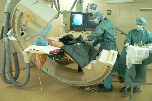 Diagnosis of coronary artery disease is often associated with a risk for the patient, the more difficult the heart examination procedure, the greater the risk. When a catheter is inserted into the heart to conduct a coronary vascular study, there is a high probability of adverse consequences in the form of a heart attack, heart attack, and stroke. The death rate with angiography is 1: 1000.
In stress tests, the probability of heart attack or death is 1 in 5000.
IN radionuclide examination the patient can receive a small dose of radioactive radiation, but not more than X-ray radiation.
In eelectrophysiological examination rhythm disturbance, the supply of nerve impulses to the heart are evaluated. The examination takes place through the introduction of microelectrodes into the chambers of the heart, arteries and veins. By the electrical signals coming from the electrodes, you can find out the location of the nerve fibers along which the impulses travel.
Sometimes during the study, the doctor may deliberately cause arrhythmia in the patient - this is necessary in order to to determine the ability of the medication to stop seizures and to ensure that it is appropriate operations. Bring the heart rate back to normal with a discharge. Despite the introduction of various instruments, this research method is safe, the death rate is 1: 5000.
In case of heart diseases, x-ray of the chest cavity is done without fail from the side, in front.
The picture allows you to study the vascular system in the lungs, assess the size of the heart, its chambers and the general structure of the organ. With the help of a snapshot, pathological conditions of the heart can be detected - a change in shape, an increased content of calcium in the vessels. X-ray examination can also detect abnormalities in the structure of the lungs and blood vessels, as well as detect fluid in the tissues of the lungs.
The size of the heart in case of impaired cardiac function, or changes in the valves may change. But there are diseases in which the size of the heart remains the same, this can be, for example, with constructive pericarditis. The heart is enveloped in an additional layer, but the dimensions remain normal.
X-ray examination characterized by the creation of a small X-ray "film" of several images, where the organs of the chest cavity and blood vessels are captured in dynamics.
This research method is used to clarify diagnoses, when identifying heart defects, acts as an auxiliary method in electrophysiological research and in vascular catheterization. Since the dose of radiation during the examination is high, it is replaced by other safer research methods (ECHO and others).
Electrocardiography - This is the most common examination method for heart disease. The main advantage of the method is the absence of radiation, as well as the ability to give a clear image of the state of the heart and blood vessels.
In diagnostics, high-frequency ultrasonic waves are used, which transmit an image through a special sensor to the screen of the device. On the screen, you can see the heartbeat and vascular circulation. During the examination, the doctor can move the sensor along the patient's chest, change the angle of inclination, which allows you to see the complete picture of the patient's cardiac activity.
The image is recorded on a cassette in order to obtain a clearer and higher quality image, as well as see small structural components, insert a special sensor into the patient's esophagus and transmit a picture through it to screen.
The examination procedure does not harm the patient, it is available for all age categories.
Magnetic tomography (MRI) - a method in which the energy of a magnetic field is used to examine the organs of the chest.
 For this, the patient is placed in a large electromagnetic box, where the atoms of the whole organism are caused to vibrate.
The atoms, in turn, transmit signals to a recording device, in which an image of all cardiac structures is formed.
This method usually does not require the introduction of additional contrast agents, but if the heart disease is associated with violation of the activity of blood vessels, then a substance with a paramagnetic property is injected to determine the place of weak circulation vessels.
MRI is the most difficult of all diagnostic methods, which is in the process of development, its disadvantage is the high cost of the examination and the difficulty in making the correct diagnosis.
At radionuclide examination radioactive substances are injected into the arteries, which are contrast agents to identify painful areas in the heart. Radioactive tracers are distributed throughout the body at a high speed; a specialized gamma camera records the radiation of substances.
The camera records the image and saves it to disk for later study. Despite the fact that during the examination the body receives a small dose of radiation, it is safe, unlike X-ray radiation.
Positron emission tomography. This method has common features with radionuclide examination, the difference is that during PET one substance is injected with a label of a radioactive agent, which moves only in the region of the heart and its structures. Sensors on the device register areas with increased agent activity, a high-quality 3D image appears on the computer screen. The selected area shows the active areas of the heart that absorb radioactive material.
PET is an expensive test method, therefore, it is used to clarify the diagnosis or when reliable data are not obtained in other diagnostic methods.
Catheterization. Such a diagnosis of coronary heart disease is carried out as follows. The catheter is inserted into the veins or arteries, and gradually moves along with the blood flow to the main areas of the heart. If it is necessary to examine the right atrium and its chambers, a catheter is inserted into a vein. To examine the left atrium and chambers, a catheter is placed in the artery.
Catheterization is a versatile means of diagnosing and treating the heart, and sometimes microscopic pressure measuring devices are installed in the catheters.
Catheters are divided into types, depending on their purpose:
- pressure measurement;
- vascular examination;
- expansion of valves;
- elimination of vascular occlusion.
The advantage of using catheters is that they allow you to avoid surgery during treatment. Catheterization takes place in a hospital using pain relievers.
Angiographic examination involves the study of the activity and structure of coronary vessels by catheterization and X-ray imaging. The catheter is inserted into a vein in the arm or groin area and advanced into the heart area, the movement of the catheter is monitored by a doctor using an X-ray.
To obtain an image of the vessels, a contrast agent is injected into the catheter and, thus, all deficiencies in the activity or structure of the coronary arteries can be seen on the screen.
This method allows detecting coronary artery disease and promptly initiating treatment.
Partial use of site materials (no more than 30% of the content of the article) is allowed only with a hyperlink on www.med88.ru, full use of the article (more than 30% of the article content) is possible only with written permission edition.