What the MRI of the abdominal organs shows: how the procedure is performed
Modern medicine offers several studies that allow visualizing the abdominal organs and diagnosing their functioning and possible disorders. One of these methods is MRI, which stands for magnetic resonance imaging. With the help of a survey, you can get data on the presence of neoplasms, organ displacement and other disorders. Magnetic resonance imaging of the abdominal cavity is performed only for clear indications.

MRI is performed only according to the testimony of a doctor
Content
- 1 General description of abdominal MRI
- 2 Types of MRI for examining the abdominal region
- 3 Indications for the study
- 4 Contraindications to the study
- 5 How to properly prepare for the procedure
- 6 How is an MRI of the abdomen performed?
-
7 Abdominal MRI Results
- 7.1 What does an MRI of the liver show?
- 7.2 What does the MRI of the gallbladder and ducts show?
- 7.3 Pancreatic MRI results
- 7.4 What does MRI of the spleen show?
- 7.5 MRI results of the kidneys and adrenal glands
- 8 Safety of magnetic resonance therapy
-
9 Average cost of MRI in Russian cities
- 9.1 Video - What does the MRI of the abdominal organs show
General description of abdominal MRI
Magnetic resonance imaging is a highly informative, as safe as possible in the absence of contraindications and non-invasive method of examining internal organs, including the abdominal cavity. The technique is used to control and confirm diseases. MRI allows avoiding the use of painful and complex diagnostic procedures, which have many complications and consequences for the patient.

MRI is a more comfortable diagnostic method than all others, therefore it is used even for children
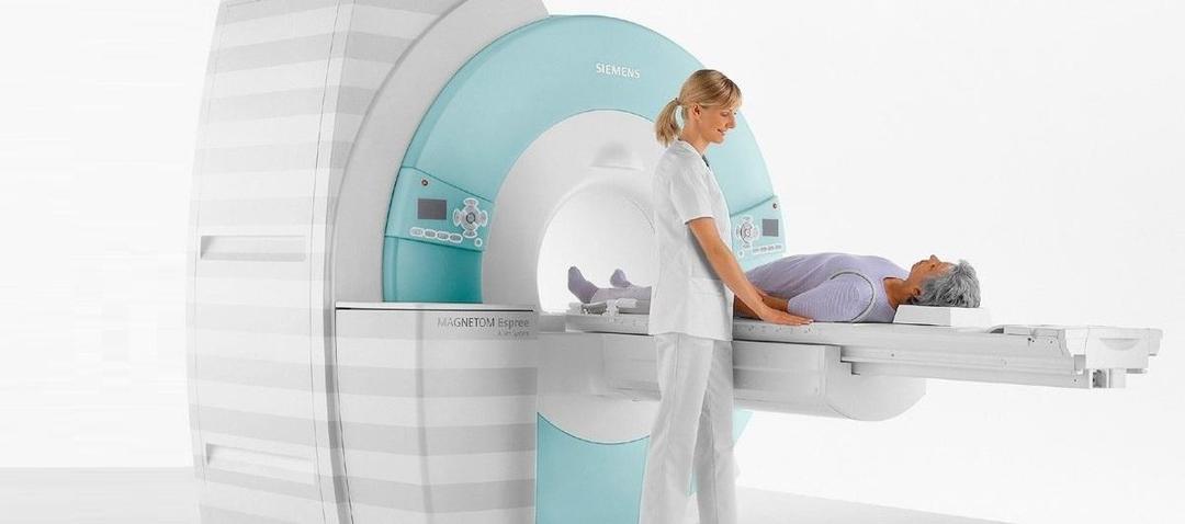
During therapy, the patient is placed inside an apparatus that looks like a large tube. The tomograph creates a moderate magnetic field, which begins to affect the hydrogen atoms in the patient's blood and organs. Due to this effect, a radio wave appears, which is captured by the device and projected onto a computer monitor in the form of images of a person's insides. As soon as the magnetic field is turned off, the cells return to their previous position. As a result of the examination, the specialist receives an image of the cavity in three projections at the highest possible resolution, which minimizes the error in the diagnosis.

How MRI works
Types of MRI for examining the abdominal region
Today, taking into account the reason for the need for an examination, doctors can offer patients several types of magnetic resonance imaging:
- general diagnostics of the peritoneum with or without contrast agent;
- magnetic resonance imaging of blood vessels;
- magnetic resonance imaging of venous and arterial communication.
Plain MRI helps to assess the condition of all internal organs located in the peritoneum
The first type of examination involves monitoring the state of functioning of the liver, spleen, stomach, intestines, kidneys, adrenal glands, urinary system, pancreas, lymphatic and circulatory systems, biliary bubble.
Attention! If the patient independently applies for an examination, he should choose an overview diagnosis. It is more informative and will allow you to coordinate further treatment.
Indications for the study
Magnetic resonance imaging is one of the most expensive procedures, therefore it cannot be used as a screening examination. Prescribing an MRI is required in the presence of questionable results of a previous diagnosis, as well as in severe conditions of the patient that require a deeper study. Often, diagnostics are prescribed to monitor the course of oncological processes, as well as the effectiveness of the treatment.

Colon cancer detected by MRI
MRI can be prescribed in the presence of the following cases:
- inaccurate results of ultrasound, computed tomography, X-ray and other manipulations;
- injuries of the peritoneal organs, requiring more detailed consideration to exclude complications of the patient's condition;
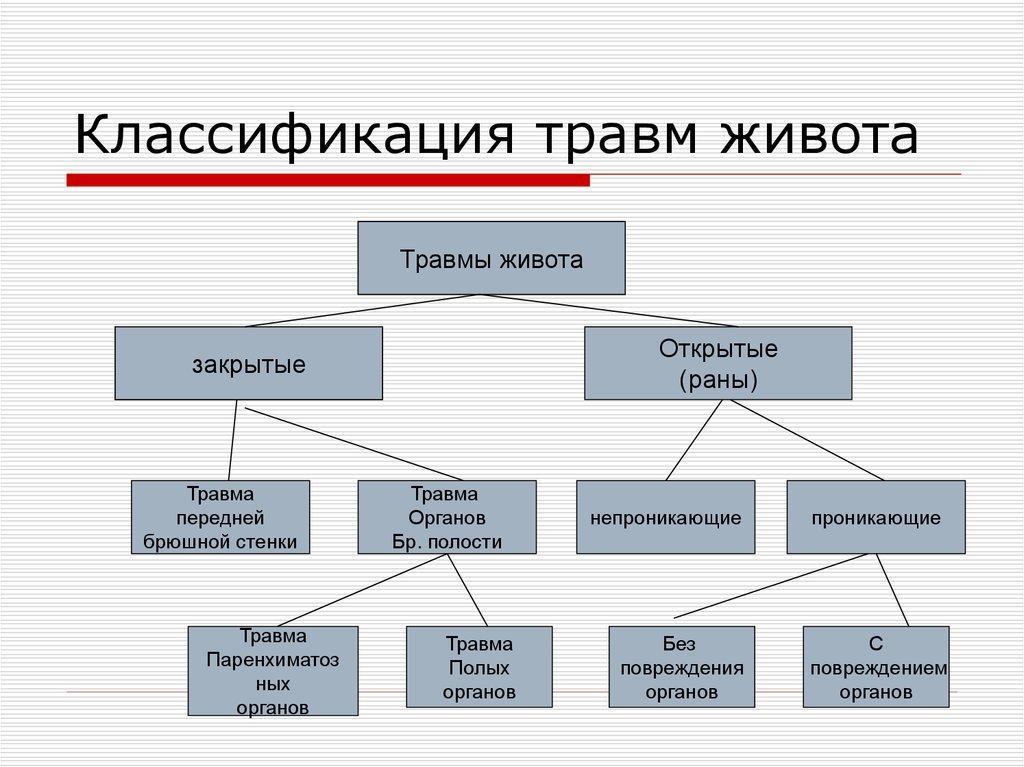
Classification of abdominal injuries
- the presence or suspicion of internal bleeding;
- ischemic disorders in the structure of the tissues of the abdominal organs;
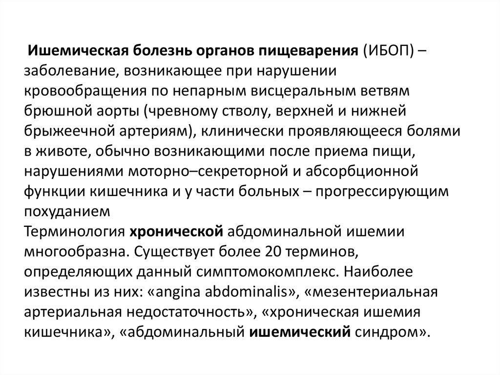
Ischemia of the digestive system
- inflammation in the area under study or fluid accumulation in organ cavities;
- visible or palpable enlargement of the spleen and liver;

MRI is used if the reason for the enlargement of the liver and spleen is not clear
- the appearance of symptoms of obstructive jaundice or other pathologies in the flow of bile;
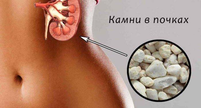
- pancreatitis of any type, the appearance of calculi;
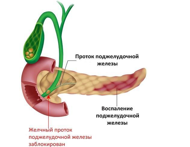
With pancreatitis, an MRI is mandatory
- suspicion of the development of oncological neoplasms or metastases;
- the appearance of cystic cavities, adenomas and other benign neoplasms;

How does a liver adenoma look in the picture?
- the presence of possible complications after a surgical intervention or an assessment of the success of the provided surgical aid;
- the impossibility of carrying out other diagnostic manipulations due to their intolerance or contraindications.
Attention! Permanent contrasts in magnetic resonance imaging are used to detect abnormalities in the circulatory system, as well as to confirm ischemic processes in tissues. With the help of contrast, the formed fractions and tumors can also be detected. MRI uses a contrast based on gadolinium metal.
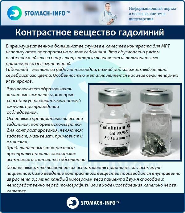
Gadolinium
Contraindications to the study
The procedure has absolute and conditional contraindications. The absolute are the following:
- the patient has implants with electronics and ferromagnetic elements;
- the patient has a mechanical heart valve implanted;

The presence of a valve in the heart is a direct contraindication to MRI
- even small tattoos, the pigment of which includes metal;
- the first 12 weeks of pregnancy;
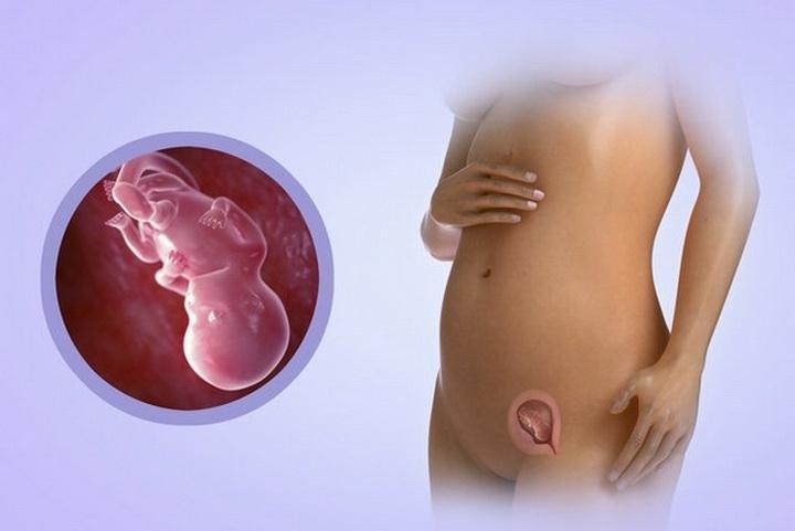
In the 1st trimester of pregnancy, MRI is contraindicated
- obesity in a patient, usually from 120 kg, when it is not possible to place the subject in the apparatus.
Attention! When performing diagnostics of the abdominal cavity, the presence of titanium structures, braces made of modern metals, prostheses or veneers in a patient is not an obstacle to diagnosis.

Metal parts in the mouth, including braces, are not a contraindication to MRI if the abdominal cavity is being diagnosed
It is undesirable to resort to using MRI in the following cases:
- second and third trimesters of pregnancy, breastfeeding;
- the appointment of hemodialysis;

If the patient is concurrently undergoing hemodialysis treatment, it is better to exclude MRI
- severe renal failure;
- possible development of an allergic reaction to the components of the contrast.

If there is a reaction of the body to the contrast agent, for example, a rash or redness, then the MRI is either not performed or is performed without the use of contrast
The decision to conduct an MRI in the last four cases can only be made by a specialist when the patient's life is in real danger. During lactation, before the diagnosis, a sufficient amount of milk should be collected for two days, since after the manipulations it is forbidden to feed the child for 48 hours.

Breastfeeding mothers need to express enough milk before MRI
Conditional contraindications to the use of diagnostics include mental disorders in the patient, fear of a confined space, as well as pain that does not allow you to spend the necessary time in a motionless condition. In the presence of claustrophobia, the patient may find a clinic that uses an open device.

It is difficult for people with cerebral palsy to undergo an MRI because they cannot control their movements
How to properly prepare for the procedure
Preparation is essential for reliable results after MRI. It is not difficult and easy to do. Taking into account the specific problem of the patient, he may be advised additional preparatory measures. Otherwise, the algorithm described below should be followed.
Algorithm of preparation
- Avoid foods that can cause gas production 72 hours before the MRI. These include legumes, raw vegetables and fruits, fresh baked goods, soda and alcohol.

Products that cause gassing must be excluded
- In case of disorders in the liver, spleen and pancreas, carbohydrates are excluded 72 hours before diagnosis.
- In the presence of flatulence and constipation, mild carminative and laxative medicines are used, it is better to use "Espumisan" or "Sorbex".

For bloating, you can use "Espumisan"
- Before an MRI scan with a contrast agent, the doctor will prescribe an allergy test 24-48 hours in advance.
- If symptoms of hypermobility are present, consultation with an anesthesiologist may be recommended in order to conduct a session under general anesthesia.

If necessary, an MRI can be done under general anesthesia
- General urine and blood tests are required to exclude renal failure.
- Women before menopause undergo ultrasound to rule out pregnancy.
Women before an MRI undergo an ultrasound scan to rule out pregnancy
- You cannot eat 8 hours before the examination, drink 4 hours before the examination. On the day of therapy, carbonated water, coffee and tea are completely excluded.
- It is recommended to take an antispasmodic half an hour before the MRI; it is enough to use drugs such as No-Shpa or Papaverine. The dose is selected separately for each patient.

Before the procedure, you can drink an antispasmodic, for example, the drug "No-Shpa"
- A full bladder session should not be performed.
- All metal objects, watches, prostheses must be removed.
Attention! In case of increased anxiety, a decision may be made to stay close to the patient's relative. But to exclude complications from the accompanying person, he also needs to remove all metal and electronic objects, if possible.

A relative may be nearby during an MRI
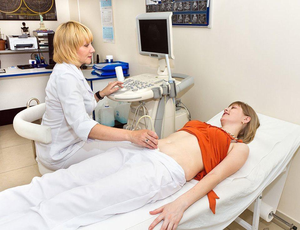
How is an MRI of the abdomen performed?
To carry out a successful procedure, you must adhere to a clear plan.
- First, the patient is changed into disposable clothing and a contrast agent is injected if necessary for an accurate examination.

Disposable MRI clothing
- Then the patient lies down on the tomograph table, puts on headphones or earplugs, this is necessary for sound insulation during the diagnosis. The sound of the tomograph is loud enough and can be uncomfortable and fearful. In some cases, the doctor can fix the patient's limbs.

To prevent the sound of the device from causing discomfort, use earplugs
- The examinee is pushed into the tube of the MRI machine. The specialist goes to his office and maintains communication with the patient through a microphone, if the situation requires it.

While the patient is in the apparatus, the doctor is in a special booth, where he captures the images.
- After turning on the tomograph, you cannot move, the examination itself takes from half an hour to an hour. Sometimes the doctor may ask the examinee to hold his breath for 3-10 seconds for a clearer picture.

Remember that an MRI scan can take up to an hour.
- At the end of the procedure, the patient is helped to get out of the tomograph and, if necessary, they are assisted in returning the normal tone of the stiff muscles by ordinary rubbing.
Attention! If the patient becomes ill during the procedure, the doctor's office has a panic button to call the emergency team.

With the help of a special panic button, the doctor can call for help if the patient suddenly becomes ill during the diagnosis
Once the session is over, it takes at least two hours to process the received data. Sometimes, for a better conclusion, the doctor engages cardiologists, neurologists and other narrow specialists. MRI results can be obtained on film or electronic media.

It may take a long time to decrypt the data
Abdominal MRI Results
Based on the results of scanning organs in this area, the following violations can be clearly identified:
- changes in their structure, shape and size;
- the presence of congenital or acquired abnormalities in the structure of tissues, including blood vessels;

MRI can detect organ abnormalities such as liver hyperplasia or hypoplasia
- the presence of inflammatory processes;
- the appearance of cysts, obstructions and degenerative processes on the tissues;

With the help of tomography, it is possible to identify cysts in the abdominal organs
- the formation of tumors of benign or malignant types;
- changes in nerve endings, the appearance of calculi and metastases;
Even small calculi can be seen on MRI scans
- problems with sufficient blood circulation to tissues and organs;
- the possible presence of aneurysms, blood clots, vascular ruptures and their deformation;
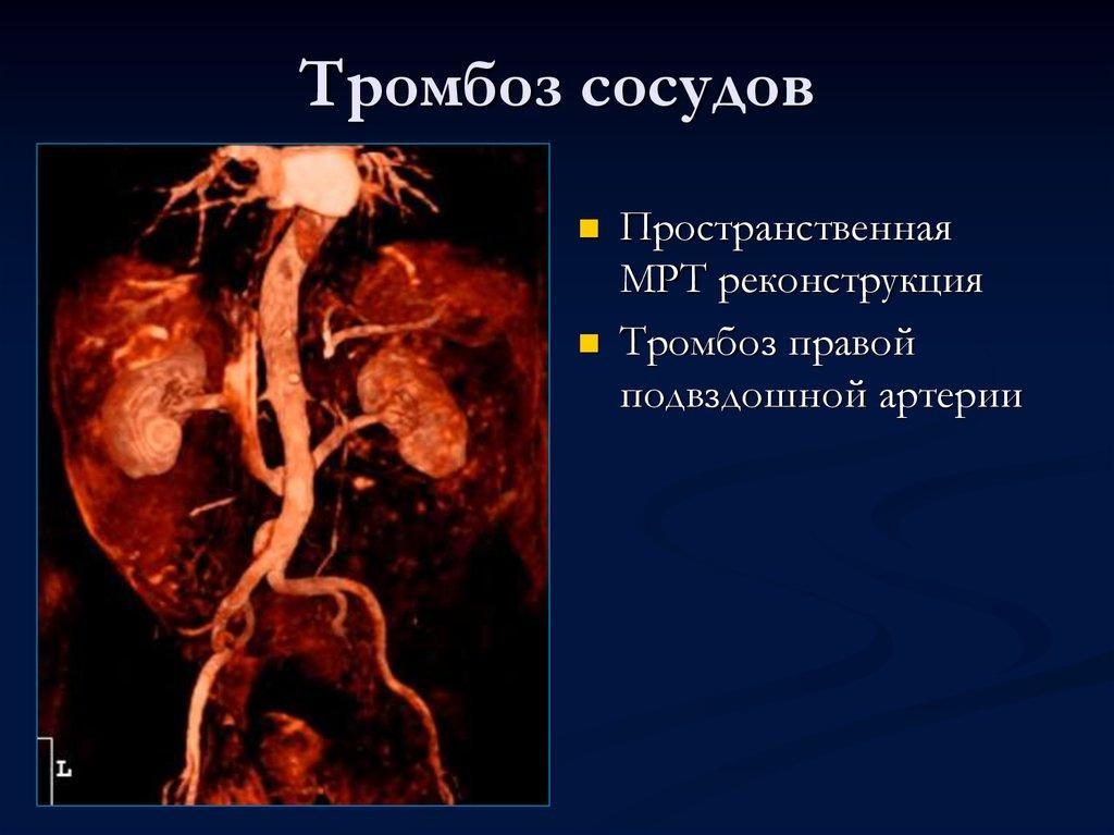
MRI performed with a contrast agent allows you to determine the pathology of blood vessels, arteries and veins
Attention! It is especially important not to give up MRI for patients suffering from cancer. The procedure allows you to control the growth of the tumor, its treatment and the possible appearance of metastases.
What does an MRI of the liver show?
When using MRI in the study of the liver, it is possible to identify such dangerous conditions as hemangiomas, organ adenomas, and hyperplasia of the frocal nodular type. With the help of diagnostics, hepatocellular carcinoma and other types of oncological neoplasms, including cysts and abscesses, are recorded in 100% of cases. Examination of the liver is carried out with a contrast method using the introduction of paramagnetic substances. With the help of such a contrast, it is also possible to identify metastases, the size of which exceeds one centimeter.

MRI of the liver
Attention! For better liver imaging three days before the MRI, a diet with a minimum amount of carbohydrates is required.
What does the MRI of the gallbladder and ducts show?
The gallbladder and organ ducts are best viewed without contrast. After receiving the results, you can be sure of the presence or absence of stones, sufficient permeability of the organ, trauma, cholangitis, choledocholithiasis, and oncological neoplasms. With the help of MRI, you can see the slightest changes in the structure of tissues and their congenital abnormalities. Such an examination of the gallbladder and bile ducts is the most gentle procedure, which, unlike endoscopic manipulations, does not give complications and does not require a recovery period.
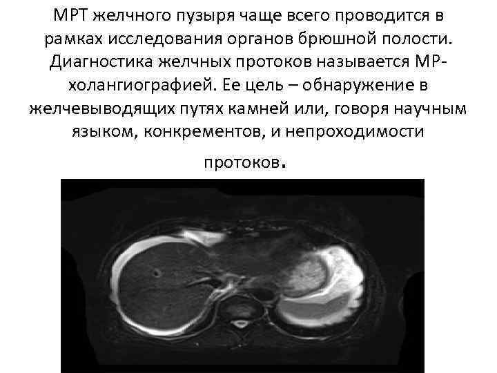
MRI of the gallbladder
Pancreatic MRI results
Such a diagnosis of the pancreas gives a clear idea whether the patient has malignant tumors, benign neoplasms and cystic structures. With the help of magnetic resonance therapy, it is possible to confirm acute and chronic pancreatitis, but this expensive service is rarely used to diagnose them. For good pictures, it takes three days to completely eliminate carbohydrates from the diet. MRI can be done with or without contrast.
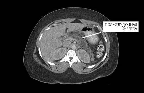
MRI scan of the pancreas
What does MRI of the spleen show?
When carrying out magnetic resonance imaging of the abdominal region, the spleen is necessarily examined, since it reacts to many pathologies of internal organs. The procedure allows you to identify the size of the organ, how much has changed its shape, tissue structure. On MRI, multiple or single cysts, tumors, abscesses, heart attack, malformations of the spleen are revealed. For the best result, it is required to give up carbohydrates two days before the diagnosis. MRI can be done with or without contrast.

Spleen infarction can be detected with MRI
MRI results of the kidneys and adrenal glands
With the help of such an examination of the abdominal cavity, it is highly likely that the doctor will be able to detect even small injuries in the kidneys and adrenal glands, the size and nature of malignant and benign neoplasms character. The results show abnormalities in the formation of the urinary system, disorders in the lymph nodes, including in the retroperitoneal space. Changes in tissue structures, including the space adjacent to them, also show themselves well in MRI scans. MRI can be done with or without contrast.

MRI of the kidneys
Attention! MRI of the abdominal region is a real opportunity for patients suffering from intolerance to iodine-containing contrasts to find out their diagnosis if there is a suspicion of kidney and adrenal gland pathology.
Safety of magnetic resonance therapy
WHO classifies MRI as an absolutely safe study, since during the procedure, a field with a force of no more than 1.5 T is used. During the session, the patient does not feel pain or discomfort if he has no contraindications. MRI does not involve the use of X-rays, so the patient does not receive even the minimum dose of radiation.

How MRI works
The gadolinium metal is also well tolerated and only in exceptional cases causes unwanted allergic reactions.
Average cost of MRI in Russian cities
Table. Estimated cost of MRI for 2018
| Town | Price in rubles |
|---|---|
| Moscow | 7000-9000 |
| Nizhny Novgorod | 3500-4000 |
| Ryazan | 3100-4000 |
| St. Petersburg | 7000-9000 |
| Novosibirsk | 3800-4500 |
| Tver | 3300-3500 |
| Volgograd | 4000-7000 |
* the price for the examination is indicative, information is of an introductory nature
MRI is one of the most informative procedures for visualizing the abdominal organs in three planes. In most cases, this expensive diagnosis is used only when there is doubt and difficulty in making an accurate diagnosis. To get reliable results, you need to go through the preparatory stage as accurately as possible and behave correctly during the procedure. As a result, the doctor will be able to prescribe the correct treatment and prevent disability and even death of the patient due to the lack of a diagnosis.