Ultrasound of the retroperitoneal space: what is included, features of the procedure
Today, instrumental diagnostics of diseases reaches incredible heights. There are a variety of tools and methods for studying the human body, which can visualize any organ, nerve plexus or vessel to the smallest detail. However, most of the state-of-the-art techniques are present only in large cities, have low throughput and high research costs. Therefore, the most popular in wide practice for screening, initial studies are the time-tested methods available. One of them is ultrasound.

Ultrasound of the retroperitoneal space - what is included
With the help of ultrasound, you can examine almost all parts of the body, the localization of some of them is more understandable to the layman, and some less. So, for example, what constitutes an abdominal ultrasound is clear to everyone. But what is ultrasound of the retroperitoneal space: what is included, how is it performed and what diseases can be detected with it?

Ultrasonic technology
Content
- 1 Ultrasound procedure
- 2 Retroperitoneal anatomy
- 3 How is ultrasound performed?
- 4 Indications and contraindications
- 5 Preparation for research
- 6 Normal indicators
-
7 What pathology can you find?
- 7.1 Renal pathology
- 7.2 Adrenal glands
- 7.3 Bladder
- 7.4 Pancreas
- 7.5 Video - Ultrasound of the abdominal cavity and retroperitoneal space
Ultrasound procedure
The method is based on the physical properties of ultrasound. Each organ and tissue, each structure has its own acoustic resistance, i.e. reflects an ultrasonic wave with a certain strength. It is these reflected waves that are visible on the monitor of the functional diagnostics doctor when he brings the sensor to the patient's body.

Types of ultrasound diagnostics
There are certain norms for each organ. In the event that its density increases or decreases, pathological foci with increased or reduced density, the waves begin to be reflected in a different way, which can also be seen on the screen or on the printed picture. It is also possible to reveal the size of the organs. Waves change when they cross the media boundary. Therefore, knowing the boundary points (cavity - organ - cavity), you can visualize the object and determine how big it is.
During the study, the gel is used in order to minimize the free surface of the sensor, to prevent image distortion by air or other elements.

Ultrasound gels
The state of which organs can be examined using ultrasound:
- organs of the abdominal space;
- small pelvis;
- organs located in the retroperitoneal space;
- thyroid gland;
- a heart;
- vessels;
- joints;
- pleural cavity;
- the location and size of the lens of the eye;
- cerebral vessels in newborns through the opening of the large fontanelle.

Ultrasound
It is important to understand that ultrasound practically does not pass through the bones, it penetrates poorly through muscle tissue, so it is impossible, for example, an ultrasound of the spinal cord or the brain.
Retroperitoneal anatomy
Many are familiar with the term "abdominal cavity". This is the space, limited from above by the diaphragm, and from below, at the level of the ridges of the pelvic bones (the most protruding parts of them are felt on the right and left slightly above the inguinal folds), passing into the pelvis. There are two sections in the abdominal cavity - the peritoneal cavity and the retroperitoneal space.
Peritoneum - This is a special tissue that lines the inside of the walls of the abdomen with one sheet and covers the internal organs with another. The leaves pass into each other completely (in men) or not completely (in women, they provide communication between the abdominal cavity and the uterus through the fallopian tubes). In fact, the peritoneum is a large serous sac, consisting of two walls, one of which encloses the walls of the abdomen, and the other encloses the internal organs.

Abdominal structure
But not all of the insides are completely covered with this shell. Some of them are completely under her cover, some are partially. There is a group of organs that are not covered at all by the peritoneum. So, between the leaf wrapping the organs and the spine, there remains another part of the abdominal cavity, which is called retroperitoneal space. Simply because it is located behind the peritoneum.

Retroperitoneal space
This space includes:
- both kidneys;
- two adrenal glands (endocrine glands);
- two ureters;
- some parts of the duodenum;
- lateral sections of the large intestine;
- abdominal aorta;
- inferior vena cava with branches;
- a few more veins;
- some nerve plexuses;
- lymph nodes;
- partly the pancreas.
Most often, together with the kidneys, the bladder is examined, although in fact it is already in the pelvis.

Kidney ultrasound
There is a lot of fatty tissue around all of these organs, and this is the tissue that transmits ultrasound best. Therefore, ultrasound of the retroperitoneal space is quite indicative.
How is ultrasound performed?
In most cases, the diagnosis of the condition of the kidneys and adrenal glands is carried out together with the examination of the peritoneal cavity. The subject lies on his back, removes clothes from the abdomen. A special gel is applied to the ultrasound sensor, and then the study begins. First, a thorough examination of the liver, gallbladder, partially - the pancreas, spleen is carried out. And then the doctor proceeds to examine the retroperitoneal space directly. The patient is sometimes asked to roll over onto the side, on the stomach or in a half-turn, and also to stand up. This is done in order to better visualize organs and tissues.

How is ultrasound performed?
A Doppler study, or USDG, can be done. Its mechanism is a combination of the properties of ultrasound and the Doppler effect. It helps to determine the speed of blood flow in the vessels, to find blood clots in them, areas of blockage or narrowing, to determine the elasticity of the wall.
Most often, an isolated ultrasound of the kidneys is prescribed, it is most in demand in routine practice and is informative. But other organs can also be carefully examined. Usually, the procedure does not take more than 20 minutes, but it can be prolonged if there is a suspicion of a tumor in the retroperitoneal space, an increase in lymph nodes.
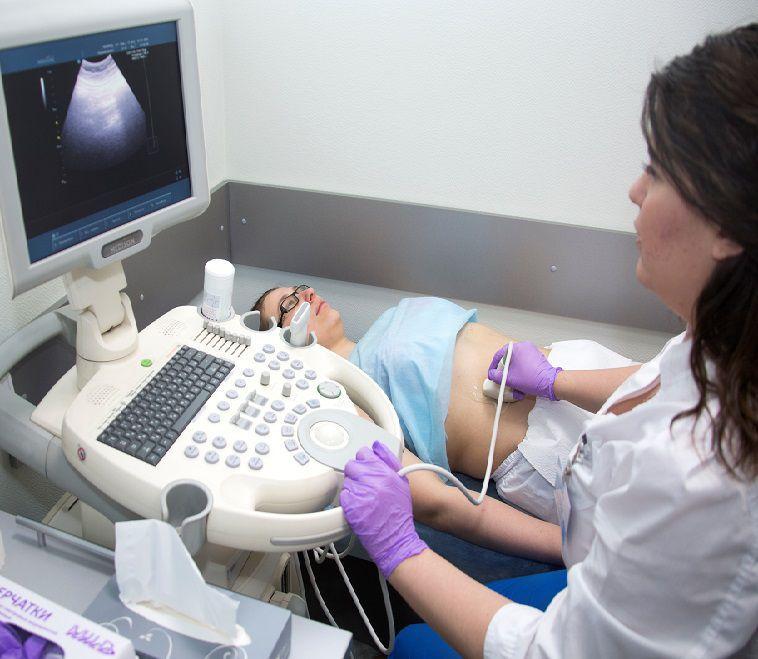
Abdominal ultrasound
Indications and contraindications
Table. For what symptoms is an ultrasound of the kidneys and retroperitoneal space prescribed?
| Organ | Symptoms |
|---|---|
 Suspected kidney disease |
Pain in the lumbar region or sides of a aching, pulling character, especially combined with a rise in temperature; · The appearance of blood in the urine; · Deviations in urine tests; • increased frequency or, on the contrary, decreased urination; · Nausea and vomiting, not associated with the pathology of the gastrointestinal tract or brain; · Rise in blood pressure not associated with essential hypertension. |
 Suspected bladder pathology |
· Increased urination; · Difficulty urinating; Soreness, cramps; · Blood in the urine. |
 Suspected adrenal pathology |
• unmotivated severe weakness; · migraine; · Convulsions; · Menstrual irregularities; · Changes in blood pressure without hypertension; · Unreasonable, insatiable thirst. |
 Suspicion of an oncological process in the retroperitoneal space |
Pain in the depths of the abdomen; · Formations that can be felt in the abdomen; · Sharp weight loss; • weakness, quickly approaching fatigue; • disorders of lymph metabolism in the lower extremities (edema, heaviness in the legs, pain, varicose veins, cyanosis); • disorders of urination and defecation (due to compression of organs by the tumor); · Unmotivated increase in body temperature. |
 Suspected pancreatic pathology |
· Pain of a girdle nature in the upper third of the abdomen, often radiating to the chest or lower back; · nausea, vomiting; • stool disorders ("fat", discolored, unformed); Belching; · loss of appetite. |
 Suspected vascular pathology |
· Venous congestion throughout the body (edema, shortness of breath, cyanosis); · Pain, swelling of the thigh and the entire lower limb; · Expansion of the saphenous veins; · Trophic ulcers on the legs. |

Stomach ache
There are no absolute contraindications to the study, i.e. no disease or condition can be an obstacle to an ultrasound scan for life or in urgent need. However, there are a number of situations in which it is better to wait with the research, if it is not urgent:
- pustular lesions of the skin of the abdomen;
- damage to the integrity of the skin;
- lack of preparation for research.

Purulent acne on the abdomen
Preparation for research
In principle, no special specific training is required. However, you need to adhere to some rules for the diagnosis to be more correct and informative:
- the last time you can eat 9 hours before the study;
- a couple of days before the procedure, products that cause increased gas formation are excluded - cabbage, black bread, freshly baked pastries, beer, kvass, soda, grapes, fatty heavy foods, peas and legumes;
- on the eve of the study in the evening, you can take Espumisan.

Espumisan
When checking the bladder, there are usually two stages - a full bladder and an empty bladder. Therefore, before the study, it is necessary to drink up to 1 liter of water, and then, after stage 1, urinate. But doctors warn about this in advance.
Usually you are asked to take a diaper with you. It's also a good idea to have wipes with you to wipe off excess gel. If there are results of previous ultrasounds, it is better to take them with you.

Dry wipes
Normal indicators
The conclusions of the diagnostic doctors are not always clear to the subjects. Only the doctor who sent them to the study can fully interpret them. However, you can do some analysis yourself.
Table. Normal readings on ultrasound.
| Index | Norm |
|---|---|
| Kidney sizes | Length 100-120 mm, thickness 40-50 mm, width 50-60 mm |
| The thickness of the parenchyma (main substance) of the kidneys | Up to 23 mm |
| Kidney contours | Smooth, clear |
| Echogenicity of the kidneys | Normal or slightly elevated |
| Kidney position | The left is slightly higher than the right (usually they write "correct" or "normal") |
| Breathing mobility | 2-3 cm |
| Pelvis, cups | Usually not visible, only visible when the bladder is full |
| Additional education | No |
| Adrenal glands | May not be visible in obese people, have a homogeneous structure, triangular (right) or crescent (left) shape |
| Bladder | Symmetrical |
| Bladder volume | 250-500 ml for women, 350-700 ml for men |
| Content | Homogeneous |
| Wall thickness | 3-5 mm |
| Circuit | Clear, smooth |
| Additional formations in the cavity | No |
| Neck | Not changed |
| Residual urine volume | No more than 50 ml |
| Thyroid length | 12-20 cm |
| Head | 30-35 mm |
| Neck | 10-15 mm |
| Body | 30 mm |
| Tail | 30-35 mm |
| Duct width | No more than 3 mm |

Image from ultrasound of the kidneys
What pathology can you find?
Ultrasound is a very revealing and informative study. Of course, only one conclusion may not always be an unconditional confirmation of the diagnosis, but it has a rather significant weight.
Renal pathology
-
Developmental anomalies. Usually, additional kidneys, the absence of one kidney, and an irregular structure are isolated. The size and position of the kidney will be outside the normal range, and the structure may be disturbed. The type of deviation will necessarily be reflected in the conclusion.

Localization of the accessory kidney
-
Hydronephrosis. This is an expansion of the calyx-pelvic system in the kidney, a violation of the outflow of urine as a result of a violation of the patency of the urinary tract. On ultrasound, it looks like the appearance of dark large triangles on one edge and a large dark oval on the other. The conclusion will reflect changes in the calyx-pelvic system, an increase (sometimes very significant) in the size of the kidneys. Allocate 4 degrees of the disease, the man in the street can already see only 3 and 4.

Hydronephrosis
-
Urolithiasis disease. Kidney stones are a very common problem in both men and women. As a result of a violation of the acidity of urine, metabolic processes in the body, an excessive amount of salts begins to accumulate in the kidneys, eventually causing the formation of stones. In this case, extraneous formations in the kidney will be described, and they look like light spots in the depths of the organ. Often due to the presence of stones, the kidney grows in size.

Types of kidney stones
- Cysts and tumors. These are pathological formations in the structure of the kidneys, and it is they who will be brought to conclusion. Cysts appear black or black with gray inclusions, usually rounded shadows. Tumors, on the other hand, are very light, usually bright, well visualized. A benign tumor has a smooth contour and deforms the organ. Oncological neoplasm is usually bumpy, uneven, the kidney can be deformed.
- Chronic diseases. In the presence of various kinds of pathologies, the echogenicity (usually upward) of the kidney may change, its structure may be disturbed, and its size may increase or decrease.

Kidney disease
Adrenal glands
- Developmental defects. These can be accessory glands, as well as underdevelopment of the organ.
- Hyperplasia. This is a significant increase in the adrenal gland in size, an increase in its echogenicity, an irregular shape is possible.
- Glandular hemorrhage. This pathology may look like a cavity - a dark spot. At the very beginning of the process, it is homogeneous, and then it becomes heterogeneous.
- Tumors. As with the kidneys, they appear as light areas of various shapes and sizes.

Classification of adrenal tumors
Bladder
- Inflammatory process. Wall thickness may increase, volume may decrease, and urine homogeneity may be impaired.
- Tumors. Increase in wall thickness, violation of the shape of the organ, the appearance of pathological formations.
- Fistulas. A fistula is a connecting passage between organs or between an organ and a cavity, which should not normally be present. In women, it can be vesicovaginal fistulas, in men, vesicovaginal fistulas.

Bladder fistula
Pancreas
- Cysts and tumors. The changes are the same as in the kidney - the appearance of a pathological light formation.
- Pancreatitis. Inflammation of the gland is accompanied by an increase in its size and impaired echogenicity due to edema, the duct can be enlarged, the structure of the tissue is disrupted.
- Bile duct stones, adhesions, head swelling accompanied by the expansion of the duct of the gland. It not only increases in size, but also acquires other pathological properties. Its walls become uneven, stones or other pathological fluid can be visualized in the duct.

Pancreatitis
Thus, a large number of pathologies can be seen on ultrasound of the retroperitoneal space. However, you should not rush to interpret the results yourself - there are many subtleties. It is impossible to rely only on the conclusion of a diagnostician, it is necessary to take into account the clinical picture, the history of the disease, as well as the individual characteristics of the patient's body. How to take flaxseed oil for constipation, read on our website.